2020年 第6卷 第11期
《工程(英文)》 >> 2020年 第6卷 第11期 doi: 10.1016/j.eng.2020.08.001
3D 打印细胞容器样支架在多细胞组织工程中的应用研究
a State Key Laboratory of High Performance Ceramics and Superfine Microstructure, Shanghai Institute of Ceramics, Chinese Academy of Sciences, Shanghai 200050, China
b Center of Materials Science and Optoelectronics Engineering, University of Chinese Academy of Sciences, Beijing 100049, China
c Center for Translational Bone, Joint and Soft Tissue Research, University Hospital Carl Gustav Carus & Faculty of Medicine, Technische Universität Dresden, Dresden 01307, Germany
# These authors contributed equally to this work.
下一篇 上一篇
摘要
人工设计的非接触式多细胞共培养模型可以模仿人体组织的细胞内微环境,但其发展始终面临各种挑战。本研究利用3D打印技术,成功制得了一种含β-磷酸三钙/羟基磷灰石(β-TCP/HA)的细胞容器样支架,该支架设计了四种不同形状的微孔结构:三角形、正方形、平行四边形和长方形微孔结构。这些支架可以在非接触的方式下同时培养四种细胞。本研究构建了一种由人骨髓间质干细胞(HBMSC)、人脐静脉内皮细胞(HUVEC)、人脐静脉平滑肌细胞(HUVSMC)、人真皮成纤维细胞(HDF)组成的3D共培养模型,用以研究这些细胞在促进骨生成和血管生成过程中细胞的个体效应与协同效应。结果表明,相较于在3D细胞容器中仅培养一种细胞,共培养三种或四种细胞展现出了更高的细胞增殖率。HBMSC与HUVEC的细胞间相互作用的研究表明,含有四种独立空间结构的3D细胞容器可以通过放大共培养细胞的旁分泌效应促进细胞的骨生成和血管生成能力。此外,在3D细胞容器中建立多细胞非接触体系,特别是含有三种或四种细胞的共培养体系,相对于单细胞培养模式与两种细胞共培养模式来说,在促进细胞成骨分化和成血管分化方面展现出明显的优势。本研究为基于支架的多细胞非接触式共培养体系的发展提供了新的研究方向。
关键词
3D细胞容器 ; 非接触式多细胞共培养 ; 相互作用 ; 血管生成 ; 骨生成
图片
图1
图2

图3

图4
图5
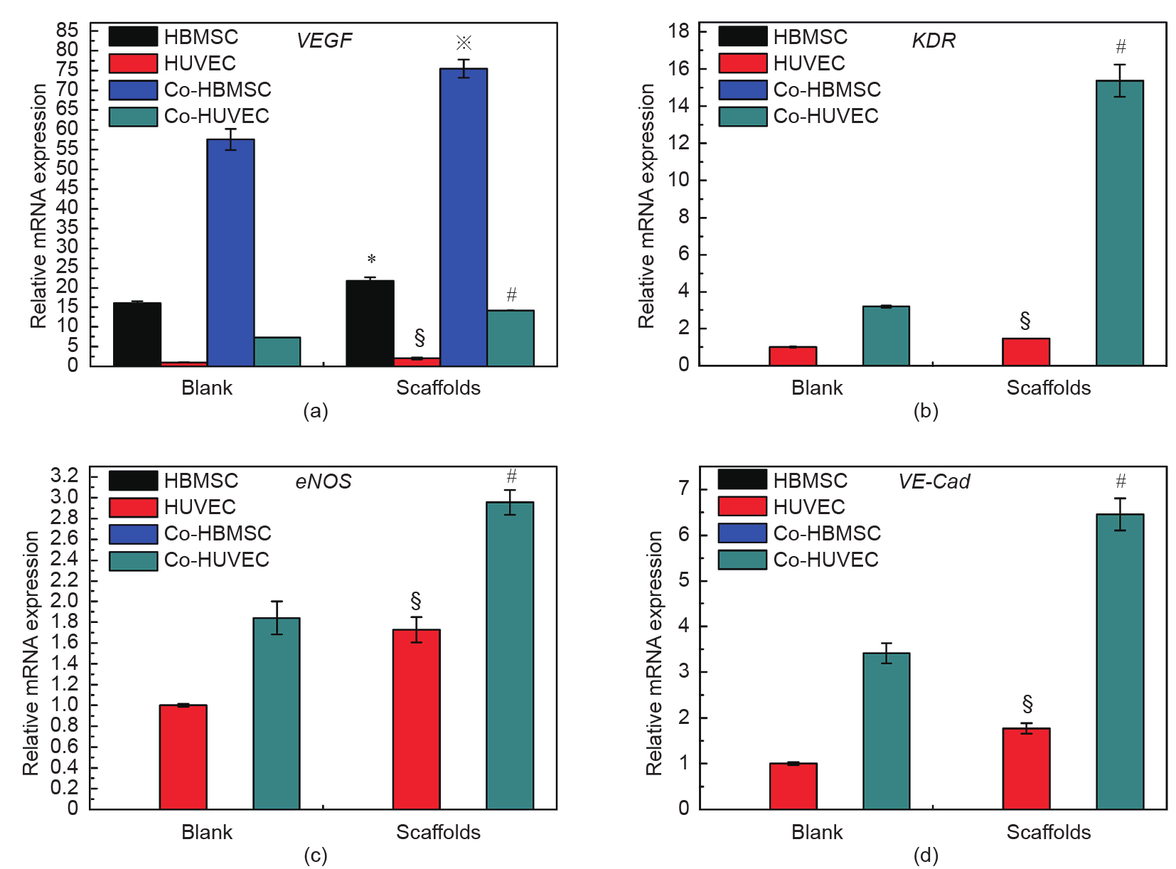
图6

图7

图8
参考文献
[ 1 ] Langer R, Vacanti J. Tissue engineering. Science 1993;260(5110):920–6. 链接1
[ 2 ] Hopkins AM, DeSimone E, Chwalek K, Kaplan DL. 3D in vitro modeling of the central nervous system. Prog Neurobiol 2015;125:1–25. 链接1
[ 3 ] Giger RJ, Hollis ER, Tuszynski MH. Guidance molecules in axon regeneration. Cold Spring Harbor Perspect Biol 2010;2(7):a001867.
[ 4 ] Annabi N, Tamayol A, Uquillas JA, Akbari M, Bertassoni LE, Cha C. 25th Anniversary Article: rational design and applications of hydrogels in regenerative medicine. Adv Mater 2014;26(1):85–124. 链接1
[ 5 ] Akbari M, Tamayol A, Bagherifard S, Serex L, Mostafalu P, Faramarzi N. Textile technologies and tissue engineering: a path toward organ weaving. Adv Healthc Mater 2016;5(7):751–66. 链接1
[ 6 ] Gu Q, Hao J, Lu Y, Wang L, Wallace GG, Zhou Q. Three-dimensional bioprinting. Sci China Life Sci 2015;58(5):411–9. 链接1
[ 7 ] Antoni D, Burckel H, Josset E, Noel G. Three-dimensional cell culture: a breakthrough in vivo. Int J Mol Sci 2015;16(3):5517–27. 链接1
[ 8 ] Park SB, Lee SY, Jung WH, Lee J, Jeong HG, Hong J. Development of in vitro three-dimensional co-culture system for metabolic syndrome therapeutic agents. Diabetes Obes Metab 2019;21(5):1146–57. 链接1
[ 9 ] Knight E, Przyborski S. Advances in 3D cell culture technologies enabling tissue-like structures to be created in vitro. J Anat 2015;227(6):746–56. 链接1
[10] Battiston KG, Cheung JWC, Jain D, Santerre JP. Biomaterials in co-culture systems: towards optimizing tissue integration and cell signaling within scaffolds. Biomaterials 2014;35(15):4465–76. 链接1
[11] Li H, Chang J. Bioactive silicate materials stimulate angiogenesis in fibroblast and endothelial cell co-culture system through paracrine effect. Acta Biomater 2013;9(6):6981–91. 链接1
[12] Li H, Xue Ke, Kong Ni, Liu K, Chang J. Silicate bioceramics enhanced vascularization and osteogenesis through stimulating interactions between endothelia cells and bone marrow stromal cells. Biomaterials 2014;35 (12):3803–18. 链接1
[13] Clarke MSF, Sundaresan A, Vanderburg CR, Banigan MG, Pellis NR. A threedimensional tissue culture model of bone formation utilizing rotational coculture of human adult osteoblasts and osteoclasts. Acta Biomater 2013;9 (8):7908–16. 链接1
[14] Kang Y, Kim S, Fahrenholtz M, Khademhosseini A, Yang Y. Osteogenic and angiogenic potentials of monocultured and co-cultured human-bone-marrowderived mesenchymal stem cells and human-umbilical-vein endothelial cells on three-dimensional porous beta-tricalcium phosphate scaffold. Acta Biomater 2013;9(1):4906–15. 链接1
[15] Kook YM, Jeong Y, Lee K, Koh WG. Design of biomimetic cellular scaffolds for co-culture system and their application. J Tissue Eng 2017;8. 204173141772464. 链接1
[16] Levorson EJ, Santoro M, Kurtis Kasper F, Mikos AG. Direct and indirect co-culture of chondrocytes and mesenchymal stem cells for the generation of polymer/ extracellular matrix hybrid constructs. Acta Biomater 2014;10(5):1824–35. 链接1
[17] Gao B, Konno T, Ishihara K. Quantitating distance-dependent, indirect cell–cell interactions with a multilayered phospholipid polymer hydrogel. Biomaterials 2014;35(7):2181–7. 链接1
[18] Feng C, Zhang W, Deng C, Li G, Chang J, Zhang Z, Jiang X, Wu C. 3D printing of lotus root-like biomimetic materials for cell delivery and tissue regeneration. Adv Sci 2017;4(12):1700401. 链接1
[19] Li H, Daculsi R, Grellier M, Bareille R, Bourget C, Amedee J. Role of neuralcadherin in early osteoblastic differentiation of human bone marrow stromal cells cocultured with human umbilical vein endothelial cells. Am J Physiol-Cell Physiol 2010;299(2):C422–30. 链接1
[20] Sorrell JM, Baber MA, Brinon L, Carrino DA, Seavolt M, Asselineau D, et al. Production of a monoclonal antibody, DF-5, that identifies cells at the epithelial-mesenchymal interface in normal human skin. APN/CD13 is an epithelial-mesenchymal marker in skin. Exp Dermatol 2003;12(3):315–23. 链接1
[21] Lai N, Jayaraman A, Lee K. Enhanced proliferation of human umbilical vein endothelial cells and differentiation of 3T3-L1 adipocytes in coculture. Tissue Eng Part A 2009;15(5):1053–61. 链接1
[22] Saiki A, Watanabe F, Murano T, Miyashita Y, Shirai K. Hepatocyte growth factor secreted by cultured adipocytes promotes tube formation of vascular endothelial cells in vitro. Int J Obes 2006;30(11):1676–84. 链接1
[23] Kang JH, Gimble JM, Kaplan DL. In vitro 3D model for human vascularized adipose tissue. Tissue Eng Part A 2009;15(8):2227–36. 链接1
[24] Borges J, Müller MC, Momeni A, Björn Stark G, Torio-Padron N. In vitro analysis of the interactions between preadipocytes and endothelial cells in a 3D fibrin matrix. Minim Invasiv Ther 2007;16(3):141–8. 链接1
[25] Choi JH, Bellas E, Gimble JM, Vunjak-Novakovic G, Kaplan DL. Lipolytic function of adipocyte/endothelial cocultures. Tissue Eng Part A 2011;17(9– 10):1437–44. 链接1
[26] Zhu W, Castro NJ, Cui H, Zhou X, Boualam B, McGrane R, et al. A 3D printed nano bone matrix for characterization of breast cancer cell and osteoblast interactions. Nanotechnology 2016;27(31):315103. 链接1
[27] Rao RB, Krafcik KL, Morales AM, Lewis JA. Microfabricated deposition nozzles for direct-write assembly of three-dimensional periodic structures. Adv Mater 2005;17(3):289–93. 链接1
[28] Therriault D, Shepherd RF, White SR, Lewis JA. Fugitive inks for direct-write assembly of three-dimensional microvascular networks. Adv Mater 2005;17 (4):395–9. 链接1
[29] Lee SH, Jeong HE, Park MC, Hur JY, Cho HS, Park SH. Fabrication of hollow polymeric microstructures for shear-protecting cell-containers. Adv Mater 2008;20(4):788–92. 链接1
[30] Truckenmüller R, Giselbrecht S, Escalante-Marun M, Groenendijk M, Papenburg B, Rivron N. Fabrication of cell container arrays with overlaid surface topographies. Biomed Microdevices 2012;14(1):95–107. 链接1
[31] Oh HH, Ko YG, Lu H, Kawazoe N, Chen G. Preparation of porous collagen scaffolds with micropatterned structures. Adv Mater 2012;24(31):4311–6. 链接1
[32] Hutmacher DW. Scaffolds in tissue engineering bone and cartilage. Biomaterials 2000;21(24):2529–43. 链接1
[33] Yun HS, Park JW, Kim SH, Kim YJ, Jang JH. Effect of the pore structure of bioactive glass balls on biocompatibility in vitro and in vivo. Acta Biomater 2011;7(6):2651–60. 链接1
[34] Cho SY, Park HH, Jin HJ. Controlling pore size of electrospun silk fibroin scaffold for tissue engineering. Polym-Korea 2012;36(5):651–5. 链接1
[35] Xu M, Zhai D, Chang J, Wu C. In vitro assessment of three-dimensionally plotted nagelschmidtite bioceramic scaffolds with varied macropore morphologies. Acta Biomater 2014;10(1):463–76. 链接1
[36] Martín-Saavedra F, Crespo L, Escudero-Duch C, Saldaña L, Gómez-Barrena E, Vilaboa N. Substrate microarchitecture shapes the paracrine crosstalk of stem cells with endothelial cells and osteoblasts. Sci Rep 2017;7(1):15182. 链接1
[37] Yu YY, Lieu S, Lu C, Colnot C. Bone morphogenetic protein 2 stimulates endochondral ossification by regulating periosteal cell fate during bone repair. Bone 2010;47(1):65–73. 链接1
[38] Zhai D, Xu M, Liu L, Chang J, Wu C. Silicate-based bioceramics regulating osteoblast differentiation through a BMP2 signalling pathway. J Mater Chem B 2017;5(35):7297–306. 链接1
[39] Dou DD, Zhou G, Liu HW, Zhang J, Liu ML, Xiao XF. Sequential releasing of VEGF and BMP-2 in hydroxyapatite collagen scaffolds for bone tissue engineering: design and characterization. Int J Biol Macromol 2019;123:622–8. 链接1
[40] Subbiah R, Du P, Hwang MP, Kim IG, Van SY, Noh YK. Dual growth factorloaded core-shell polymer microcapsules can promote osteogenesis and angiogenesis. Macromol Res 2014;22(12):1320–9. 链接1
[41] Kim M, Jung WK, Kim G. Bio-composites composed of a solid free-form fabricated polycaprolactone and alginate-releasing bone morphogenic protein and bone formation peptide for bone tissue regeneration. Bioprocess Biosyst Eng 2013;36(11):1725–34. 链接1
[42] Han Y, Li Y, Zeng Q, Li H, Peng J, Xu Y. Injectable bioactive akermanite/alginate composite hydrogels for in situ skin tissue engineering. J Mater Chem B 2017;5 (18):3315–26. 链接1
[43] Mitchell AC, Briquez PS, Hubbell JA, Cochran JR. Engineering growth factors for regenerative medicine applications. Acta Biomater 2016;30:1–12. 链接1
[44] Matsumoto T, Goto D, Sato S. Subtraction micro-computed tomography of angiogenesis and osteogenesis during bone repair using synchrotron radiation with a novel contrast agent. Lab Invest 2013;93(9):1054–63. 链接1
[45] Kanczler JM, Oreffo ROC. Osteogenesis and angiogenesis: the potential for engineering bone. Eur Cells Mater 2008;15:100–14. 链接1
[46] Mantella LE, Quan A, Verma S. Variability in vascular smooth muscle cell stretch-induced responses in 2D culture. Vasc cell 2015;7(1):7–16. 链接1
[47] Anwar MA, Shalhoub J, Lim CS, Gohel MS, Davies AH. The effect of pressureinduced mechanical stretch on vascular wall differential gene expression. J Vasc Res 2012;49(6):463–78. 链接1
[48] Lemarié CA, Tharaux PL, Lehoux S. Extracellular matrix alterations in hypertensive vascular remodeling. J Mol Cell Cardiol 2010;48(3):433–9. 链接1


 京公网安备 11010502051620号


