2022年 第13卷 第6期
《工程(英文)》 >> 2022年 第13卷 第6期 doi: 10.1016/j.eng.2021.01.010
明胶调控异种复合植骨材料的降解速率及成骨效应
a Department of Orthopedic Surgery, Tongji Hospital, Tongji Medical College, Huazhong University of Science and Technology, Wuhan 430030, China
b Department of Biomaterials, Institute of Clinical Dentistry, University of Oslo, Oslo 0317, Norway
c Industrie Biomediche Insubri SA, Mezzovico-Vira 6805, Switzerland
d Ludwig Boltzmann Institute for Experimental and Clinical Traumatology, Donaueschingenstrasse 13, 1200 Vienna, Austria
e Faculty of Biomedical Sciences, University of Southern Switzerland, Lugano 6900, Switzerland
f Faculty of Veterinary, University of Santiago de Compostela, Lugo 27002, Spain
下一篇 上一篇
摘要
由外伤、手术、先天性畸形和其他因素导致的骨缺损是当今最常见的健康问题之一。尽管自体骨移植和同种异体骨移植等策略是目前促进骨再生最成功的治疗方法,但仍存在移植物来源有限和并发症等局限性。Smartbone®是一种异种复合植骨材料(由牛骨基质、聚L-乳酸-己内酯和明胶制成),具有良好的临床骨再生效果。在这项研究中,我们研究了使用不同来源的明胶(牛和猪来源)制备的异种复合植骨材料(分别命名为SBN和SPK),并在体外和体内评估了其生物学效应。结果表明,来自牛和猪的明胶都可以成功且安全负载于Smartbone®上,并且能够承受苛刻制备过程。SBN和SPK在体外显示出不同的成骨细胞效应。SBN可促进人成骨细胞的骨钙蛋白分泌,而SPK可上调骨桥蛋白的表达。在体内实验中,两种植骨材料都促进了成骨,但SPK比SBN更早降解。我们的研究结果表明,SBN和SPK为优化骨修复植入物的吸收和再生平衡提供了不同的解决方案,这些异种复合植骨材料具有应用于骨缺损修复领域的巨大潜力。
补充材料
图片

图1

图2

图3
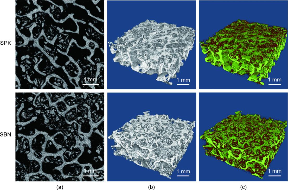
图4
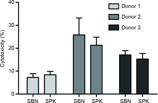
图5

图6
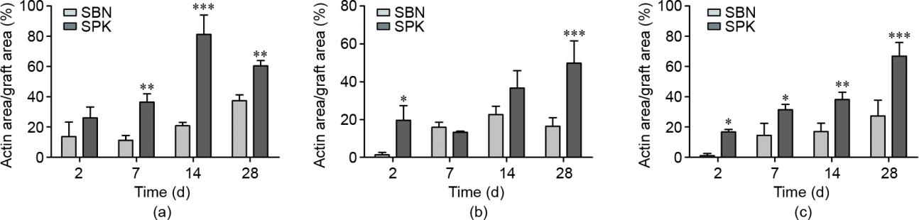
图7

图8
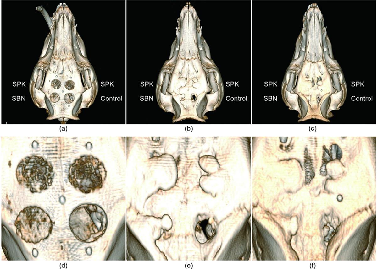
图9

图10

图11
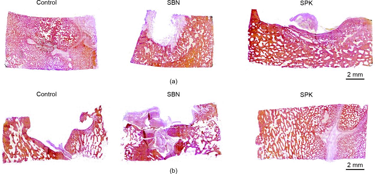
图12
参考文献
[ 1 ] Wang W, Yeung KWK. Bone grafts and biomaterials substitutes for bone defect repair: a review. Bioact Mater 2017;2(4):224‒47. 链接1
[ 2 ] Winkler T, Sass FA, Duda GN, Schmidt-Bleek K. A review of biomaterials in bone defect healing, remaining shortcomings and future opportunities for bone tissue engineering. Bone Joint Res 2018;7(3):232‒43. 链接1
[ 3 ] Kaur M, Singh K. Review on titanium and titanium based alloys as biomaterials for orthopaedic applications. Mater Sci Eng C 2019;102:844‒62. 链接1
[ 4 ] Pierannunzii L, Zagra L. Bone grafts, bone graft extenders, substitutes and enhancers for acetabular reconstruction in revision total hip arthroplasty. EFORT Open Rev 2016;1(12):431‒9. 链接1
[ 5 ] Fillingham Y, Jacobs J. Bone grafts and their substitutes. Bone Joint J 2016;98-B (1 Suppl A):6‒9. 链接1
[ 6 ] Sakkas A, Wilde F, Heufelder M, Winter K, Schramm A. Autogenous bone grafts in oral implantology—is it still a “gold standard”? A consecutive review of 279 patients with 456 clinical procedures. Int J Implant Dent 2017;3(1):23. 链接1
[ 7 ] Roseti L, Parisi V, Petretta M, Cavallo C, Desando G, Bartolotti I, et al. Scaffolds for bone tissue engineering: state of the art and new perspectives. Mater Sci Eng C 2017;78:1246‒62. 链接1
[ 8 ] Sanz M, Dahlin C, Apatzidou D, Artzi Z, Bozic D, Calciolari E, et al. Biomaterials and regenerative technologies used in bone regeneration in the craniomaxillofacial region: consensus report of group 2 of the 15th European Workshop on Periodontology on Bone Regeneration. J Clin Periodontol 2019;46:82‒91. 链接1
[ 9 ] Agarwal R, García AJ. Biomaterial strategies for engineering implants for enhanced osseointegration and bone repair. Adv Drug Deliv Rev 2015;94:53‒62. 链接1
[10] Skaggs DL, Samuelson MA, Hale JM, Kay RM, Tolo VT. Complications of posterior iliac crest bone grafting in spine surgery in children. Spine 2000;25(18):2400‒2. 链接1
[11] Colo E, Rijnen WHC, Schreurs BW. The biological approach in acetabular revision surgery: impaction bone grafting and a cemented cup. Hip Int 2015;25(4):361‒7. 链接1
[12] Issack PS, Nousiainen M, Beksac B, Helfet DL, Sculco TP, Buly RL. Acetabular component revision in total hip arthroplasty. Part II: management of major bone loss and pelvic discontinuity. Am J Orthop 2009;38(11):550‒6.
[13] Greenwald AS, Boden SD, Goldberg VM, Khan Y, Laurencin CT, Rosier RN; American Academy of Orthopaedic Surgeons, The Committee on Biological Implants. Bone-graft substitutes: facts, fictions, and applications. J Bone Joint Surg Am 2001;83-A(Suppl 2 Pt 2):98‒103. 链接1
[14] Sang T, Li S, Ting HK, Stevens MM, Becer CR, Jones JR. Hybrids of silica/poly (caprolactone coglycidoxypropyl trimethoxysilane) as biomaterials. Chem Mater 2018;30(11):3743‒51. 链接1
[15] Saigo L, Kumar V, Liu Y, Lim J, Teoh SH, Goh BT. A pilot study: clinical efficacy of novel polycaprolactone‒tricalcium phosphate membrane for guided bone regeneration in rabbit calvarial defect model. J Oral Max Surg Med 2018;30(3):212‒9. 链接1
[16] Nasajpour A, Ansari S, Rinoldi C, Rad AS, Aghaloo T, Shin SR, et al. A multifunctional polymeric periodontal membrane with osteogenic and antibacterial characteristics. Adv Funct Mater 2018;28(3):1703437. 链接1
[17] Huang B, Caetano G, Vyas C, Blaker JJ, Diver C, Bártolo P. Polymer-ceramic composite scaffolds: the effect of hydroxyapatite and β-tri-calcium phosphate. Materials 2018;11(1):129. 链接1
[18] Cristofaro F, Gigli M, Bloise N, Chen H, Bruni G, Munari A, et al. Influence of the nanofiber chemistry and orientation of biodegradable poly(butylene succinate)-based scaffolds on osteoblast differentiation for bone tissue regeneration. Nanoscale 2018;10(18):8689‒703. 链接1
[19] Domingos M, Gloria A, Coelho J, Bartolo P, Ciurana J. Three-dimensional printed bone scaffolds: the role of nano-/micro-hydroxyapatite particles on the adhesion and differentiation of human mesenchymal stem cells. Proc Inst Mech Eng H 2017;231(6):555‒64. 链接1
[20] Haugen HJ, Lyngstadaas SP, Rossi F, Perale G. Bone grafts: which is the ideal biomaterial? J Clin Periodontol 2019;46(Suppl 21):92‒102. 链接1
[21] Amini AR, Laurencin CT, Nukavarapu SP. Bone tissue engineering: recent advances and challenges. Crit Rev Biomed Eng 2012;40(5):363‒408. 链接1
[22] Athanasiou VT, Papachristou DJ, Panagopoulos A, Saridis A, Scopa CD, Megas P. Histological comparison of autograft, allograft-DBM, xenograft, and synthetic grafts in a trabecular bone defect: an experimental study in rabbits. Med Sci Monit 2010;16(1):BR24‒31.
[23] Meloni SM, Jovanovic SA, Pisano M, Xhanari E, De Riu G, Tullio A, et al. Sinus lift grafting with anorganic bovine bone vs 50% autologous bone mixed with 50% anorganic bovine bone: 2 years after loading results from a randomized controlled trial. Eur J Oral Implantol 2017;10(4):425‒32.
[24] Cheng L, Wang Yi, Sun G, Wen S, Deng L, Zhang H, et al. Hydration-enhanced lubricating electrospun nanofibrous membranes prevent tissue adhesion. Research 2020;2020:1‒12. 链接1
[25] Chen W, Tian X, He W, Li J, Feng Y, Pan G. Emerging functional materials based on chemically designed molecular recognition. BMC Materials 2020;2(1):1. 链接1
[26] Qian Y, Shen Y, Deng S, Liu T, Qi F, Lu Z, et al. Dual functional β-peptide polymer-modified resin beads for bacterial killing and endotoxin adsorption. BMC Materials 2019;1(1):5. 链接1
[27] D’Alessandro D, Perale G, Milazzo M, Moscato S, Stefanini C, Pertici G, et al. Bovine bone matrix/poly(L-lactic-co-ε-caprolactone)/gelatin hybrid scaffold (SmartBone®) for maxillary sinus augmentation: a histologic study on bone regeneration. Int J Pharm 2017;523(2):534‒44. 链接1
[28] Facciuto E, Grottoli CF, Mattarocci M, Illiano F, Compagno M, Ferracini R, et al. Three-dimensional craniofacial bone reconstruction with SmartBone® on demand. J Craniofac Surg 2019;30(3):739‒41. 链接1
[29] Grottoli CF, Cingolani A, Zambon F, Ferracini R, Villa T, Perale G. Simulated performance of a xenohybrid bone graft (SmartBone®) in the treatment of acetabular prosthetic reconstruction. J Funct Biomater 2019;10(4):53. 链接1
[30] Cingolani A, Grottoli CF, Esposito R, Villa T, Rossi F, Perale G. Improving bovine bone mechanical characteristics for the development of xenohybrid bone grafts. Curr Pharm Biotechnol 2019;19(12):1005‒13. 链接1
[31] van den Bosch E, Gielens C. Gelatin degradation at elevated temperature. Int J Biol Macromol 2003;32(3-5):129‒38. 链接1
[32] Esposito M, Grusovin MG, Papanikolaou N, Coulthard P, Worthington HV. Enamel matrix derivative (Emdogain®) for periodontal tissue regeneration in intrabony defects. Eur J Oral Implantol 2009;2(4):247‒66.
[33] Leijten J, Chai YC, Papantoniou I, Geris L, Schrooten J, Luyten FP. Cell based advanced therapeutic medicinal products for bone repair: keep it simple? Adv Drug Deliv Rev 2015;84:30‒44. 链接1
[34] Gallop PM, Lian JB, Hauschka PV. Carboxylated calcium-binding proteins and vitamin K. N Engl J Med 1980;302(26):1460‒6. 链接1
[35] McKee MD, Cole WG. Bone matrix and mineralization. In: Glorieux FH, Pettifor JM, Jüppner H, editors. Pediatric bone. San Diego: Academic Press; 2012. p.9‒37. 链接1
[36] Price PA, Otsuka AA, Poser JW, Kristaponis J, Raman N. Characterization of a ccarboxyglutamic acid-containing protein from bone. Proc Natl Acad Sci USA 1976;73(5):1447‒51. 链接1
[37] Eastell R, Hannon RA. Biochemical markers of bone turnover. In: Lobo RA, editor. Treatment of the postmenopausal woman. St. Louis: Academic Press; 2007. p. 337‒49. 链接1
[38] Hall BK, Part I. Vertebrate skeletal tissues. In: Hall BK, editor. Bones and cartilage. San Diego: Academic Press; 2015. p. 1. 链接1
[39] Maeda H, Wada N, Tomokiyo A, Monnouchi S, Akamine A. Prospective potency of TGF-a1 on maintenance and regeneration of periodontal tissue. In: Jeon KW, editor. International review of cell and molecular biology. New York: Academic Press; 2013. p. 283‒367. 链接1
[40] Butler WT. The nature and significance of osteopontin. Connect Tissue Res 1989;23(2‒3):123‒36.
[41] Sodek J, Ganss B, McKee MD. Osteopontin. Crit Rev Oral Biol Med 2000;11(3):279‒303. 链接1
[42] Chabas D. L’ostéopontine, une molécule aux multiples facettes. Med Sci 2005;21(10):832‒8. French. 链接1
[43] Giachelli CM, Steitz S. Osteopontin: a versatile regulator of inflammation and biomineralization. Matrix Biol 2000;19(7):615‒22. 链接1
[44] Nikel O, Laurencin D, McCallum SA, Gundberg CM, Vashishth D. NMR investigation of the role of osteocalcin and osteopontin at the organic‒inorganic interface in bone. Langmuir 2013;29(45):13873‒82. 链接1
[45] Dashdulam D, Kim ID, Lee H, Lee HK, Kim SW, Lee JK. Osteopontin heptamer peptide containing the RGD motif enhances the phagocytic function of microglia. Biochem Biophys Res Commun 2020;524(2):371‒7. 链接1
[46] Denhardt DT, Guo X. Osteopontin: a protein with diverse functions. FASEB J 1993;7(15):1475‒82. 链接1
[47] Kaminska B. Role of osteopontin‒integrin signalling in glioma‒microglia crosstalk. FEBS Open Bio 2019;9:33.
[48] Schett G, Redlich K, Smolen JS. The role of osteoprotegerin in arthritis. Arthritis Res Ther 2003;5(5):239‒45. 链接1
[49] Lories RJU, Luyten FP. Osteoprotegerin and osteoprotegerin-ligand balance: a new paradigm in bone metabolism providing new therapeutic targets. Clin Rheumatol 2001;20(1):3‒9. 链接1
[50] Yeung RSM. The osteoprotegerin/osteoprotegerin ligand family: role in inflammation and bone loss. J Rheumatol 2004;31(5):844‒6.
[51] Kurban S, Mehmetoglu I. Osteoprotegerin, RANK and RANK ligand. Turk J Biochem 2007;32(4):178‒84.
[52] Martin TJ, Romas E, Gillespie MT. Interleukins in the control of osteoclast differentiation. Crit Rev Eukaryot Gene Expr 1998;8(2):107‒23. 链接1
[53] Wang T, He C. TNF-a and IL-6: the link between immune and bone system. Curr Drug Targets 2020;21(3):213‒27. 链接1
[54] Williams DF. On the mechanisms of biocompatibility. Biomaterials 2008;29(20):2941‒53. 链接1
[55] Murphy CM, Haugh MG, O’Brien FJ. The effect of mean pore size on cell attachment, proliferation and migration in collagen‒glycosaminoglycan scaffolds for bone tissue engineering. Biomaterials 2010;31(3):461‒6. 链接1


 京公网安备 11010502051620号


