2021年 第7卷 第7期
《工程(英文)》 >> 2021年 第7卷 第7期 doi: 10.1016/j.eng.2021.04.015
新冠病毒肺炎抗体应答、细胞因子的动态变化及其与生存状况的关联 ——一项回顾性队列研究
a Department of Epidemiology and Biostatistics, Ministry of Education Key Laboratory of Environment and Health, School of Public Health, Tongji Medical College, Huazhong University of Science and Technology, Wuhan 430030, China
b Department of Laboratory Medicine, Tongji Hospital, Tongji Medical College, Huazhong University of Science and Technology, Wuhan 430030, China
c Department of Neurology, Tongji Hospital, Tongji Medical College, Huazhong University of Science and Technology, Wuhan 430030, China
d Department of Otolaryngology–Head and Neck Surgery, Tongji Hospital, Tongji Medical College, Huazhong University of Science and Technology, Wuhan 430030, China
下一篇 上一篇
摘要
目前,患者感染严重急性呼吸综合征冠状病毒2型(SARS-CoV-2)后,机体免疫状况随时间的纵向变化及其与临床结局的关联尚不明确。因此,我们致力于研究新冠病毒特异性抗体[免疫球蛋白G(IgG)和免疫球蛋白M(IgM)]随时间变化的特征,并分析特异性抗体、炎性细胞因子及其与新冠病毒肺炎(COVID-19,简称新冠肺炎)患者的生存状况之间的关联。研究共招募了1830例实验室确诊的新冠肺炎感染病例。利用局部加权回归散点平滑法(LOWESS)拟合患者自发病以来直至12周的病毒载量、特异性抗体及细胞因子水平随时间的变化谱。通过中介分析,探究细胞因子在抗体应答与生存状况之间的中介效应。在1830例患者中,新冠病毒核酸阳性患者共1435例,新冠病毒特异性IgG和(或)IgM抗体阳性患者为395例。在1435例患者中,2.4%的患者在住院期间既未出现IgG也未出现IgM的血清学转变。特异性IgG和IgM的血清阳性率在发病后的第1周分别为29.6%和48.1%,并在5周内达到峰值。对于痊愈出院患者组,在发病后的12周内,IgM水平缓慢下降,而IgG水平基本维持在188 AU· mL−1左右。反之,对于最终进展为死亡的患者,其IgM水平迅速下降,IgG水平在第12周也下降至87 AU· mL−1。与出院患者组相比,病亡患者组的白细胞介素6(IL-6)、白细胞介素8(IL-8)、白细胞介素10(IL-10)、白细胞介素1β(IL-1β)、白细胞介素2受体(IL-2R)及肿瘤坏死因子-α(TNF-α)水平均较高,IgG水平与死亡风险之间12.5%的关联由上述细胞因子介导。本研究阐明了新冠病毒特异性抗体自发病以来12周内的实时变化特征,并表明了抗体应答对生存结局的积极作用,相关发现对新冠肺炎患者的预后评估可能有所帮助。
补充材料
图片

图1
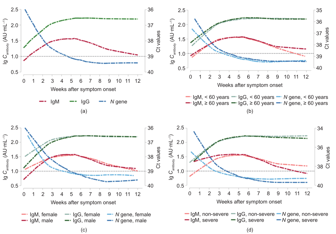
图2

图3
参考文献
[ 1 ] Weekly operational update on COVID-19 [Internet]. Geneva: World Health Organization; 2020 Sep 4 [cited 2020 Sep 4]. Available from: https://www. who.int/docs/default-source/coronaviruse/situation-reports/wou-4-september2020-approved.pdf?sfvrsn=91215c78_2. 链接1
[ 2 ] Wang D, Hu B, Hu C, Zhu F, Liu X, Zhang J, et al. Clinical characteristics of 138 hospitalized patients with 2019 novel coronavirus-infected pneumonia in Wuhan, China. JAMA 2020;323(11):1061–9. 链接1
[ 3 ] Chen N, Zhou M, Dong X, Qu J, Gong F, Han Y, et al. Epidemiological and clinical characteristics of 99 cases of 2019 novel coronavirus pneumonia in Wuhan, China: a descriptive study. Lancet 2020;395(10223):507–13. 链接1
[ 4 ] Zhou F, Yu T, Du R, Fan G, Liu Y, Liu Z, et al. Clinical course and risk factors for mortality of adult inpatients with COVID-19 in Wuhan, China: a retrospective cohort study. Lancet 2020;395(10229):1054–62. 链接1
[ 5 ] Guan WJ, Ni ZY, Hu Y, Liang WH, Ou CQ, He JX, et al. China Medical Treatment Expert Group for COVID-19. Clinical characteristics of coronavirus disease 2019 in China. N Engl J Med 2020;382(18):1708–20. 链接1
[ 6 ] Zheng S, Fan J, Yu F, Feng B, Lou B, Zou Q, et al. Viral load dynamics and disease severity in patients infected with SARS-CoV-2 in Zhejiang Province, China, January–March 2020: retrospective cohort study. BMJ 2020;369:m1443.
[ 7 ] Zou L, Ruan F, Huang M, Liang L, Huang H, Hong Z, et al. SARS-CoV-2 viral load in upper respiratory specimens of infected patients. N Engl J Med 2020;382 (12):1177–9. 链接1
[ 8 ] Zaki AM, van Boheemen S, Bestebroer TM, Osterhaus AD, Fouchier RA. Isolation of a novel coronavirus from a man with pneumonia in Saudi Arabia. N Engl J Med 2012;367(19):1814–20. 链接1
[ 9 ] Memish ZA, Al-Tawfiq JA, Makhdoom HQ, Assiri A, Alhakeem RF, Albarrak A, et al. Respiratory tract samples, viral load, and genome fraction yield in patients with Middle East respiratory syndrome. J Infect Dis 2014;210(10):1590–4. 链接1
[10] To KKW, Tsang OTY, Leung WS, Tam AR, Wu TC, Lung DC, et al. Temporal profiles of viral load in posterior oropharyngeal saliva samples and serum antibody responses during infection by SARS-CoV-2: an observational cohort study. Lancet Infect Dis 2020;20(5):565–74. 链接1
[11] Long QX, Liu BZ, Deng HJ, Wu GC, Deng K, Chen YK, et al. Antibody responses to SARS-CoV-2 in patients with COVID-19. Nat Med 2020;26(6):845–8. 链接1
[12] Moore JB, June CH. Cytokine release syndrome in severe COVID-19. Science 2020;368(6490):473–4. 链接1
[13] Mehta P, McAuley DF, Brown M, Sanchez E, Tattersall RS, Manson JJ; HLH Across Speciality Collaboration, UK. COVID-19: consider cytokine storm syndromes and immunosuppression. Lancet 2020;395(10229):1033–4. 链接1
[14] Chen X, Zhao B, Qu Y, Chen Y, Xiong J, Feng Y, et al. Detectable serum severe acute respiratory syndrome coronavirus 2 viral load (RNAemia) is closely correlated with drastically elevated interleukin 6 level in critically Ill patients with coronavirus disease 2019. Clin Infect Dis 2020;71(8):1937–42. 链接1
[15] National Health Commission of the Peoples’s Republic of China; National Administration of Traditional Chiese Medicine. [Diagnosis and treatment protocol for novel coronavirus pneumonia (trial version 7)]. Report. Beijing: The State Council for the Peoples’s Republic of China; 2020 Mar 3. Chinese.
[16] Wang X, Tan Li, Wang Xu, Liu W, Lu Y, Cheng L, et al. Comparison of nasopharyngeal and oropharyngeal swabs for SARS-CoV-2 detection in 353 patients received tests with both specimens simultaneously. Int J Infect Dis 2020;94:107–9. 链接1
[17] Cleveland WS. Robust locally weighted regression and smoothing scatterplots. J Am Stat Assoc 1979;74(368):829–36. 链接1
[18] McKinnon LR, Liebenberg LJ, Yende-Zuma N, Archary D, Ngcapu S, Sivro A, et al. Genital inflammation undermines the effectiveness of tenofovir gel in preventing HIV acquisition in women. Nat Med 2018;24(4):491–6. 链接1
[19] Lee H, Herbert RD, McAuley JH. Mediation analysis. JAMA 2019;321(7):697–8. 链接1
[20] Xiang F, Wang X, He X, Peng Z, Yang B, Zhang J, et al. Antibody detection and dynamic characteristics in patients with coronavirus disease 2019. Clin Infect Dis 2020;71(8):1930–4. 链接1
[21] Zhao J, Yuan Q, Wang H, Liu W, Liao X, Su Y, et al. Antibody responses to SARSCoV-2 in patients with novel coronavirus disease 2019. Clin Infect Dis 2020;71 (16):2027–34. 链接1
[22] Röltgen K, Powell AE, Wirz OF, Stevens BA, Hogan CA, Najeeb J, et al. Defining the features and duration of antibody responses to SARS-CoV-2 infection associated with disease severity and outcome. Sci Immunol 2020;5(54): eabe0240. 链接1
[23] Pollán M, Pérez-Gómez B, Pastor-Barriuso R, Oteo J, Hernán MA, Pérez-Olmeda M, et al. ENE-COVID Study Group. Prevalence of SARS-CoV-2 in Spain (ENECOVID): a nationwide, population-based seroepidemiological study. Lancet 2020;396(10250):535–44. 链接1
[24] Hallal PC, Hartwig FP, Horta BL, Silveira MF, Struchiner CJ, Vidaletti LP, et al. SARS-CoV-2 antibody prevalence in Brazil: results from two successive nationwide serological household surveys. Lancet Glob Health 2020;8(11): e1390–8. 链接1
[25] Long QX, Tang XJ, Shi QL, Li Q, Deng HJ, Yuan J, et al. Clinical and immunological assessment of asymptomatic SARS-CoV-2 infections. Nat Med 2020;26(8):1200–4. 链接1
[26] Shi Y, Wang Y, Shao C, Huang J, Gan J, Huang X, et al. COVID-19 infection: the perspectives on immune responses. Cell Death Differ 2020;27(5):1451–4. 链接1
[27] Chu CM, Poon LLM, Cheng VCC, Chan KS, Hung IFN, Wong MML, et al. Initial viral load and the outcomes of SARS. CMAJ 2004;171(11):1349–52. 链接1
[28] Di Mauro G, Scavone C, Rafaniello C, Rossi F, Capuano A. SARS-CoV-2 infection: response of human immune system and possible implications for the rapid test and treatment. Int Immunopharmacol 2020;84:106519. 链接1
[29] Wan Y, Shang J, Sun S, Tai W, Chen J, Geng Q, et al. Molecular mechanism for antibody-dependent enhancement of coronavirus entry. J Virol 2020;94(5): e02015–9. 链接1
[30] Cao X. COVID-19: immunopathology and its implications for therapy. Nat Rev Immunol 2020;20(5):269–70. 链接1
[31] Zeng F, Huang Y, Guo Y, Yin M, Chen X, Xiao L, et al. Association of inflammatory markers with the severity of COVID-19: a meta-analysis. Int J Infect Dis 2020;96:467–74. 链接1
[32] Huang C, Wang Y, Li X, Ren L, Zhao J, Hu Yi, et al. Clinical features of patients infected with 2019 novel coronavirus in Wuhan, China. Lancet 2020;395 (10223):497–506. 链接1
[33] Shen C, Wang Z, Zhao F, Yang Y, Li J, Yuan J, et al. Treatment of 5 critically ill patients with COVID-19 with convalescent plasma. JAMA 2020;323 (16):1582–9. 链接1
[34] Duan K, Liu B, Li C, Zhang H, Yu T, Qu J, et al. Effectiveness of convalescent plasma therapy in severe COVID-19 patients. Proc Natl Acad Sci USA 2020;117 (17):9490–6. 链接1
[35] Ahn JY, Sohn Y, Lee SH, Cho Y, Hyun JH, Baek YJ, et al. Use of convalescent plasma therapy in two COVID-19 patients with acute respiratory distress syndrome in Korea. J Korean Med Sci 2020;35(14):e149. 链接1
[36] Peng H, Gong T, Huang X, Sun X, Luo H, Wang W, et al. A synergistic role of convalescent plasma and mesenchymal stem cells in the treatment of severely Ill COVID-19 patients: a clinical case report. Stem Cell Res Ther 2020;11:291. 链接1
[37] Hadjadj J, Yatim N, Barnabei L, Corneau A, Boussier J, Smith N, et al. Impaired type I interferon activity and inflammatory responses in severe COVID-19 patients. Science 2020;369(6504):718–24. 链接1


 京公网安备 11010502051620号


