2015年 第1卷 第3期
《工程(英文)》 >> 2015年 第1卷 第3期 doi: 10.15302/J-ENG-2015079
超声背散射松质骨诊断仪及其在新生儿骨质评价中的应用
1 Department of Electronic Engineering, Fudan University, Shanghai 200433, China
2 Department of Neonatology, Children's Hospital of Fudan University, Shanghai 201102, China
3 Key Laboratory of Medical Imaging Computing and Computer Assisted Intervention (MICCAI) of Shanghai, Shanghai 200032, China
下一篇 上一篇
摘要
超声背散射技术在松质骨骨质无损评价中极具前景。本文介绍了一种新型超声背散射骨质诊断仪 (UBBD) 及其在新生儿骨质状况评价中的应用。超声背散射骨质诊断仪具有无损、无电离辐射、便携及操作简便等优点,可以在短时间内 (5 s) 获得人体松质骨背散射信号。笔者共采集了467例新生儿 (268男/199女) 左侧跟骨处的背散射信号,所用探头中心频率为3.5 MHz。逐步调节背散射有效信号 (SOI) 的延迟 (T1) 和长度 (T2),计算表观积分背散射 (AIB)、表观背散射频率斜率 (FSAB) 及零频率截距 (FIAB) 和频谱质心偏移量 (SCS) 等参数。结果表明,背散射有效信号的选取对背散射测量有直接的影响。当T1较短时 (< 8 μs),AIB和FIAB与新生儿胎龄有显著的正相关性(|R|max = 0.45, P < 0.001);而当T1较长时 (> 10 μs),AIB和FIAB负相关于新生儿胎龄 (|R|max = 0.56, P < 0.001),还可以观察到FSAB和SCS与新生儿胎龄有中度的正相关性 (|R|max = 0.45, P < 0.001)。T2对背散射测量的影响较小,引起相关系数的波动较小。本文通过自制的松质骨超声诊断仪证明超声背散射信号用于评价新生儿骨质状况的可行性。本文还提出了明确的背散射有效信号选取规则及新生儿骨质评价标准。
图片
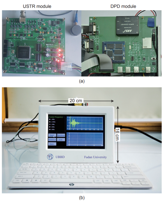
图1

图2
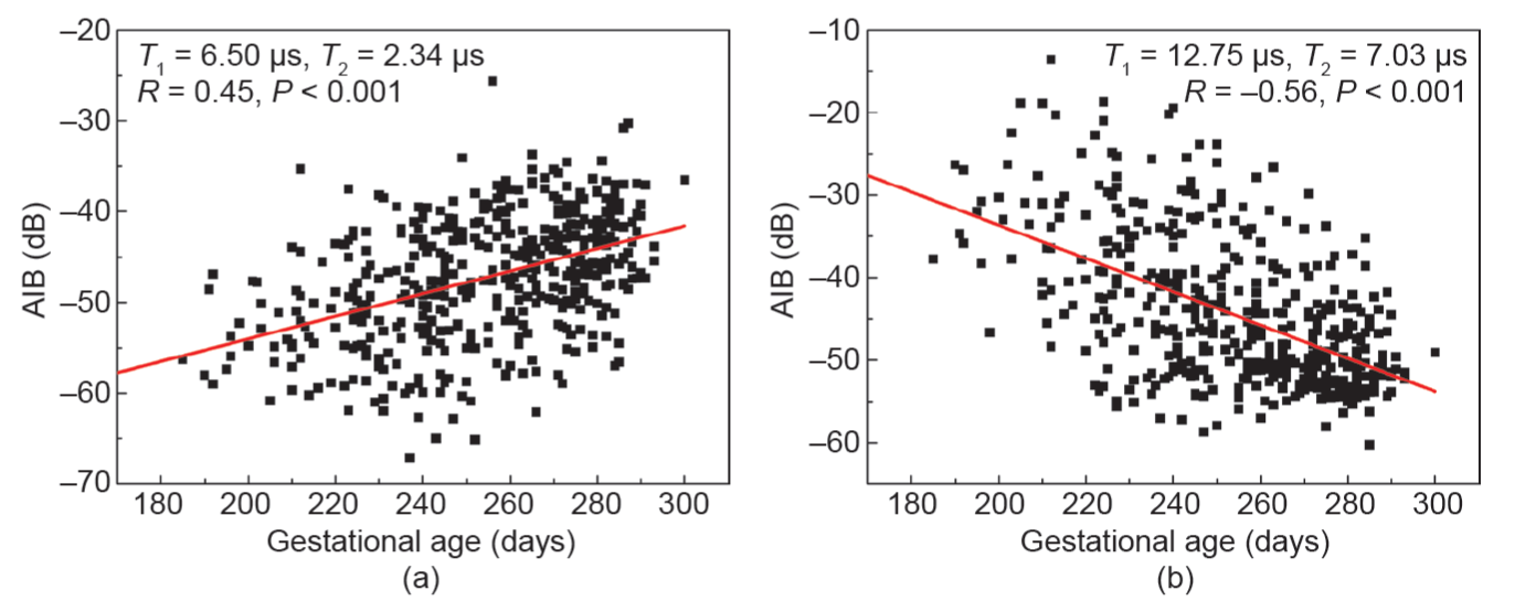
图3

图4

图5
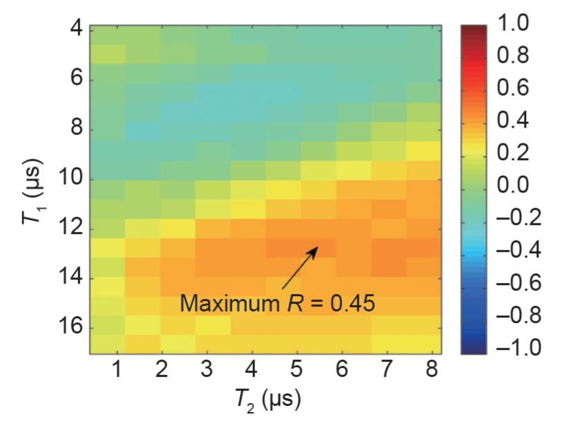
图6
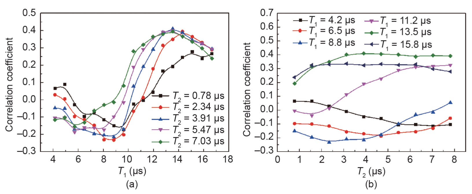
图7
参考文献
[ 1 ] K. Engelke, Clinical use of quantitative computed tomography and peripheral quantitative computed tomography in the management of osteoporosis in adults: The 2007 ISCD Official Positions. J. Clin. Densitom., 2008, 11(1): 123–162 链接1
[ 2 ] R. Lorente-Ramos, J. Azpeitia-Armán, A. Muñoz-Hernández, J. M. García-Gómez, P. Díez-Martínez, M. Grande-Bárez. Dual-energy X-ray absorptiometry in the diagnosis of osteoporosis: A practical guide. AJR Am. J. Roentgenol., 2011, 196(4): 897–904 链接1
[ 3 ] P. Andreopoulou, R. S. Bockman. Management of postmenopausal osteoporosis. Annu. Rev. Med., 2015, 66: 329–342 链接1
[ 4 ] C. B. Becker. Sclerostin inhibition for osteoporosis—A new approach. N. Engl. J. Med., 2014, 370(5): 476–477
[ 5 ] T. D. Rachner, S. Khosla, L. C. Hofbauer. Osteoporosis: Now and the future. Lancet, 2011, 377(9773): 1276–1287 链接1
[ 6 ] P. Laugier. Quantitative ultrasound of bone: Looking ahead. Joint Bone Spine, 2006, 73(2): 125–128
[ 7 ] D. Mulleman, I. Legroux-Gerot, B. Duquesnoy, X. Marchandise, B. Delcambre, B. Cortet. Quantitative ultrasound of bone in male osteoporosis. Osteoporos. Int., 2002, 13(5): 388–393 链接1
[ 8 ] P. H. Nicholson, R. Alkalay. Quantitative ultrasound predicts bone mineral density and failure load in human lumbar vertebrae. Clin. Biomech. (Bristol, Avon), 2007, 22(6): 623–629 链接1
[ 9 ] F. Padilla, F. Jenson, V. Bousson, F. Peyrin, P. Laugier. Relationships of trabecular bone structure with quantitative ultrasound parameters: In vitro study on human proximal femur using transmission and backscatter measurements. Bone, 2008, 42(6): 1193–1202 链接1
[10] K. A. Wear, S. Nagaraja, M. L. Dreher, S. L. Gibson. Relationships of quantitative ultrasound parameters with cancellous bone microstructure in human calcaneus in vitro. J. Acoust. Soc. Am., 2012, 131(2): 1605–1612
[11] D. Ta, W. Wang, K. Huang, Y. Wang, L. H. Le. Analysis of frequency dependence of ultrasonic backscatter coefficient in cancellous bone. J. Acoust. Soc. Am., 2008, 124(6): 4083–4090
[12] C. Chappard, P. Laugier, B. Fournier, C. Roux, G. Berger. Assessment of the relationship between broadband ultrasound attenuation and bone mineral density at the calcaneus using BUA imaging and DXA. Osteoporos. Int., 1997, 7(4): 316–322 链接1
[13] G. Haïat, In vitro speed of sound measurement at intact human femur specimens. Ultrasound Med. Biol., 2005, 31(7): 987–996 链接1
[14] D. Hans, Ultrasound velocity of trabecular cubes reflects mainly bone density and elasticity. Calcif. Tissue Int., 1999, 64(1): 18–23 链接1
[15] S. Mészáros, E. Tóth, V. Ferencz, E. Csupor, E. Hosszú, C. Horváth. Calcaneous quantitative ultrasound measurements predicts vertebral fractures in idiopathic male osteoporosis. Joint Bone Spine, 2007, 74(1): 79–84
[16] W. Pluskiewicz, B. Drozdzowska. Ultrasonic measurement of the calcaneus in Polish normal and osteoporotic women and men. Bone, 1999, 24(6): 611–617 链接1
[17] P. Laugier. An overview of bone sonometry. Int. Congr. Ser., 2004, 1274: 23–32 链接1
[18] K. A. Wear. Ultrasonic scattering from cancellous bone: A review. IEEE Trans. Ultrason. Ferroelectr. Freq. Control, 2008, 55(7): 1432–1441 链接1
[19] B. K. Hoffmeister, A. P. Holt, S. C. Kaste. Effect of the cortex on ultrasonic backscatter measurements of cancellous bone. Phys. Med. Biol., 2011, 56(19): 6243–6255 链接1
[20] B. K. Hoffmeister, Ultrasonic characterization of human cancellous bone in vitro using three different apparent backscatter parameters in the frequency range 0.6−15.0 MHz. IEEE Trans. Ultrason. Ferroelectr. Freq. Control, 2008, 55(7): 1442–1452 链接1
[21] K. Il Lee, M. Joo Choi. Frequency-dependent attenuation and backscatter coefficients in bovine trabecular bone from 0.2 to 1.2 MHz. J. Acoust. Soc. Am., 2012, 131(1): EL67–EL73
[22] C. C. Liu, H. J. Han, D. A. Ta, W. Q. Wang. Effect of selected signals of interest on ultrasonic backscattering measurement in cancellous bones. Sci. China Phys. Mech., 2013, 56(7): 1310–1316
[23] C. C. Liu, D. Ta, B. Hu, L. H. Le, W. Wang. The analysis and compensation of cortical thickness effect on ultrasonic backscatter signals in cancellous bone. J. Appl. Phys., 2014, 116(12): 124903
[24] C. C. Liu, The relationship between ultrasonic backscatter and trabecular anisotropic microstructure in cancellous bone. J. Appl. Phys., 2014, 115(6): 064906
[25] F. Padilla, F. Peyrin, P. Laugier. Prediction of backscatter coefficient in trabecular bones using a numerical model of three-dimensional microstructure. J. Acoust. Soc. Am., 2003, 113(2): 1122–1129
[26] K. A. Wear, A. Laib. The dependence of ultrasonic backscatter on trabecular thickness in human calcaneus: Theoretical and experimental results. IEEE Trans. Ultrason. Ferroelectr. Freq. Control, 2003, 50(8): 979–986 链接1
[27] K. A. Wear, A. P. Stuber, J. C. Reynolds. Relationships of ultrasonic backscatter with ultrasonic attenuation, sound speed and bone mineral density in human calcaneus. Ultrasound Med. Biol., 2000, 26(8): 1311–1316 链接1
[28] B. S. Garra, M. Locher, S. Felker, K. A. Wear. Measurements of ultrasonic backscattered spectral centroid shift from spine in vivo: Methodology and preliminary results. Ultrasound Med. Biol., 2009, 35(1): 165–168 链接1
[29] K. Huang, D. Ta, W. Wang, L. H. Le. Simplified inverse filter tracking algorithm for estimating the mean trabecular bone spacing. IEEE Trans. Ultrason. Ferroelectr. Freq. Control, 2008, 55(7): 1453–1464 链接1
[30] W. C. Pereira, S. L. Bridal, A. Coron, P. Laugier. Singular spectrum analysis applied to backscattered ultrasound signals from in vitro human cancellous bone specimens. IEEE Trans. Ultrason. Ferroelectr. Freq. Control, 2004, 51(3): 302–312 链接1
[31] Y. Q. Jiang, Analysis of apparent integrated backscatter coefficient and backscattered spectral centroid shift in Calcaneus in vivo for the ultrasonic evaluation of osteoporosis. Ultrasound Med. Biol., 2014, 40(6): 1307–1317 链接1
[32] R. Zhang, D. Ta, C. Liu, C. Chen. Feasibility of bone assessment with ultrasonic backscatter signals in neonates. Ultrasound Med. Biol., 2013, 39(10): 1751–1759 链接1
[33] J. Litniewski, L. Cieslik, M. Lewandowski, R. Tymkiewicz, B. Zienkiewicz, A. Nowicki. Ultrasonic scanner for in vivo measurement of cancellous bone properties from backscattered data. IEEE Trans. Ultrason. Ferroelectr. Freq. Control, 2012, 59(7): 1470–1477 链接1
[34] J. P. Karjalainen, Multi-site bone ultrasound measurements in elderly women with and without previous hip fractures. Osteoporos. Int., 2012, 23(4): 1287–1295 链接1
[35] C. Liu, Signal of interest selection standard for ultrasonic backscatter in cancellous bone evaluation. Ultrasound Med. Biol., 2015, 41(10): 2714–2721 链接1
[36] M. S. Fewtrell, T. J. Cole, N. J. Bishop, A. Lucas. Neonatal factors predicting childhood height in preterm infants: Evidence for a persisting effect of early metabolic bone disease? J. Pediatr., 2000, 137(5): 668–673
[37] M. C. Backström, A. L. Kuusela, R. Mäki. Metabolic bone disease of prematurity. Ann. Med., 1996, 28(4): 275–282 链接1
[38] A. Lucas, O. G. Brooke, B. A. Baker, N. Bishop, R. Morley. High alkaline phosphatase activity and growth in preterm neonates. Arch. Dis. Child., 1989, 64(7 Spec No): 902–909 链接1
[39] J. E. Teitelbaum, Quantitative ultrasound in the evaluation of bone status in premature and full-term infants. J. Clin. Densitom., 2006, 9(3): 358–362 链接1
[40] M. Catache, C. R. Leone. Role of plasma and urinary calcium and phosphorus measurements in early detection of phosphorus deficiency in very low birthweight infants. Acta Paediatr., 2003, 92(1): 76–80
[41] J. Faerk, B. Peitersen, S. Petersen, K. F. Michaelsen. Bone mineralisation in premature infants cannot be predicted from serum alkaline phosphatase or serum phosphate. Arch. Dis. Child. Fetal Neonatal Ed., 2002, 87(2): F133–F136 链接1
[42] W. W. K. Koo, J. Walters, A. J. Bush, R. W. Chesney, S. E. Carlson. Dual-energy X-ray absorptiometry studies of bone mineral status in newborn infants. J. Bone Miner. Res., 1996, 11(7): 997–1002
[43] H. McDevitt, S. F. Ahmed. Quantitative ultrasound assessment of bone health in the neonate. Neonatology, 2007, 91(1): 2–11 链接1
[44] L. Pereda, T. Ashmeade, J. Zaritt, J. D. Carver. The use of quantitative ultrasound in assessing bone status in newborn preterm infants. J. Perinatol., 2003, 23(8): 655–659 链接1
[45] A. Omar, S. Turan, A. Bereket. Reference data for bone speed of sound measurement by quantitative ultrasound in healthy children. Arch. Osteoporos., 2006, 1(1−2): 37–41
[46] P. A. Narayana, J. Ophir. A closed form method for the measurement of attenuation in nonlinearly dispersive media. Ultrason. Imaging, 1983, 5(1): 17–21 链接1
[47] B. Rack, Ultrasound for the assessment of bone quality in preterm and term infants. J. Perinatol., 2012, 32(3): 218–226 链接1


 京公网安备 11010502051620号


