
2021, Volume 7, Issue 11
Engineering >> 2021, Volume 7, Issue 11 doi: 10.1016/j.eng.2020.04.017
Nanotechnology and Nanomedicine: A Promising Avenue for Lung Cancer Diagnosis and Therapy
a Key Laboratory of Oral Biomedical Engineering (Wuhan University), Ministry of Education, Hospital of Stomatology, School of Stomatology, Wuhan University, Wuhan 430079, China
b Department of Biomedical Data Science, Geisel School of Medicine, Dartmouth College, Hanover, NH, 03756, United States
c Department of Thoracic Surgery, Shanghai Pulmonary Hospital, School of Medicine, Tongji University, Shanghai 200433, China
d Tus-Medical Health Technology Investment (Jiaxing) Co., Ltd., Jiaxing 314033, China
e Minimally Invasive Thoracic Surgery Unit (UCTMI), Hospital San Rafael, Coruña 15006, Spain
f Osaka Toneyama Medical Center, Osaka 560-8552, Japan
g Key Laboratory of Nanosystem and Hierarchical Fabrication, CAS, National Center for Nanoscience and Technology, Beijing 100190, China
Next Previous
Abstract
Lung cancer is a leading cause of cancer-related death worldwide, with a very poor overall five-year survival rate. The intrinsic limitations associated with the conventional diagnosis and therapeutic strategies used for lung cancer have motivated the development of nanotechnology and nanomedicine approaches, in order to improve early diagnosis rate and develop more effective and safer therapeutic options for lung cancer. Cancer nanomedicines aim to individualize drug delivery, diagnosis, and therapy by tailoring them to each patient's unique physiology and pathological features—on both the genomic and proteomic levels—and have attracted widespread attention in this field. Despite the successful application of nanomedicine techniques in lung cancer research, the clinical translation of nanomedicine approaches remains challenging due to the limited understanding of the interactions that occur between nanotechnology and biology, and the challenges posed by the toxicology, pharmacology, immunology, and large-scale manufacturing of nanoparticles. In this review, we highlight the progress and opportunities associated with nanomedicine use for lung cancer treatment and discuss the prospects of this field, together with the challenges associated with clinical translation.
Keywords
Figures
Fig. 1
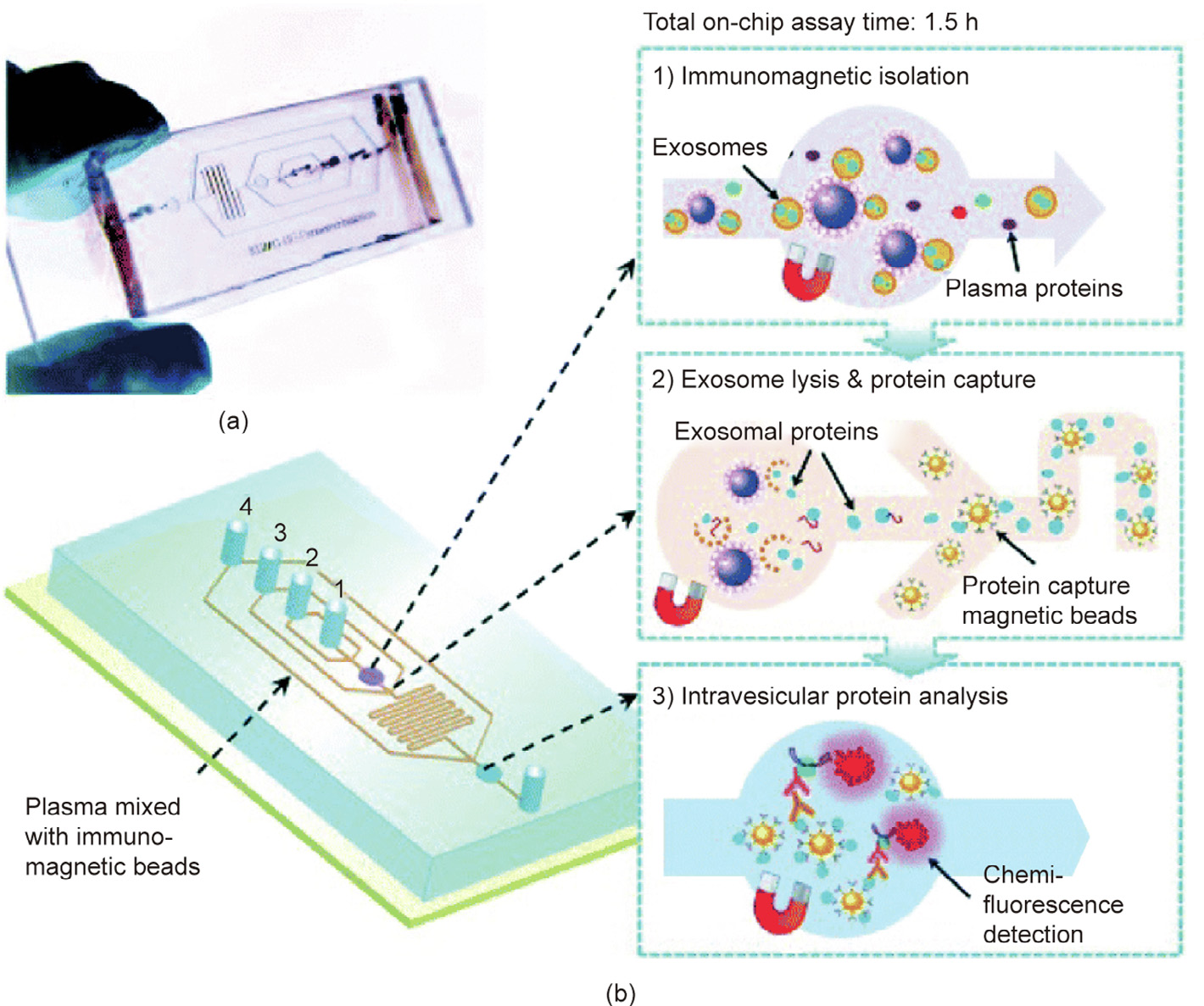
Fig. 2

Fig. 3

Fig. 4
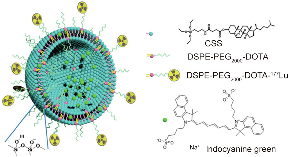
Fig. 5
References
[ 1 ] Siegel RL, Miller KD, Jemal A. Cancer statistics, 2019. CA Cancer J Clin 2019;69 (1):7–34. link1
[ 2 ] Chen W, Zheng R, Baade PD, Zhang S, Zeng H, Bray F, et al. Cancer statistics in China, 2015. CA Cancer J Clin 2016;66(2):115–32. link1
[ 3 ] Sun IC, Ahn CH, Kim K, Emelianov S. Photoacoustic imaging of cancer cells with glycol–chitosan-coated gold nanoparticles as contrast agents. J Biomed Opt 2019;24(12):1–5. link1
[ 4 ] Garcia VB, de Carvalho TG, da Silva Gasparotto LH, da Silva HFO, de Araújo AA, Guerra GCB, et al. Environmentally compatible bioconjugated gold nanoparticles as efficient contrast agents for inflammation-induced cancer imaging. Nanoscale Res Lett 2019;14(1):166. link1
[ 5 ] Tang CK, Vaze A, Shen M, Rusling JF. High-throughput electrochemical microfluidic immunoarray for multiplexed detection of cancer biomarker proteins. ACS Sens 2016;1(8):1036–43. link1
[ 6 ] Munge BS, Stracensky T, Gamez K, DiBiase D, Rusling JF. Multiplex immunosensor arrays for electrochemical detection of cancer biomarker proteins. Electroanalysis 2016;28(11):2644–58. link1
[ 7 ] Cheng Z, Yan X, Sun X, Shen B, Gambhir SS. Tumor molecular imaging with nanoparticles. Engineering 2016;2(1):132–40. link1
[ 8 ] Hood RL, Andriani RT, Ecker TE, Robertson JL, Rylander CG. Characterizing thermal augmentation of convection-enhanced drug delivery with the fiberoptic microneedle device. Engineering 2015;1(3):344–50. link1
[ 9 ] Tang T, Azuma T, Iwahashi T, Takeuchi H, Kobayashi E, Sakuma I. A highprecision US-guided robot-assisted HIFU treatment system for breast cancer. Engineering 2018;4(5):702–13. link1
[10] Hu G, Guan K, Lu L, Zhang J, Lu N, Guan Y. Engineered functional surfaces by laser microprocessing for biomedical applications. Engineering 2018;4 (6):822–30. link1
[11] He W. Cell therapy: pharmacological intervention enters a third era. Engineering 2019;5(1):5–9. link1
[12] Tonelli MR, Shirts BH. Knowledge for precision medicine: mechanistic reasoning and methodological pluralism. JAMA 2017;318(17):1649–50. link1
[13] Badrzadeh F, Rahmati-Yamchi M, Badrzadeh K, Valizadeh A, Zarghami N, Farkhani SM, et al. Drug delivery and nanodetection in lung cancer. Artif Cells Nanomed Biotechnol 2016;44(2):618–34. link1
[14] Wang Z, Qiao R, Tang N, Lu Z, Wang H, Zhang Z, et al. Active targeting theranostic iron oxide nanoparticles for MRI and magnetic resonanceguided focused ultrasound ablation of lung cancer. Biomaterials 2017;127:25–35. link1
[15] Ghasemi Y, Peymani P, Afifi S. Quantum dot: magic nanoparticle for imaging, detection and targeting. Acta Biomed 2009;80(2):156–65. link1
[16] Papagiannaros A, Upponi J, Hartner W, Mongayt D, Levchenko T, Torchilin V. Quantum dot loaded immunomicelles for tumor imaging. BMC Med Imaging 2010;10(1):22. link1
[17] Jin Y, Jia C, Huang SW, O’Donnell M, Gao X. Multifunctional nanoparticles as coupled contrast agents. Nat Commun 2010;1(1):41. link1
[18] Xiao L, Tian X, Harihar S, Li Q, Li L, Welch DR, et al. Gd2O3-doped silica @ Au nanoparticles for in vitro imaging cancer biomarkers using surface-enhanced Raman scattering. Spectrochim Acta A 2017;181:218–25. link1
[19] Neoh KH, Hassan AA, Chen A, Sun Y, Liu P, Xu KF, et al. Rethinking liquid biopsy: microfluidic assays for mobile tumor cells in human body fluids. Biomaterials 2018;150:112–24. link1
[20] Ooki A, Maleki Z, Tsay JC, Goparaju C, Brait M, Turaga N, et al. A panel of novel detection and prognostic methylated DNA markers in primary non-small cell lung cancer and serum DNA. Clin Cancer Res 2017;23(22):7141–52. link1
[21] Campbell JD, Alexandrov A, Kim J, Wala J, Berger AH, Pedamallu CS, et al.; Cancer Genome Atlas Research Network. Distinct patterns of somatic genome alterations in lung adenocarcinomas and squamous cell carcinomas. Nat Genet 2016;48(6):607–16. link1
[22] Mandel P, Métais P. Les acides nucléiques du plasma sanguin chez l’Homme. CR Seances Soc Biol Fil 1948;142(3–4):241–3. French.
[23] Jahr S, Hentze H, Englisch S, Hardt D, Fackelmayer FO, Hesch RD, et al. DNA fragments in the blood plasma of cancer patients: quantitations and evidence for their origin from apoptotic and necrotic cells. Cancer Res 2001;61 (4):1659–65. link1
[24] Leon SA, Shapiro B, Sklaroff DM, Yaros MJ. Free DNA in the serum of cancer patients and the effect of therapy. Cancer Res 1977;37(3):646–50. link1
[25] Chaudhuri AA, Chabon JJ, Lovejoy AF, Newman AM, Stehr H, Azad TD, et al. Early detection of molecular residual disease in localized lung cancer by circulating tumor DNA profiling. Cancer Discov 2017;7(12):1394–403. link1
[26] Liang Z, Cheng Y, Chen Y, Hu Y, Liu WP, Lu Y, et al. EGFR T790M ctDNA testing platforms and their role as companion diagnostics: correlation with clinical outcomes to EGFR-TKIs. Cancer Lett 2017;403:186–94. link1
[27] Nguyen AH, Sim SJ. Nanoplasmonic biosensor: detection and amplification of dual bio-signatures of circulating tumor DNA. Biosens Bioelectron 2015;67:443–9. link1
[28] Dianat S, Bordbar AK, Tangestaninejad S, Yadollahi B, Amiri R, ZarkeshEsfahani SH, et al. In vitro antitumor activity of free and nano-encapsulated Na5[PMo10V2O40]nH2O and its binding properties with ctDNA by using combined spectroscopic methods. J Inorg Biochem 2015;152:74–81. link1
[29] Makarovskiy AN, Ackerley W, Wojcik L, Halpert GK, Stein BS, Carreiro MP, et al. Application of immunomagnetic beads in combination with RT-PCR for the detection of circulating prostate cancer cells. J Clin Lab Anal 1997;11 (6):346–50. link1
[30] Simitzi C, Efstathopoulos P, Kourgiantaki A, Ranella A, Charalampopoulos I, Fotakis C, et al. Laser fabricated discontinuous anisotropic microconical substrates as a new model scaffold to control the directionality of neuronal network outgrowth. Biomaterials 2015;67:115–28. link1
[31] Sonnenberg A, Marciniak JY, McCanna J, Krishnan R, Rassenti L, Kipps TJ, et al. Dielectrophoretic isolation and detection of cfc-DNA nanoparticulate biomarkers and virus from blood. Electrophoresis 2013;34(7):1076–84. link1
[32] Jeon SH, Hong WY, Lee ES, Cho Y. High-purity isolation and recovery of circulating tumor cells using conducting polymer-deposited microfluidic device. Theranostics 2014;4(11):1123–32. link1
[33] Jeon SH, Lee HJ, Bae K, Yoon KA, Lee ES, Cho Y. Efficient capture and isolation of tumor-related circulating cell-free DNA from cancer patients using electroactive conducting polymer nanowire platforms. Theranostics 2016;6 (6):828–36. link1
[34] Bernabé R, Hickson N, Wallace A, Blackhall FH. What do we need to make circulating tumour DNA (ctDNA) a routine diagnostic test in lung cancer? Eur J Cancer 2017;81:66–73. link1
[35] Xu T, Kang X, You X, Dai L, Tian D, Yan W, et al. Cross-platform comparison of four leading technologies for detecting EGFR mutations in circulating tumor DNA from non-small cell lung carcinoma patient plasma. Theranostics 2017;7 (6):1437–46. link1
[36] Zhao L, Tang C, Xu L, Zhang Z, Li X, Hu H, et al. Enhanced and differential capture of circulating tumor cells from lung cancer patients by microfluidic assays using aptamer cocktail. Small 2016;12(8):1072–81. link1
[37] Zhou B, Nie J, Yang W, Huang C, Huang Y, Zhao H. Effect of hydrothorax EGFR gene mutation and EGFR-TKI targeted therapy on advanced non-small cell lung cancer patients. Oncol Lett 2016;11(2):1413–7. link1
[38] Roscilli G, De Vitis C, Ferrara FF, Noto A, Cherubini E, Ricci A, et al. Human lung adenocarcinoma cell cultures derived from malignant pleural effusions as model system to predict patients chemosensitivity. J Transl Med 2016;14 (1):61. link1
[39] Vallée A, Marcq M, Bizieux A, Kouri CE, Lacroix H, Bennouna J, et al. Plasma is a better source of tumor-derived circulating cell-free DNA than serum for the detection of EGFR alterations in lung tumor patients. Lung Cancer 2013;82 (2):373–4. link1
[40] Latifi Z, Fattahi A, Ranjbaran A, Nejabati HR, Imakawa K. Potential roles of metalloproteinases of endometrium–derived exosomes in embryo–maternal crosstalk during implantation. J Cell Physiol 2018;233(6):4530–45. link1
[41] Yang MQ, Du Q, Varley PR, Goswami J, Liang Z, Wang R, et al. Interferon regulatory factor 1 priming of tumour-derived exosomes enhances the antitumour immune response. Br J Cancer 2018;118(1):62–71. link1
[42] Sansone P, Savini C, Kurelac I, Chang Q, Amato LB, Strillacci A, et al. Packaging and transfer of mitochondrial DNA via exosomes regulate escape from dormancy in hormonal therapy-resistant breast cancer. Proc Natl Acad Sci USA 2017;114(43):E9066–75. Corrected in: Proc Natl Acad Sci USA 2017;114 (47):E10255.
[43] Son KJ, Rahimian A, Shin DS, Siltanen C, Patel T, Revzin A. Microfluidic compartments with sensing microbeads for dynamic monitoring of cytokine and exosome release from single cells. Analyst 2016;141 (2):679–88. link1
[44] He M, Crow J, Roth M, Zeng Y, Godwin AK. Integrated immunoisolation and protein analysis of circulating exosomes using microfluidic technology. Lab Chip 2014;14(19):3773–80. link1
[45] Mansur N, Raziul Hasan M, Kim YT, Iqbal SM. Functionalization of nanotextured substrates for enhanced identification of metastatic breast cancer cells. Nanotechnology 2017;28(38):385101. link1
[46] Wan Y, Cheng G, Liu X, Hao SJ, Nisic M, Zhu CD, et al. Rapid magnetic isolation of extracellular vesicles via lipid-based nanoprobes. Nat Biomed Eng 2017; 1(4):0058. link1
[47] Zhang P, He M, Zeng Y. Ultrasensitive microfluidic analysis of circulating exosomes using a nanostructured graphene oxide/polydopamine coating. Lab Chip 2016;16(16):3033–42. link1
[48] In GK, Nieva J. Emerging chemotherapy agents in lung cancer: nanoparticle therapeutics for non-small cell lung cancer. Transl Cancer Res 2015;4 (4):340–55. link1
[49] Nazir S, Hussain T, Ayub A, Rashid U, MacRobert AJ. Nanomaterials in combating cancer: therapeutic applications and developments. NanomedNanotechnol 2014;10(1):19–34. link1
[50] Wicki A, Witzigmann D, Balasubramanian V, Huwyler J. Nanomedicine in cancer therapy: challenges, opportunities, and clinical applications. J Control Release 2015;200:138–57. link1
[51] Kumar D, Mutreja I, Chitcholtan K, Sykes P. Cytotoxicity and cellular uptake of different sized gold nanoparticles in ovarian cancer cells. Nanotechnology 2017;28(47):475101. link1
[52] Chen YH, Tsai CY, Huang PY, Chang MY, Cheng PC, Chou CH, et al. Methotrexate conjugated to gold nanoparticles inhibits tumor growth in a syngeneic lung tumor model. Mol Pharmaceut 2007;4(5):713–22. link1
[53] Yokoyama T, Tam J, Kuroda S, Scott AW, Aaron J, Larson T, et al. EGFR-targeted hybrid plasmonic magnetic nanoparticles synergistically induce autophagy and apoptosis in non-small cell lung cancer cells. PLoS ONE 2011;6(11): e25507. link1
[54] Kao HW, Lin YY, Chen CC, Chi KH, Tien DC, Hsia CC, et al. Evaluation of EGFRtargeted radioimmuno-gold-nanoparticles as a theranostic agent in a tumor animal model. Bioorg Med Chem Lett 2013;23(11):3180–5. link1
[55] Bernardes VHF, Qu Y, Du Z, Beaton J, Vargas MD, Farrell NP. Interaction of the HIV NCp7 protein with platinum(II) and gold(III) complexes containing tridentate ligands. Inorg Chem 2016;55(21):11396–407. link1
[56] Li Q, Wang Q, Yang X, Wang K, Zhang H, Nie W. High sensitivity surface plasmon resonance biosensor for detection of microRNA and small molecule based on graphene oxide–gold nanoparticles composites. Talanta 2017;174:521–6. link1
[57] Knezˇevic´ NZˇ, Durand JO. Targeted treatment of cancer with nanotherapeutics based on mesoporous silica nanoparticles. ChemPlusChem 2015;80 (1):26–36. link1
[58] Bardhan M, Majumdar A, Jana S, Ghosh T, Pal U, Swarnakar S, et al. Mesoporous silica for drug delivery: interactions with model fluorescent lipid vesicles and live cells. J Photochem Photobiol B 2018;178:19–26. link1
[59] Zhang Z, Wang L, Wang J, Jiang X, Li X, Hu Z, et al. Mesoporous silica-coated gold nanorods as a light-mediated multifunctional theranostic platform for cancer treatment. Adv Mater 2012;24(11):1418–23. link1
[60] Liu J, Luo Z, Zhang J, Luo T, Zhou J, Zhao X, et al. Hollow mesoporous silica nanoparticles facilitated drug delivery via cascade pH stimuli in tumor microenvironment for tumor therapy. Biomaterials 2016;83:51–65. link1
[61] Liu D, Kramer SA, Huxford-Phillips RC, Wang S, Della Rocca J, Lin W. Coercing bisphosphonates to kill cancer cells with nanoscale coordination polymers. Chem Commun 2012;48(21):2668–70. link1
[62] Liu D, Poon C, Lu K, He C, Lin W. Self-assembled nanoscale coordination polymers with trigger release properties for effective anticancer therapy. Nat Commun 2014;5(1):4182. link1
[63] Au KM, Satterlee A, Min Y, Tian X, Kim YS, Caster JM, et al. Folate-targeted pHresponsive calcium zoledronate nanoscale metal–organic frameworks: turning a bone antiresorptive agent into an anticancer therapeutic. Biomaterials 2016;82:178–93. link1
[64] Muthu MS, Mei L, Feng SS. Nanotheranostics: advanced nanomedicine for the integration of diagnosis and therapy. Nanomedicine 2014;9(9):1277–80. link1
[65] Mura S, Couvreur P. Nanotheranostics for personalized medicine. Adv Drug Deliv Rev 2012;64(13):1394–416. link1
[66] Cheng FY, Su CH, Wu PC, Yeh CS. Multifunctional polymeric nanoparticles for combined chemotherapeutic and near-infrared photothermal cancer therapy in vitro and in vivo. Chem Commun 2010;46(18):3167–9. link1
[67] Jing L, Shi J, Fan D, Li Y, Liu R, Dai Z, et al. 177Lu-labeled cerasomes encapsulating indocyanine green for cancer theranostics. ACS Appl Mater Interfaces 2015;7(39):22095–105. link1
[68] Zhang L, Li J, Liu K. Recent advances in gadolinium-based MRI metal responsive agent. Sci China Technol Sci 2018;61(9):1329–33. link1
[69] Anselmo AC, Mitragotri S. Nanoparticles in the clinic. Bioeng Transl Med 2016;1(1):10–29. link1
[70] Knights OB, McLaughlan JR. Gold nanorods for light-based lung cancer theranostics. Int J Mol Sci 2018;19(11):3318. link1
[71] He Y, Du Z, Ma S, Liu Y, Li D, Huang H, et al. Effects of green-synthesized silver nanoparticles on lung cancer cells in vitro and grown as xenograft tumors in vivo. Int J Nanomed 2016;11:1879–87.
[72] Svechkarev D, Mohs AM. Organic fluorescent dye-based nanomaterials: advances in the rational design for imaging and sensing applications. Curr Med Chem 2019;26(21):4042–64. link1
[73] Le Duc G, Roux S, Paruta-Tuarez A, Dufort S, Brauer E, Marais A, et al. Advantages of gadolinium based ultrasmall nanoparticles vs molecular gadolinium chelates for radiotherapy guided by MRI for glioma treatment. Cancer Nanotechnol 2014;5(1):4. link1
[74] Ivashchenko O, Peplin´ ska B, Gapin´ ski J, Flak D, Jarek M, Załe˛ski K, et al. Silver and ultrasmall iron oxides nanoparticles in hydrocolloids: effect of magnetic field and temperature on self-organization. Sci Rep 2018;8(1):4041. link1
[75] Nguyen H, Tinet E, Chauveau T, Geinguenaud F, Lalatonne Y, Michel A, et al. Bimodal fucoidan-coated zinc oxide/iron oxide-based nanoparticles for the imaging of atherothrombosis. Molecules 2019;24(5):962. link1
[76] Gonzalez-Rivas D, Yang Y, Ng C. Advances in uniportal video-assisted thoracoscopic surgery: pushing the envelope. Thorac Surg Clin 2016;26 (2):187–201. link1
[77] Ban I, Markuš S, Gyergyek S, Drofenik M, Korenak J, Helix-Nielsen C, et al. Synthesis of poly-sodium-acrylate (PSA)-coated magnetic nanoparticles for use in forward osmosis draw solutions. Nanomaterials 2019;9(9):1238. link1
[78] Edelman R, Assaraf YG, Slavkin A, Dolev T, Shahar T, Livney YD. Developing body-components-based theranostic nanoparticles for targeting ovarian cancer. Pharmaceutics 2019;11(5):216. link1
[79] Ahlawat J, Henriquez G, Narayan M. Enhancing the delivery of chemotherapeutics: role of biodegradable polymeric nanoparticles. Molecules 2018;23(9):2157. link1
[80] Muhamad N, Plengsuriyakarn T, Na-Bangchang K. Application of active targeting nanoparticle delivery system for chemotherapeutic drugs and traditional/herbal medicines in cancer therapy: a systematic review. Int J Nanomed 2018;13:3921–35. link1
[81] Zylberberg C, Gaskill K, Pasley S, Matosevic S. Engineering liposomal nanoparticles for targeted gene therapy. Gene Ther 2017;24(8):441–52. link1
[82] Mayer LD, Tardi P, Louie AC. CPX-351: a nanoscale liposomal co-formulation of daunorubicin and cytarabine with unique biodistribution and tumor cell uptake properties. Int J Nanomed 2019;14:3819–30. link1
[83] Caracciolo G, Palchetti S, Digiacomo L, Chiozzi RZ, Capriotti AL, Amenitsch H, et al. Human biomolecular corona of liposomal doxorubicin: the overlooked factor in anticancer drug delivery. ACS Appl Mater Interfaces 2018;10 (27):22951–62. link1
[84] Fathi S, Oyelere AK. Liposomal drug delivery systems for targeted cancer therapy: is active targeting the best choice? Future Med Chem 2016;8 (17):2091–112. link1
[85] Hwang H, Jeong HS, Oh PS, Kim M, Lee TK, Kwon J, et al. PEGylated nanoliposomes encapsulating angiogenic peptides improve perfusion defects: radionuclide imaging-based study. Nucl Med Biol 2016;43(9):552–8. link1
[86] Wagner U, Marth C, Largillier R, Kaern J, Brown C, Heywood M, et al. Final overall survival results of phase III GCIG CALYPSO trial of pegylated liposomal doxorubicin and carboplatin vs paclitaxel and carboplatin in platinumsensitive ovarian cancer patients. Br J Cancer 2012;107(4):588–91. link1
[87] Nehate C, Jain S, Saneja A, Khare V, Alam N, Dubey RD, et al. Paclitaxel formulations: challenges and novel delivery options. Curr Drug Deliv 2014;11 (6):666–86. link1
[88] Pujade-Lauraine E, Hilpert F, Weber B, Reuss A, Poveda A, Kristensen G, et al. Bevacizumab combined with chemotherapy for platinum-resistant recurrent ovarian cancer: the AURELIA open-label randomized phase III trial. J Clin Oncol 2014;32(13):1302–8. link1
[89] Rajendrakumar SK, Uthaman S, Cho CS, Park IK. Nanoparticle-based phototriggered cancer immunotherapy and its domino effect in the tumor microenvironment. Biomacromolecules 2018;19(6):1869–87. link1
[90] Dougan M, Dougan SK. Targeting immunotherapy to the tumor microenvironment. J Cell Biochem 2017;118(10):3049–54. link1
[91] Riley RS, June CH, Langer R, Mitchell MJ. Delivery technologies for cancer immunotherapy. Nat Rev Drug Discov 2019;18(3):175–96. link1
[92] Cisterna BA, Kamaly N, Choi WI, Tavakkoli A, Farokhzad OC, Vilos C. Targeted nanoparticles for colorectal cancer. Nanomedicine 2016;11(18):2443–56. link1
[93] Geiger BC, Wang S, Padera RF, Grodzinsky AJ, Hammond PT. Cartilagepenetrating nanocarriers improve delivery and efficacy of growth factor treatment of osteoarthritis. Sci Transl Med 2018;10(469):eaat8800. link1
[94] Battogtokh G, Ko YT. Mitochondrial-targeted photosensitizer-loaded folate– albumin nanoparticle for photodynamic therapy of cancer. Nanomedicine 2017;13(2):733–43. link1
[95] Sivasubramanian M, Chuang YC, Lo LW. Evolution of nanoparticle-mediated photodynamic therapy: from superficial to deep-seated cancers. Molecules 2019;24(3):520. link1
[96] Ghalandari B, Asadollahi K, Shakerizadeh A, Komeili A, Riazi G, Kamrava SK, et al. Microtubule network as a potential candidate for targeting by gold nanoparticleassisted photothermal therapy. J Photochem Photobiol B 2019;192:131–40. link1
[97] Sweeney EE, Cano-Mejia J, Fernandes R. Photothermal therapy generates a thermal window of immunogenic cell death in neuroblastoma. Small 2018;14 (20):1800678. link1
[98] Daraee H, Eatemadi A, Abbasi E, Fekri Aval S, Kouhi M, Akbarzadeh A. Application of gold nanoparticles in biomedical and drug delivery. Artif Cells Nanomed Biotechnol 2016;44(1):410–22. link1
[99] Kruger CA, Abrahamse H. Utilisation of targeted nanoparticle photosensitiser drug delivery systems for the enhancement of photodynamic therapy. Molecules 2018;23(10):2628. link1
[100] Kim T, Zhang Q, Li J, Zhang L, Jokerst JV. A gold/silver hybrid nanoparticle for treatment and photoacoustic imaging of bacterial infection. ACS Nano 2018;12(6):5615–25. link1
[101] Donnelly EM, Kubelick KP, Dumani DS, Emelianov SY. Photoacoustic imageguided delivery of plasmonic-nanoparticle-labeled mesenchymal stem cells to the spinal cord. Nano Lett 2018;18(10):6625–32. link1


 京公网安备 11010502051620号


