2015, Volume 1, Issue 3
Engineering >> 2015, Volume 1, Issue 3 doi: 10.15302/J-ENG-2015078
Systems Neuroengineering: Understanding and Interacting with the Brain
1 Department of Biomedical Engineering, University of Minnesota, Minneapolis, MN 55455, USA
2 School of Biomedical Engineering, Shanghai Jiao Tong University, Shanghai 200030, China
3 Department of Biomedical Engineering, Johns Hopkins University, Baltimore, MD 21205, USA
4 SINAPSE Institute, National University of Singapore 119077, Singapore
5 Institute for Engineering in Medicine, University of Minnesota, Minneapolis, MN 55455, USA
# These authors contributed equally.
Next Previous
Abstract
In this paper, we review the current state-of-the-art techniques used for understanding the inner workings of the brain at a systems level. The neural activity that governs our everyday lives involves an intricate coordination of many processes that can be attributed to a variety of brain regions. On the surface, many of these functions can appear to be controlled by specific anatomical structures; however, in reality, numerous dynamic networks within the brain contribute to its function through an interconnected web of neuronal and synaptic pathways. The brain, in its healthy or pathological state, can therefore be best understood by taking a systems-level approach. While numerous neuroengineering technologies exist, we focus here on three major thrusts in the field of systems neuroengineering: neuroimaging, neural interfacing, and neuromodulation. Neuroimaging enables us to delineate the structural and functional organization of the brain, which is key in understanding how the neural system functions in both normal and disease states. Based on such knowledge, devices can be used either to communicate with the neural system, as in neural interface systems, or to modulate brain activity, as in neuromodulation systems. The consideration of these three fields is key to the development and application of neuro-devices. Feedback-based neuro-devices require the ability to sense neural activity (via a neuroimaging modality) through a neural interface (invasive or noninvasive) and ultimately to select a set of stimulation parameters in order to alter neural function via a neuromodulation modality. Systems neuroengineering refers to the use of engineering tools and technologies to image, decode, and modulate the brain in order to comprehend its functions and to repair its dysfunction. Interactions between these fields will help to shape the future of systems neuroengineering—to develop neurotechniques for enhancing the understanding of whole-brain function and dysfunction, and the management of neurological and mental disorders.
Keywords
systems neuroengineering ; neuroimaging ; neural interface ; neuromodulation ; neurotechnology ; brain-computer interface ; brain-machine interface ; neural stimulation
Figures
Fig. 1
Fig. 2
Fig. 3
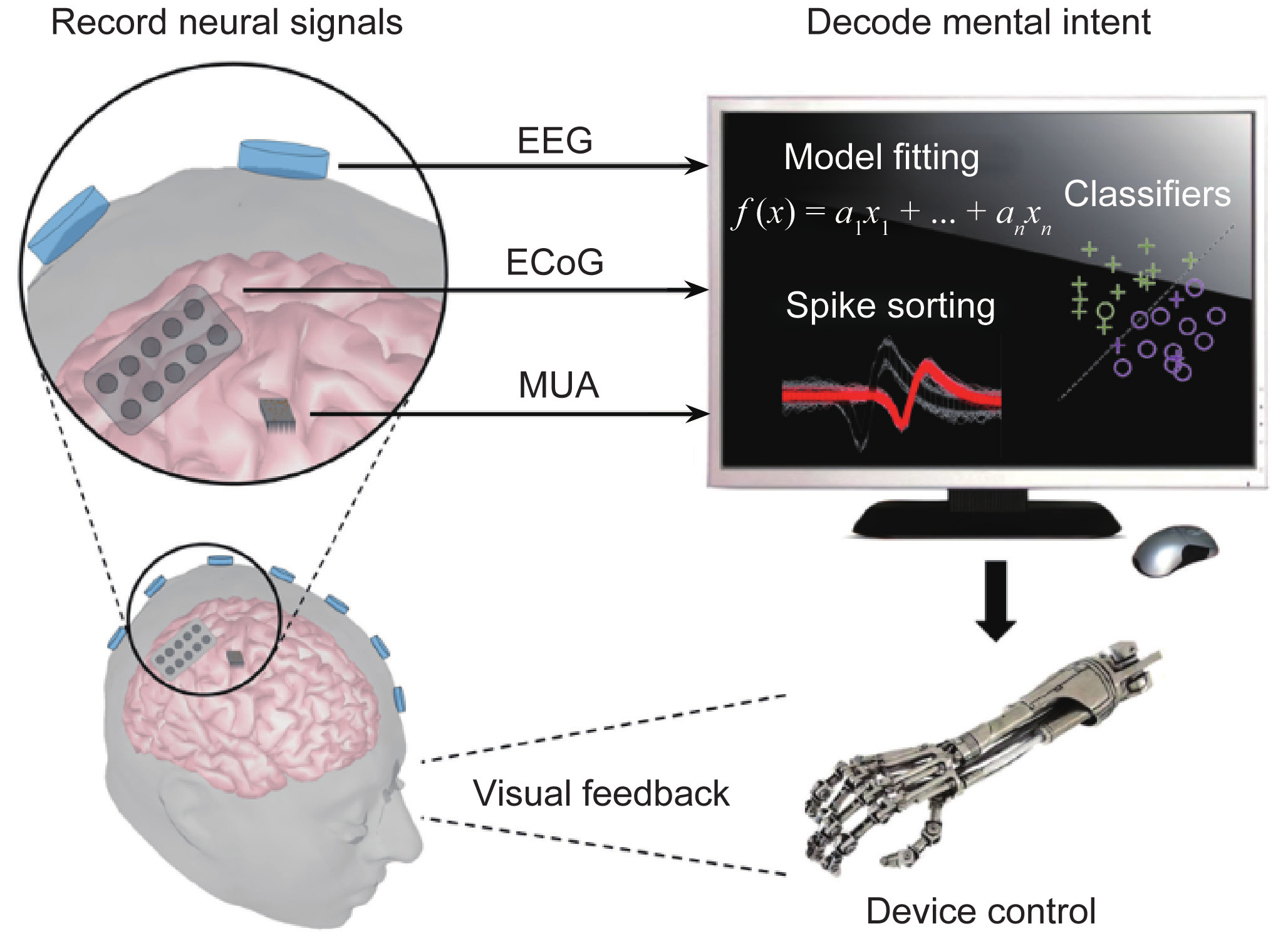
Fig. 4
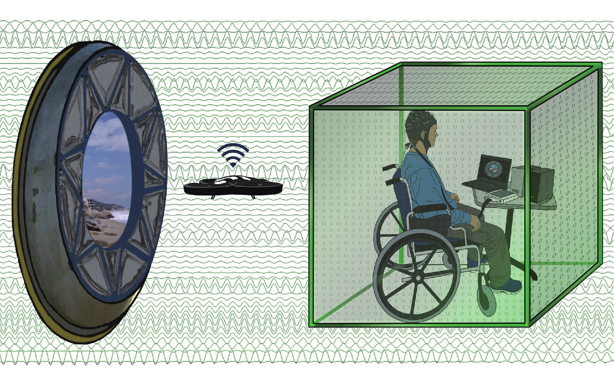
Fig. 5
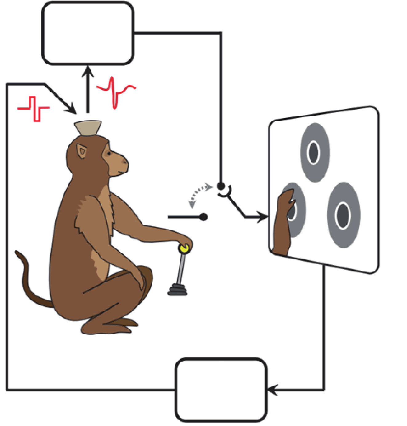
Fig. 6
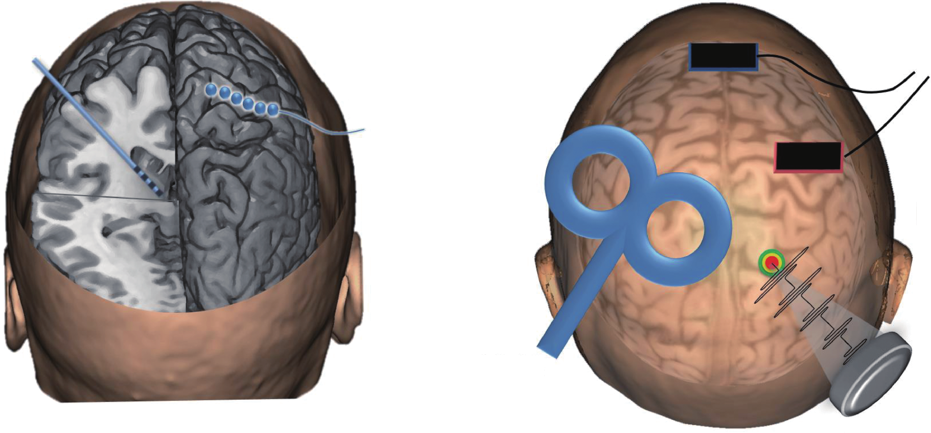
Fig. 7
Fig. 8
References
[ 1 ] Z. Liu, B. He. fMRI-EEG integrated cortical source imaging by use of time-variant spatial constraints. Neuroimage, 2008, 39(3): 1198–1214 link1
[ 2 ] F. Babiloni, Estimation of the cortical functional connectivity with the multimodal integration of high-resolution EEG and fMRI data by directed transfer function. Neuroimage, 2005, 24(1): 118–131 link1
[ 3 ] G. T. Herman. Fundamentals of Computerized Tomography: Image Reconstruction from Projections. London: Springer Science & Business Media, 2009
[ 4 ] U. Bogdahn, G. Becker, J. Winkler, K. Greiner, J. Perez, B. Meurers. Transcranial color-coded real-time sonography in adults. Stroke, 1990, 21(12): 1680–1688 link1
[ 5 ] P. J. Basser, J. Mattiello, D. LeBihan. MR diffusion tensor spectroscopy and imaging. Biophys. J., 1994, 66(1): 259–267 link1
[ 6 ] K. Uğurbil, ; WU-Minn HCP Consortium. Pushing spatial and temporal resolution for functional and diffusion MRI in the Human Connectome Project. Neuroimage, 2013, 80: 80–104 link1
[ 7 ] K. Setsompop, Pushing the limits of in vivo diffusion MRI for the Human Connectome Project. Neuroimage, 2013, 80: 220–233 link1
[ 8 ] E. M. Sevick-Muraca. Translation of near-infrared fluorescence imaging technologies: Emerging clinical applications. Annu. Rev. Med., 2012, 63: 217–231 link1
[ 9 ] G. J. Tearney, In vivo endoscopic optical biopsy with optical coherence tomography. Science, 1997, 276(5321): 2037–2039 link1
[10] W. Lau, S. Tong, N. V. Thakor. Spatiotemporal characteristics of low-frequency functional activation measured by laser speckle imaging. IEEE Trans. Neural Syst. Rehabil. Eng., 2005, 13(2): 179–185 link1
[11] A. Rege, J. Senarathna, N. Li, N. V. Thakor. Anisotropic processing of laser speckle images improves spatiotemporal resolution. IEEE Trans. Biomed. Eng., 2012, 59(5): 1272–1280 link1
[12] J. Senarathna, A. Rege, N. Li, N. V. Thakor. Laser Speckle Contrast Imaging: Theory, instrumentation and applications. IEEE Rev. Biomed. Eng., 2013, 6: 99–110
[13] B. Horwitz, K. J. Friston, J. G. Taylor. Neural modeling and functional brain imaging: An overview. Neural Netw., 2000, 13(8−9): 829–846 link1
[14] B. He, L. Yang, C. Wilke, H. Yuan. Electrophysiological imaging of brain activity and connectivity-challenges and opportunities. IEEE Trans. Biomed. Eng., 2011, 58(7): 1918–1931 link1
[15] B. He, Grand challenges in mapping the human brain: NSF workshop report. IEEE Trans. Biomed. Eng., 2013, 60(11): 2983–2992 link1
[16] M. M. Ter-Pogossian, M. E. Phelps, E. J. Hoffman, N. A. Mullani. A positron-emission transaxial tomograph for nuclear imaging (PETT). Radiology, 1975, 114(1): 89–98 link1
[17] P. H. Murphy, J. A. Burdine, M. Moore, R. J. Jaszcak, W. Thompson, G. DuPuey. Single photon emission computed tomography (SPECT) of the body. J. Nucl. Med., 1978, 19: 683
[18] N. K. Logothetis. What we can do and what we cannot do with fMRI. Nature, 2008, 453(7197): 869–878 link1
[19] K. J. Friston. Modalities, modes, and models in functional neuroimaging. Science, 2009, 326(5951): 399–403 link1
[20] P. A. Bandettini, E. C. Wong, R. S. Hinks, R. S. Tikofsky, J. S. Hyde. Time course EPI of human brain function during task activation. Magn. Reson. Med., 1992, 25(2): 390–397 link1
[21] K. K. Kwong, Dynamic magnetic resonance imaging of human brain activity during primary sensory stimulation. Proc. Natl. Acad. Sci. U.S.A., 1992, 89(12): 5675–5679 link1
[22] S. Ogawa, Intrinsic signal changes accompanying sensory stimulation: Functional brain mapping with magnetic resonance imaging. Proc. Natl. Acad. Sci. U.S.A., 1992, 89(13): 5951–5955 link1
[23] H. Blumenfeld, Cortical and subcortical networks in human secondarily generalized tonic-clonic seizures. Brain, 2009, 132(4): 999–1012
[24] E. Yacoub, N. Harel, K. Ugurbil. High-field fMRI unveils orientation columns in humans. Proc. Natl. Acad. Sci. U.S.A., 2008, 105(30): 10607–10612 link1
[25] C. M. Michel, M. M. Murray, G. Lantz, S. Gonzalez, L. Spinelli, R. Grave de Peralta. EEG source imaging. Clin. Neurophysiol., 2004, 115(10): 2195–2222 link1
[26] B. He, T. Musha, Y. Okamoto, S. Homma, Y. Nakajima, T. Sato. Electric dipole tracing in the brain by means of the boundary element method and its accuracy. IEEE Trans. Biomed. Eng., 1987, 34(6): 406–414
[27] A. M. Dale, M. I. Sereno. Improved localizadon of cortical activity by combining EEG and MEG with MRI cortical surface reconstruction: A linear approach. J. Cogn. Neurosci., 1993, 5(2): 162–176 link1
[28] R. D. Pascual-Marqui, C. M. Michel, D. Lehmann. Low resolution electromagnetic tomography: A new method for localizing electrical activity in the brain. Int. J. Psychophysiol., 1994, 18(1): 49–65 link1
[29] C. M. Michel, B. He. EEG mapping and source imaging. In: D. L. Schomer, F. H. Lopes da Silva, eds. Niedermeyer’s Electroencephalography: Basic Principles, Clinical Applications, and Related Fields. 6th ed. New York: Lippincott Williams & Wilkins, 2011: 1179–1202
[30] B. L. P. Cheung, B. A. Riedner, G. Tononi, B. D. Van Veen. Estimation of cortical connectivity from EEG using state-space models. IEEE Trans. Biomed. Eng., 2010, 57(9): 2122–2134 link1
[31] L. Qin, L. Ding, B. He. Motor imagery classification by means of source analysis for brain-computer interface applications. J. Neural Eng., 2004, 1(3): 135–141
[32] B. Edelman, B. Baxter, B. He. EEG source imaging enhances the decoding of complex right hand motor imagery tasks. IEEE Trans. Biomed. Eng., 2015 (in press)
[33] M. Ferrari, V. Quaresima. A brief review on the history of human functional near-infrared spectroscopy (fNIRS) development and fields of application. Neuroimage, 2012, 63(2): 921–935 link1
[34] J. D. Briers. Laser speckle contrast imaging for measuring blood flow. Opt. Appl., 2007, 37(1−2): 139–152
[35] A. M. Dale, Dynamic statistical parametric mapping: Combining fMRI and MEG for high-resolution imaging of cortical activity. Neuron, 2000, 26(1): 55–67 link1
[36] Z. Liu, N. Zhang, W. Chen, B. He. Mapping the bilateral visual integration by EEG and fMRI. Neuroimage, 2009, 46(4): 989–997 link1
[37] J. Gotman, C. Grova, A. Bagshaw, E. Kobayashi, Y. Aghakhani, F. Dubeau. Generalized epileptic discharges show thalamocortical activation and suspension of the default state of the brain. Proc. Natl. Acad. Sci. U.S.A., 2005, 102(42): 15236–15240 link1
[38] B. Horwitz. The elusive concept of brain connectivity. Neuroimage, 2003, 19(2): 466–470 link1
[39] G. Pfurtscheller, F. H. Lopes da Silva. Event-related EEG/MEG synchronization and desynchronization: Basic principles. Clin. Neurophysiol., 1999, 110(11): 1842–1857 link1
[40] C. W. J. Granger. Investigating causal relations by econometric models and cross-spectral methods. Econometrica, 1969, 37(3): 424–438 link1
[41] M. J. Kamiński, K. J. Blinowska. A new method of the description of the information flow in the brain structures. Biol. Cybern., 1991, 65(3): 203–210 link1
[42] L. A. Baccalá, K. Sameshima. Partial directed coherence: A new concept in neural structure determination. Biol. Cybern., 2001, 84(6): 463–474 link1
[43] M. S. Roulston. Estimating the errors on measured entropy and mutual information. Physica D, 1999, 125(3−4): 285–294 link1
[44] C. J. Stam, B. W. van Dijk. Synchronization likelihood: An unbiased measure of generalized synchronization in multivariate data sets. Physica D, 2002, 163(3−4): 236–251 link1
[45] L. Astolfi, Comparison of different cortical connectivity estimators for high-resolution EEG recordings. Hum. Brain Mapp., 2007, 28(2): 143–157 link1
[46] O. David, D. Cosmelli, K. J. Friston. Evaluation of different measures of functional connectivity using a neural mass model. Neuroimage, 2004, 21(2): 659–673 link1
[47] A. R. McLntosh, F. Gonzalez-Lima. Structural equation modeling and its application to network analysis in functional brain imaging. Hum. Brain Mapp., 1994, 2(1−2): 2–22 link1
[48] K. J. Friston, L. Harrison, W. Penny. Dynamic causal modelling. Neuroimage, 2003, 19(4): 1273–1302 link1
[49] J. Daunizeau, O. David, K. E. Stephan. Dynamic causal modelling: A critical review of the biophysical and statistical foundations. Neuroimage, 2011, 58(2): 312–322 link1
[50] M. H. Lee, C. D. Smyser, J. S. Shimony. Resting-state fMRI: A review of methods and clinical applications. AJNR Am. J. Neuroradiol., 2013, 34(10): 1866–1872 link1
[51] L. Ding, G. A. Worrell, T. D. Lagerlund, B. He. Ictal source analysis: Localization and imaging of causal interactions in humans. Neuroimage, 2007, 34(2): 575–586 link1
[52] C. Wilke, G. Worrell, B. He. Graph analysis of epileptogenic networks in human partial epilepsy. Epilepsia, 2011, 52(1): 84–93 link1
[53] L. Yang, C. Wilke, B. Brinkmann, G. A. Worrell, B. He. Dynamic imaging of ictal oscillations using non-invasive high-resolution EEG. Neuroimage, 2011, 56(4): 1908–1917 link1
[54] L. Lemieux, Event-related fMRI with simultaneous and continuous EEG: Description of the method and initial case report. Neuroimage, 2001, 14(3): 780–787 link1
[55] M. S. Judenhofer, Simultaneous PET-MRI: A new approach for functional and morphological imaging. Nat. Med., 2008, 14(4): 459–465 link1
[56] L. V. Wang, S. Hu. Photoacoustic tomography: In vivo imaging from organelles to organs. Science, 2012, 335(6075): 1458–1462 link1
[57] Y. Xu, B. He. Magnetoacoustic tomography with magnetic induction (MAT-MI). Phys. Med. Biol., 2005, 50(21): 5175–5187 link1
[58] J. Liu, X. Zhang, S. Schmitter, P. F. van de Moortele, B. He. Gradient-based electrical properties tomography (gEPT): A robust method for mapping electrical properties of biological tissues in vivo using magnetic resonance imaging. Magn. Reson. Med., 2015, 74(3): 634–646 link1
[59] D. A. Boas, C. E. Elwell, M. Ferrari, G. Taga. Twenty years of functional near-infrared spectroscopy: Introduction for the special issue. Neuroimage, 2014, 85(1): 1–5 link1
[60] A. K. Dunn. Laser speckle contrast imaging of cerebral blood flow. Ann. Biomed. Eng., 2012, 40(2): 367–377 link1
[61] Q. Liu, Y. Li, H. Lu, S. Tong. Real-time high resolution laser speckle imaging of cerebral vascular changes in a rodent photothrombosis model. Biomed. Opt. Express, 2014, 5(5): 1483–1493 link1
[62] S. Moeller, Multiband multislice GE-EPI at 7 tesla, with 16-fold acceleration using partial parallel imaging with application to high spatial and temporal whole-brain fMRI. Magn. Reson. Med., 2010, 63(5): 1144–1153 link1
[63] S. Haufe, V. V. Nikulin, A. Ziehe, K. R. Müller, G. Nolte. Combining sparsity and rotational invariance in EEG/MEG source reconstruction. Neuroimage, 2008, 42(2): 726–738 link1
[64] A. Gramfort, D. Strohmeier, J. Haueisen, M. S. Hämäläinen, M. Kowalski. Time-frequency mixed-norm estimates: Sparse M/EEG imaging with non-stationary source activations. Neuroimage, 2013, 70: 410–422 link1
[65] L. Ding, B. He. Sparse source imaging in electroencephalography with accurate field modeling. Hum. Brain Mapp., 2008, 29(9): 1053–1067 link1
[66] A. Ale, V. Ermolayev, E. Herzog, C. Cohrs, M. H. de Angelis, V. Ntziachristos. FMT-XCT: In vivo animal studies with hybrid fluorescence molecular tomography-X-ray computed tomography. Nat. Methods, 2012, 9(6): 615–620 link1
[67] K. Deisseroth, G. Feng, A. K. Majewska, G. Miesenböck, A. Ting, M. J. Schnitzer. Next-generation optical technologies for illuminating genetically targeted brain circuits. J. Neurosci., 2006, 26(41): 10380–10386
[68] E. S. Boyden, F. Zhang, E. Bamberg, G. Nagel, K. Deisseroth. Millisecond-timescale, genetically targeted optical control of neural activity. Nat. Neurosci., 2005, 8(9): 1263–1268 link1
[69] J. H. Lee, Global and local fMRI signals driven by neurons defined optogenetically by type and wiring. Nature, 2010, 465(7299): 788–792 link1
[70] J. R. Wolpaw, N. Birbaumer, D. J. McFarland, G. Pfurtscheller, T. M. Vaughan. Brain-computer interfaces for communication and control. Clin. Neurophysiol., 2002, 113(6): 767–791 link1
[71] B. He, S. Gao, H. Yuan, J. R. Wolpaw. Brain-computer interface. In: B. He, ed. Neural Engineering. 2nd ed. Boston, MA: Springer US, 2013: 87–151
[72] B. He, B. Baxter, B. J. Edelman, C. C. Cline, W. W. Ye. Noninvasive brain-computer interfaces based on sensorimotor rhythms. P. IEEE, 2015, 103(6): 907–925
[73] H. Yuan, B. He. Brain-computer interfaces using sensorimotor rhythms: Current state and future perspectives. IEEE Trans. Biomed. Eng., 2014, 61(5): 1425–1435 link1
[74] J. R. Wolpaw, D. J. McFarland, G. W. Neat, C. A. Forneris. An EEG-based brain-computer interface for cursor control. Electroencephalogr. Clin. Neurophysiol., 1991, 78(3): 252–259 link1
[75] A. J. Doud, J. P. Lucas, M. T. Pisansky, B. He. Continuous three-dimensional control of a virtual helicopter using a motor imagery based brain-computer interface. PLoS ONE, 2011, 6(10): e26322 link1
[76] D. J. McFarland, W. A. Sarnacki, J. R. Wolpaw. Electroencephalographic (EEG) control of three-dimensional movement. J. Neural Eng., 2010, 7(3): 036007
[77] K. LaFleur, K. Cassady, A. Doud, K. Shades, E. Rogin, B. He. Quadcopter control in three-dimensional space using a noninvasive motor imagery-based brain-computer interface. J. Neural Eng., 2013, 10(4): 046003
[78]
D. Morrison. Mind over mechanics. Minneapolis, MN: University of Minnesota. 2013-
[79] T. Carlson, J. del R. Millan. Brain-controlled wheelchairs: A robotic architecture. IEEE Robot. Autom. Mag., 2013, 20(1): 65–73 link1
[80] H. Ramoser, J. Müller-Gerking, G. Pfurtscheller. Optimal spatial filtering of single trial EEG during imagined hand movement. IEEE Trans. Rehabil. Eng., 2000, 8(4): 441–446 link1
[81] R. Leeb, L. Tonin, M. Rohm, L. Desideri, T. Carlson, J. D. R. Millan. Towards independence: A BCI telepresence robot for people with severe motor disabilities. P. IEEE, 2015, 103(6): 969–982 link1
[82] L. A. Farwell, E. Donchin. Talking off the top of your head: Toward a mental prosthesis utilizing event-related brain potentials. Electroencephalogr. Clin. Neurophysiol., 1988, 70(6): 510–523 link1
[83] M. Middendorf, G. McMillan, G. Calhoun, K. S. Jones. Brain-computer interfaces based on the steady-state visual-evoked response. IEEE Trans. Rehabil. Eng., 2000, 8(2): 211–214 link1
[84] I. Volosyak. SSVEP-based Bremen-BCI interface—Boosting information transfer rates. J. Neural Eng., 2011, 8(3): 036020
[85] G. Pfurtscheller, T. Solis-Escalante, R. Ortner, P. Linortner, G. R. Müller-Putz. Self-paced operation of an SSVEP-based orthosis with and without an imagery-based “brain switch:” A feasibility study towards a hybrid BCI. IEEE Trans. Neural Syst. Rehabil. Eng., 2010, 18(4): 409–414 link1
[86] G. Pfurtscheller, The hybrid BCI. Front. Neurosci., 2010, 4: 30
[87] A. P. Georgopoulos, A. B. Schwartz, R. E. Kettner. Neuronal population coding of movement direction. Science, 1986, 233(4771): 1416–1419 link1
[88] M. D. Serruya, N. G. Hatsopoulos, L. Paninski, M. R. Fellows, J. P. Donoghue. Brain-machine interface: Instant neural control of a movement signal. Nature, 2002, 416(6877): 141–142 link1
[89] D. M. Taylor, S. I. H. Tillery, A. B. Schwartz. Direct cortical control of 3D neuroprosthetic devices. Science, 2002, 296(5574): 1829–1832 link1
[90] J. M. Carmena, Learning to control a brain-machine interface for reaching and grasping by primates. PLoS Biol., 2003, 1(2): e42
[91] M. Velliste, S. Perel, M. C. Spalding, A. S. Whitford, A. B. Schwartz. Cortical control of a prosthetic arm for self-feeding. Nature, 2008, 453(7198): 1098–1101 link1
[92] K. V. Shenoy, J. M. Carmena. Combining decoder design and neural adaptation in brain-machine interfaces. Neuron, 2014, 84(4): 665–680 link1
[93] A. L. Orsborn, H. G. Moorman, S. A. Overduin, M. M. Shanechi, D. F. Dimitrov, J. M. Carmena. Closed-loop decoder adaptation shapes neural plasticity for skillful neuroprosthetic control. Neuron, 2014, 82(6): 1380–1393 link1
[94] R. D. Flint, Z. A. Wright, M. R. Scheid, M. W. Slutzky. Long term, stable brain machine interface performance using local field potentials and multiunit spikes. J. Neural Eng., 2013, 10(5): 056005
[95] S. D. Stavisky, J. C. Kao, P. Nuyujukian, S. I. Ryu, K. V. Shenoy. A high performing brain-machine interface driven by low-frequency local field potentials alone and together with spikes. J. Neural Eng., 2015, 12(3): 036009
[96] E. C. Leuthardt, G. Schalk, J. R. Wolpaw, J. G. Ojemann, D. W. Moran. A brain-computer interface using electrocorticographic signals in humans. J. Neural Eng., 2004, 1(2): 63–71
[97] M. S. Fifer, Simultaneous neural control of simple reaching and grasping with the modular prosthetic limb using intracranial EEG. IEEE Trans. Neural Syst. Rehabil. Eng., 2014, 22(3): 695–705 link1
[98] D. P. McMullen, Demonstration of a semi-autonomous hybrid brain-machine interface using human intracranial EEG, eye tracking, and computer vision to control a robotic upper limb prosthetic. IEEE Trans. Neural Syst. Rehabil. Eng., 2014, 22(4): 784–796
[99] J. L. Collinger, Collaborative approach in the development of high-performance brain-computer interfaces for a neuroprosthetic arm: Translation from animal models to human control. Clin. Transl. Sci., 2014, 7(1): 52–59 link1
[100] L. M. McCane, P300-based brain-computer interface (BCI) event-related potentials (ERPs): People with amyotrophic lateral sclerosis (ALS) vs. age-matched controls. Clin. Neurophysiol., 2015 (in press)
[101] N. J. Hill, A practical, intuitive brain-computer interface for communicating ‘yes’ or ‘no’ by listening. J. Neural Eng., 2014, 11(3): 035003
[102] M. Severens, M. van der Waal, J. Farquhar, P. Desain. Comparing tactile and visual gaze-independent brain-computer interfaces in patients with amyotrophic lateral sclerosis and healthy users. Clin. Neurophysiol., 2014, 125(11): 2297–2304 link1
[103] A. Ramos-Murguialday, Brain-machine interface in chronic stroke rehabilitation: A controlled study. Ann. Neurol., 2013, 74(1): 100–108 link1
[104] K. K. Ang, C. Guan. Brain-computer interface for neurorehabilitation of upper limb after stroke. P. IEEE, 2015, 103(6): 944–953 link1
[105] F. Pichiorri, Brain-computer interface boosts motor imagery practice during stroke recovery. Ann. Neurol., 2015, 77(5): 851–865 link1
[106] J. L. Collinger, High-performance neuroprosthetic control by an individual with tetraplegia. Lancet, 2013, 381(9866): 557–564 link1
[107] L. R. Hochberg, Reach and grasp by people with tetraplegia using a neurally controlled robotic arm. Nature, 2012, 485(7398): 372–375 link1
[108] B. Wodlinger, J. E. Downey, E. C. Tyler-Kabara, A. B. Schwartz, M. L. Boninger, J. L. Collinger. Ten-dimensional anthropomorphic arm control in a human brain-machine interface: Difficulties, solutions, and limitations. J. Neural Eng., 2015, 12(1): 016011
[109] C. Ethier, E. R. Oby, M. J. Bauman, L. E. Miller. Restoration of grasp following paralysis through brain-controlled stimulation of muscles. Nature, 2012, 485(7398): 368–371 link1
[110] C. T. Moritz, S. I. Perlmutter, E. E. Fetz. Direct control of paralysed muscles by cortical neurons. Nature, 2008, 456(7222): 639–642 link1
[111] N. V. Thakor. Translating the brain-machine interface. Sci. Transl. Med., 2013, 5(210): 210ps17
[112] M. Gomez-Rodriguez, J. Peters, J. Hill, B. Schölkopf, A. Gharabaghi, M. Grosse-Wentrup. Closing the sensorimotor loop: Haptic feedback facilitates decoding of motor imagery. J. Neural Eng., 2011, 8(3): 036005
[113] A. Ramos-Murguialday, Proprioceptive feedback and brain computer interface (BCI) based neuroprostheses. PLoS ONE, 2012, 7(10): e47048 link1
[114] R. Romo, A. Hernández, A. Zainos, E. Salinas. Somatosensory discrimination based on cortical microstimulation. Nature, 1998, 392(6674): 387–390 link1
[115] J. E. O’Doherty, M. A. Lebedev, T. L. Hanson, N. A. Fitzsimmons, M. A. L. Nicolelis. A brain-machine interface instructed by direct intracortical microstimulation. Front. Integr. Neurosci., 2009, 3: 20
[116] J. E. O’Doherty, Active tactile exploration using a brain-machine-brain interface. Nature, 2011, 479(7372): 228–231 link1
[117] D. H. Kim, Epidermal electronics. Science, 2011, 333(6044): 838–843 link1
[118] B. Blankertz, Neurophysiological predictor of SMR-based BCI performance. Neuroimage, 2010, 51(4): 1303–1309 link1
[119] K. Cassady, A. You, A. Doud, B. He. The impact of mind-body awareness training on the early learning of a brain-computer interface. Technology (Singap. World Sci.), 2014, 2(3): 254–260
[120] L. Yao, J. Meng, D. Zhang, X. Sheng, X. Zhu. Combining motor imagery with selective sensation toward a hybrid-modality BCI. IEEE Trans. Biomed. Eng., 2014, 61(8): 2304–2312 link1
[121] L. R. Hochberg, Neuronal ensemble control of prosthetic devices by a human with tetraplegia. Nature, 2006, 442(7099): 164–171 link1
[122] V. Aggarwal, M. Mollazadeh, A. G. Davidson, M. H. Schieber, N. V. Thakor. State-based decoding of hand and finger kinematics using neuronal ensemble and LFP activity during dexterous reach-to-grasp movements. J. Neurophysiol., 2013, 109(12): 3067–3081 link1
[123] S. Schaffelhofer, A. Agudelo-Toro, H. Scherberger. Decoding a wide range of hand configurations from macaque motor, premotor, and parietal cortices. J. Neurosci., 2015, 35(3): 1068–1081
[124] T. Aflalo, Decoding motor imagery from the posterior parietal cortex of a tetraplegic human. Science, 2015, 348(6237): 906–910 link1
[125] W. M. Grill, S. E. Norman, R. V. Bellamkonda. Implanted neural interfaces: Biochallenges and engineered solutions. Annu. Rev. Biomed. Eng., 2009, 11: 1–24 link1
[126] R. Hassler, T. Riechert, F. Mundinger, W. Umbach, J. A. Ganglberger. Physiological observations in stereotaxic operations in extrapyramidal motor disturbances. Brain, 1960, 83(2): 337–350 link1
[127] M. D. Johnson, Neuromodulation for brain disorders: Challenges and opportunities. IEEE Trans. Biomed. Eng., 2013, 60(3): 610–624 link1
[128] A. L. Benabid, Long-term suppression of tremor by chronic stimulation of the ventral intermediate thalamic nucleus. Lancet, 1991, 337(8738): 403–406 link1
[129] M. S. Okun. Deep-brain stimulation for Parkinson’s disease. N. Engl. J. Med., 2012, 367(16): 1529–1538
[130] J. A. Hyam, M. L. Kringelbach, P. A. Silburn, T. Z. Aziz, A. L. Green. The autonomic effects of deep brain stimulation—A therapeutic opportunity. Nat. Rev. Neurol., 2012, 8(7): 391–400 link1
[131] W. Penfield, H. Jasper. Epilepsy and the Functional Anatomy of the Human Brain. Boston: Little, Brown and Company, 1954
[132] J. P. Lefaucheur. Neurophysiology of cortical stimulation. Int. Rev. Neurobiol., 2012, 107: 57–85 link1
[133] S. Morris, Patient-specific cortical electrodes for sulcal and gyral implantation. IEEE Trans. Biomed. Eng., 2015, 62(4): 1034–1041 link1
[134] M. J. Morrell; RNS System in Epilepsy Study Group. Responsive cortical stimulation for the treatment of medically intractable partial epilepsy. Neurology, 2011, 77(13): 1295–1304 link1
[135] T. Wagner, A. Valero-Cabre, A. Pascual-Leone. Noninvasive human brain stimulation. Annu. Rev. Biomed. Eng., 2007, 9(1): 527–565 link1
[136] T. Krieg, D. J. Mogul. Transcranial magnetic stimulation. In: B. He, ed. Neural Engineering. 2nd ed. Boston, MA: Springer US, 2013: 405–453
[137] A. T. Barker, R. Jalinous, I. L. Freeston. Non-invasive magnetic stimulation of human motor cortex. Lancet, 1985, 325(8437): 1106–1107 link1
[138] S. Ueno, T. Tashiro, K. Harada. Localized stimulation of neural tissues in the brain by means of a paired configuration of time-varying magnetic fields. J. Appl. Phys., 1988, 64(10): 5862–5864
[139] Y. Roth, A. Zangen, M. Hallett. A coil design for transcranial magnetic stimulation of deep brain regions. J. Clin. Neurophysiol., 2002, 19(4): 361–370 link1
[140] T. Fadini, H-coil: Induced electric field properties and input/output curves on healthy volunteers, comparison with a standard figure-of-eight coil. Clin. Neurophysiol., 2009, 120(6): 1174–1182 link1
[141] Y. Roth, G. S. Pell, A. V. Chistyakov, A. Sinai, A. Zangen, M. Zaaroor. Motor cortex activation by H-coil and figure-8 coil at different depths. Combined motor threshold and electric field distribution study. Clin. Neurophysiol., 2014, 125(2): 336–343 link1
[142] J. P. O’Reardon, Efficacy and safety of transcranial magnetic stimulation in the acute treatment of major depression: A multisite randomized controlled trial. Biol. Psychiatry, 2007, 62(11): 1208–1216 link1
[143] F. Fregni, A. Pascual-Leone. Technology insight: Noninvasive brain stimulation in neurology-perspectives on the therapeutic potential of rTMS and tDCS. Nat. Clin. Pract. Neurol., 2007, 3(7): 383–393
[144] A. Priori, A. Berardelli, S. Rona, N. Accornero, M. Manfredi. Polarization of the human motor cortex through the scalp. Neuroreport, 1998, 9(10): 2257–2260 link1
[145] M. A. Nitsche, W. Paulus. Excitability changes induced in the human motor cortex by weak transcranial direct current stimulation. J. Physiol., 2000, 527(3): 633–639 link1
[146] A. Datta, V. Bansal, J. Diaz, J. Patel, D. Reato, M. Bikson. Gyri-precise head model of transcranial direct current stimulation: Improved spatial focality using a ring electrode versus conventional rectangular pad. Brain Stimul., 2009, 2(4): 201–207 link1
[147] E. N. Harvey. The effect of high frequency sound waves on heart muscle and other irritable tissues. Am. J. Physiol., 1929, 91: 284–290
[148] Y. Tufail, Transcranial pulsed ultrasound stimulates intact brain circuits. Neuron, 2010, 66(5): 681–694 link1
[149] W. Legon, Transcranial focused ultrasound modulates the activity of primary somatosensory cortex in humans. Nat. Neurosci., 2014, 17(2): 322–329 link1
[150] S. S. Yoo, Focused ultrasound modulates region-specific brain activity. Neuroimage, 2011, 56(3): 1267–1275 link1
[151] T. Guo, Neuroprotective pulsed transcranial ultrasound stimulation in ischemic brain injury after distal middle cerebral artery occlusion. IEEE Trans. Biomed. Eng., 2015 (in press)
[152] H. C. F. Martens, Spatial steering of deep brain stimulation volumes using a novel lead design. Clin. Neurophysiol., 2011, 122(3): 558–566 link1
[153] Z. D. Deng, S. H. Lisanby, A. V. Peterchev. Electric field depth-focality tradeoff in transcranial magnetic stimulation: Simulation comparison of 50 coil designs. Brain Stimul., 2013, 6(1): 1–13 link1
[154] L. I. Navarro de Lara, A novel coil array for combined TMS/fMRI experiments at 3 T. Magn. Reson. Med., 2015 (in press)
[155] A. Roy, B. Baxter, B. He. High-definition transcranial direct current stimulation induces both acute and persistent changes in broadband cortical synchronization: A simultaneous tDCS-EEG study. IEEE Trans. Biomed. Eng., 2014, 61(7): 1967–1978 link1
[156] D. W. Carmichael, Functional MRI with active, fully implanted, deep brain stimulation systems: Safety and experimental confounds. Neuroimage, 2007, 37(2): 508–517 link1
[157] A. A. Kühn, High-frequency stimulation of the subthalamic nucleus suppresses oscillatory beta activity in patients with Parkinson’s disease in parallel with improvement in motor performance. J. Neurosci., 2008, 28(24): 6165–6173 link1
[158] K. Deisseroth. Optogenetics. Nat. Methods, 2011, 8(1): 26–29
[159] K. M. Tye, K. Deisseroth. Optogenetic investigation of neural circuits underlying brain disease in animal models. Nat. Rev. Neurosci., 2012, 13(4): 251–266 link1
[160] A. S. Chuong, Noninvasive optical inhibition with a red-shifted microbial rhodopsin. Nat. Neurosci., 2014, 17(8): 1123–1129 link1
[161] R. Pashaie, R. Falk. Single optical fiber probe for fluorescence detection and optogenetic stimulation. IEEE Trans. Biomed. Eng., 2013, 60(2): 268–280 link1
[162] M. L. Kringelbach, N. Jenkinson, S. L. F. Owen, T. Z. Aziz. Translational principles of deep brain stimulation. Nat. Rev. Neurosci., 2007, 8(8): 623–635 link1
[163] B. Cheeran, A common polymorphism in the brain-derived neurotrophic factor gene (BDNF) modulates human cortical plasticity and the response to rTMS. J. Physiol., 2008, 586(23): 5717–5725 link1


 京公网安备 11010502051620号


