
2020, Volume 6, Issue 10
Engineering >> 2020, Volume 6, Issue 10 doi: 10.1016/j.eng.2020.05.013
Clinical Significance of the Correlation between Changes in the Major Intestinal Bacteria Species and COVID-19 Severity
a Department of Infectious Diseases, Shulan (Hangzhou) Hospital Affiliated to Zhejiang Shuren University Shulan International Medical College, Hangzhou 310003, China
b State Key Laboratory for Diagnosis and Treatment of Infectious Diseases, National Clinical Research Center for Infectious Diseases, Collaborative Innovation Center for Diagnosis and Treatment of Infectious Diseases, The First Affiliated Hospital, College of Medicine, Zhejiang University, Hangzhou 310003, China
c Shaoxing Tongchuang Medical Equipment Co., LTD, Shaoxing 312000, China
d Renmin Hospital of Wuhan University, Wuhan 430200, China
# These authors contributed equally to this work.
Next Previous
Abstract
Coronavirus disease 2019 (COVID-19) is a highly contagious infectious disease. Similar to H7N9 infection, pneumonia and cytokine storm are typical clinical manifestations of COVID-19. Our previous studies found that H7N9 patients had intestinal dysbiosis. However, the relationship between the gut microbiome and COVID-19 has not been determined. This study recruited a cohort of 57 patients with either general (n = 20), severe (n = 19), or critical (n = 18) disease. The objective of this study was to investigate changes in the abundance of ten predominant intestinal bacterial groups in COVID-19 patients using quantitative polymerase chain reaction (q-PCR), and to establish a correlation between these bacterial groups and clinical indicators of pneumonia in these patients. The results indicated that dysbiosis occurred in COVID-19 patients and changes in the gut microbial community were associated with disease severity and hematological parameters. The abundance of butyrate-producing bacteria, such as Faecalibacterium prausnitzii, Clostridium butyricum, Clostridium leptum, and Eubacterium rectale, decreased significantly, and this shift in bacterial community may help discriminate critical patients from general and severe patients. Moreover, the number of common opportunistic pathogens Enterococcus (Ec) and Enterobacteriaceae (E) increased, especially in critically ill patients with poor prognosis. The results suggest that these bacterial groups can serve as diagnostic biomarkers for COVID-19, and that the Ec/E ratio can be used to predict death in critically ill patients.
Keywords
SupplementaryMaterials
Figures
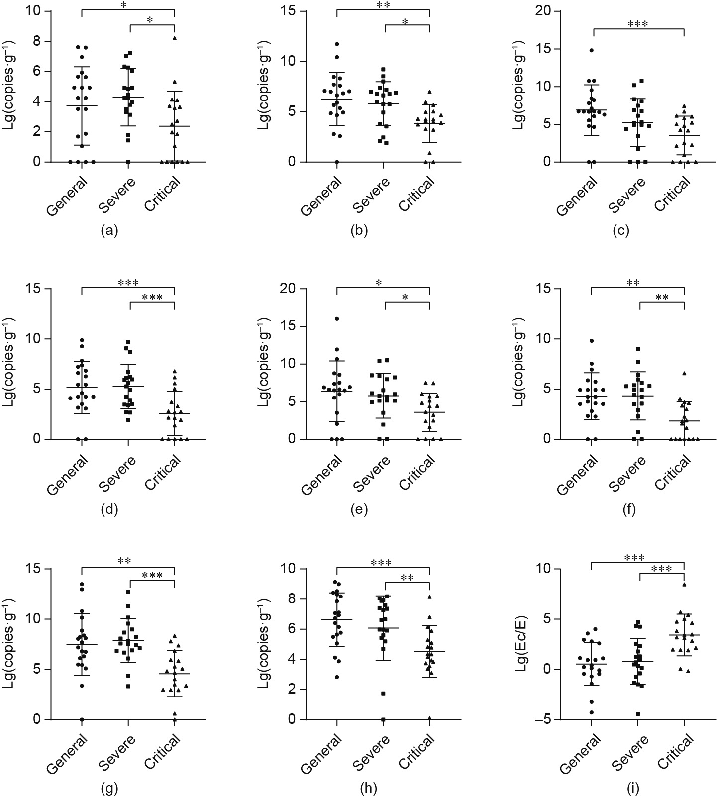
Fig. 1
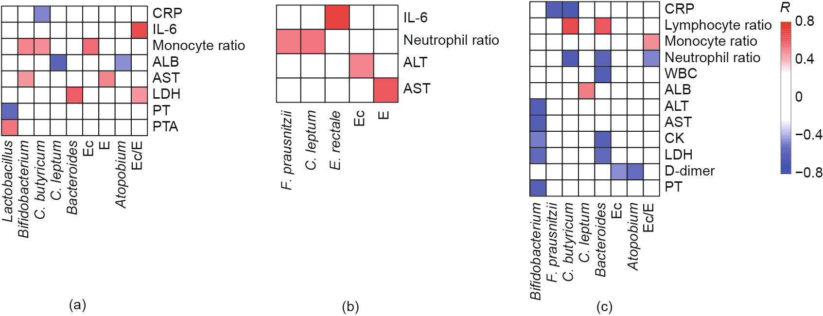
Fig. 2
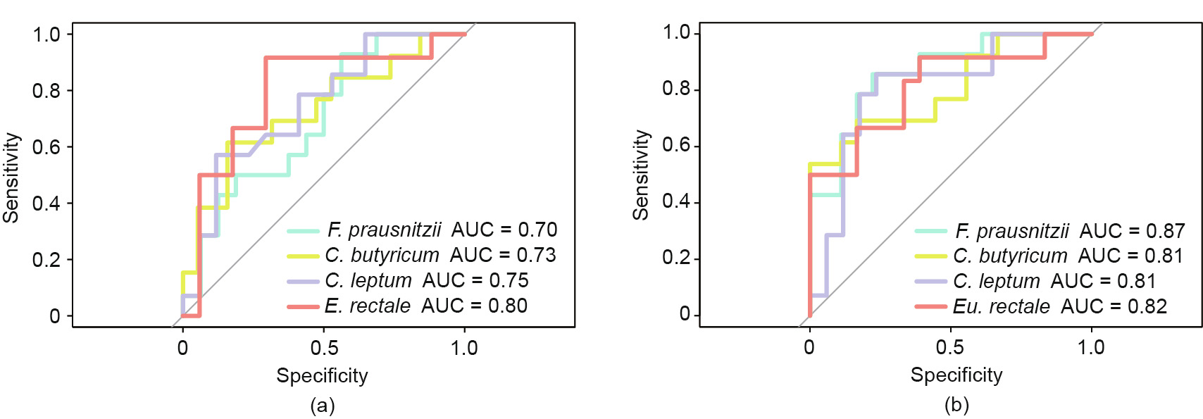
Fig. 3
References
[ 1 ] Lu H, Zhang C, Qian G, Hu X, Zhang H, Chen C, et al. An analysis of microbiotatargeted therapies in patients with avian influenza virus subtype H7N9 infection. BMC Infect Dis 2014;14:359. link1
[ 2 ] Qin N, Zheng B, Yao J, Guo L, Zuo J, Wu L, et al. Influence of H7N9 virus infection and associated treatment on human gut microbiota. Sci Rep 2015;5(1):14771. link1
[ 3 ] Belkaid Y, Harrison OJ. Homeostatic immunity and the microbiota. Immunity 2017;46(4):562–76. link1
[ 4 ] Sanaie S, Ebrahimi-Mameghani M, Hamishehkar H, Mojtahedzadeh M, Mahmoodpoor A. Effect of a multispecies probiotic on inflammatory markers in critically ill patients: a randomized, double-blind, placebo-controlled trial. J Res Med Sci 2014;19(9):827–33. link1
[ 5 ] Hanada S, Pirzadeh M, Carver KY, Deng JC. Respiratory viral infection-induced microbiome alterations and secondary bacterial pneumonia. Front Immunol 2018;9:2640. link1
[ 6 ] Deitch EA. Gut-origin sepsis: evolution of a concept. Surgeon 2012;10 (6):350–6. link1
[ 7 ] Zhou Q, Verne GN. Intestinal hyperpermeability: a gateway to multi-organ failure? J Clin Invest 2018;128(11):4764–6. link1
[ 8 ] Sanders ME, Merenstein DJ, Reid G, Gibson GR, Rastall RA. Author correction: probiotics and prebiotics in intestinal health and disease: from biology to the clinic. Nat Rev Gastroenterol Hepatol 2019;16(10):642. link1
[ 9 ] Xu K, Cai H, Shen Y, Ni Q, Chen Y, Hu S, et al. Management of corona virus disease-19 (COVID-19): the Zhejiang experience. J Zhejiang Univ Med Sci 2020;49(1):147–57. link1
[10] Wu C, Chen X, Cai Y, Xia J, Zhou X, Xu S, et al. Risk factors associated with acute respiratory distress syndrome and death in patients with coronavirus disease 2019 pneumonia in Wuhan, China. JAMA Intern Med 2020;180(7):934–43.
[11] National Health Commission of the People’s Republic of China, National Administration of Traditional Medicine. Diagnosis and treatment protocol for novel coronavirus pneumonia (trail version 7) [Internet]. Beijing: The State Council of the People’s Republic of China; 2020 Mar 3 [cited 2020 Mar 12]. Available from: http:// www.nhc.gov.cn/yzygj/s7653p/202003/46c9294a7dfe4cef80dc7f5912eb1989/ files/ce3e6945832a438eaae415350a8ce964.pdf. Chinese. link1
[12] Declaration of Helsinki—recommendations guiding medical doctors in biomedical research involving human subjects [Internet]. Ferney-Voltaire: The World Medical Assembly; c2020 [adopted 1964 Jun; revised 1975 Oct; cited 2020 Mar 12]. Available from: https://www.wma.net/wp-content/ uploads/2018/07/DoH-Oct1975.pdf.
[13] Qin J, Li R, Raes J, Arumugam M, Burgdorf KS, Manichanh C, et al. A human gut microbial gene catalogue established by metagenomic sequencing. Nature 2010;464(7285):59–65. link1
[14] D’Argenio V, Salvatore F. The role of the gut microbiome in the healthy adult status. Clin Chim Acta 2015;451(Pt A):97–102. link1
[15] Hooper LV, Macpherson AJ. Immune adaptations that maintain homeostasis with the intestinal microbiota. Nat Rev Immunol 2010;10(3):159–69. link1
[16] O’Hara AM, Shanahan F. The gut flora as a forgotten organ. EMBO Rep 2006;7 (7):688–93. link1
[17] Yu Q, Yuan L, Deng J, Yang Q. Lactobacillus protects the integrity of intestinal epithelial barrier damaged by pathogenic bacteria. Front Cell Infect Microbiol 2015;5:26. link1
[18] Salazar N, Gueimonde M, de CG, Los Reyes-Gavilán, Ruas-Madiedo P. Exopolysaccharides produced by lactic acid bacteria and bifidobacteria as fermentable substrates by the intestinal microbiota. Crit Rev Food Sci Nutr 2016;56(9):1440–53. link1
[19] Andoh A, Tsujikawa T, Fujiyama Y. Role of dietary fiber and short-chain fatty acids in the colon. Curr Pharm Des 2003;9(4):347–58. link1
[20] Louis P, Flint HJ. Diversity, metabolism and microbial ecology of butyrateproducing bacteria from the human large intestine. FEMS Microbiol Lett 2009;294(1):1–8. link1
[21] Park J, Kim M, Kang SG, Jannasch AH, Cooper B, Patterson J, et al. Short-chain fatty acids induce both effector and regulatory T cells by suppression of histone deacetylases and regulation of the mTOR-S6K pathway. Mucosal Immunol 2015;8(1):80–93. link1
[22] Mehta P, McAuley DF, Brown M, Sanchez E, Tattersall RS, Manson JJ, et al. COVID-19: consider cytokine storm syndromes and immunosuppression. Lancet 2020;395(10229):1033–4. link1
[23] Swank GM, Deitch EA. Role of the gut in multiple organ failure: bacterial translocation and permeability changes. World J Surg 1996;20(4):411–7. link1
[24] Bäumler AJ, Sperandio V. Interactions between the microbiota and pathogenic bacteria in the gut. Nature 2016;535(7610):85–93. link1
[25] Ubeda C, Taur Y, Jenq RR, Equinda MJ, Son T, Samstein M, et al. Vancomycinresistant Enterococcus domination of intestinal microbiota is enabled by antibiotic treatment in mice and precedes bloodstream invasion in humans. J Clin Invest 2010;120(12):4332–41. link1
[26] Lemon KP, Armitage GC, Relman DA, Fischbach MA. Microbiota-targeted therapies: an ecological perspective. Sci Transl Med 2012;4(137):137rv5. link1
[27] Wexler HM. Bacteroides: the good, the bad, and the nitty-gritty. Clin Microbiol Rev 2007;20(4):593–621. link1
[28] Koh A, De Vadder F, Kovatcheva-Datchary P, Bäckhed F. From dietary fiber to host physiology: short-chain fatty acids as key bacterial metabolites. Cell 2016;165(6):1332–45. link1
[29] Choi VM, Herrou J, Hecht AL, Teoh WP, Turner JR, Crosson S, et al. Activation of Bacteroides fragilis toxin by a novel bacterial protease contributes to anaerobic sepsis in mice. Nat Med 2016;22(5):563–7. link1
[30] Haak BW, Wiersinga WJ. The role of the gut microbiota in sepsis. Lancet Gastroenterol Hepatol 2017;2(2):135–43. link1
[31] Bartosch S, Fite A, Macfarlane GT, McMurdo ME. Characterization of bacterial communities in feces from healthy elderly volunteers and hospitalized elderly patients by using real-time PCR and effects of antibiotic treatment on the fecal microbiota. Appl Environ Micro 2004;70(6):3575–81. link1
[32] Yan R, Zhang Y, Li Y, Xia L, Guo Y, Zhou Q. Structural basis for the recognition of the SARS-CoV-2 by full-length human ACE2. Science 2020;367 (6485):1444–8. link1
[33] Zhou P, Yang XL, Wang XG, Hu B, Zhang L, Zhang W, et al. A pneumonia outbreak associated with a new coronavirus of probable bat origin. Nature 2020;579(7798):270–3. link1


 京公网安备 11010502051620号


