
2021年 第7卷 第12期
《工程(英文)》 >> 2021年 第7卷 第12期 doi: 10.1016/j.eng.2021.08.019
脂肪变性诱发肝癌发生机制——HCV核心基因转基因小鼠的经验教训
a Department of Metabolic Regulation, Shinshu University School of Medicine, Matsumoto 390-8621, Japan
b State Key Laboratory for Diagnosis and Treatment of Infectious Diseases, National Clinical Research Center for Infectious Diseases, Collaborative Innovation Center for Diagnosis and Treatment of Infectious Diseases, Department of Infectious Diseases, The First Affiliated Hospital, College of Medicine, Zhejiang University, Hangzhou 310003, China
c Department of Gastroenterology, Lishui Hospital, Zhejiang University School of Medicine, Lishui 323000, China
d Department of Pathophysiology, Hebei Medical University, Shijiazhuang 050017, China
e Department of Gastroenterology, Shinshu University School of Medicine, Matsumoto 390-8621, Japan
f Molecular Signaling Section, Laboratory of Bioorganic Chemistry, National Institute of Diabetes and Digestive and Kidney Diseases, National Institutes of Health, Bethesda, Bethesda, MD, 20892, United States
g Department of Infection Control and Prevention, The University of Tokyo, Tokyo 113-0033, Japan
h Department of Gastroenterology, The University of Tokyo, Tokyo 113-0033, Japan
i International Relations Office, Shinshu University School of Medicine, Matsumoto 390-8621, Japan
j Research Center for Social Systems, Shinshu University, Matsumoto 390-8621, Japan
下一篇 上一篇
摘要
丙型肝炎病毒(HCV)感染是全球慢性肝炎、肝硬化和肝细胞癌(HCC)的主要病因。在HCV的结构蛋白中,HCV核心蛋白具有调控基因转录、脂质代谢、细胞增殖、细胞凋亡和自噬的能力,所有这些都与HCC的发展密切相关。携带HCV核心基因的转基因小鼠表现出与慢性丙型肝炎患者的临床特征相似的年龄依赖性胰岛素抵抗、肝脂肪变性和HCC。一些饮食习惯的调整,包括限制热量和富含饱和脂肪酸(SFA)、反式脂肪酸(TFA)或胆固醇的饮食,被证明会影响HCV核心基因转基因小鼠的肝脏脂肪生成和肿瘤形成。这些饮食的改变除了调节肝纤维化过程和微环境外,还调节了肝细胞的应激和增殖,从而证实了饮
食习惯与脂肪变性相关的肝癌发生之间的密切联系。本文综述了HCV基因组转基因小鼠模型的研究结果,重点介绍了HCV核心基因转基因小鼠的研究结果,并讨论了HCV核心蛋白诱导脂肪变性和肝癌发生的机制,以及饮食习惯对脂肪变性所致肝癌的影响。
图片
图1

图2

图3
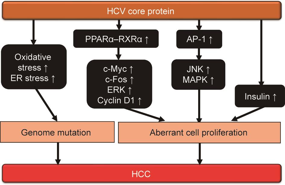
图4
参考文献
[ 1 ] Pradat P, Virlogeux V, Trépo E. Epidemiology and elimination of HCV-related liver disease. Viruses 2018;10(10):545. 链接1
[ 2 ] Kiyosawa K, Sodeyama T, Tanaka E, Gibo Y, Yoshizawa K, Nakano Y, et al. Interrelationship of blood transfusion, non-A, non-B hepatitis and hepatocellular carcinoma: analysis by detection of antibody to hepatitis C virus. Hepatology 1990;12(4):671–5. 链接1
[ 3 ] Moradpour D, Penin F. Hepatitis C virus proteins: from structure to function. Curr Top Microbiol Immunol 2013;369:113–42. 链接1
[ 4 ] Gawlik K, Gallay PA. HCV core protein and virus assembly: what we know without structures. Immunol Res 2014;60(1):1–10. 链接1
[ 5 ] Khaliq S, Jahan S, Pervaiz A. Sequence variability of HCV core region: important predictors of HCV induced pathogenesis and viral production. Infect Genet Evol 2011;11(3):543–56. 链接1
[ 6 ] Hino K, Nishina S, Sasaki K, Hara Y. Mitochondrial damage and iron metabolic dysregulation in hepatitis C virus infection. Free Radical Biol Med 2019;133:193–9. 链接1
[ 7 ] Sevastianos VA, Voulgaris TA, Dourakis SP. Hepatitis C, systemic inflammation and oxidative stress: correlations with metabolic diseases. Expert Rev Gastroenterol Hepatol 2020;14(1):27–37. 链接1
[ 8 ] Wang X, Tanaka N, Hu X, Kimura T, Lu Y, Jia F, et al. A high-cholesterol diet promotes steatohepatitis and liver tumorigenesis in HCV core gene transgenic mice. Arch Toxicol 2019;93(6):1713–25. 链接1
[ 9 ] Diao P, Wang X, Jia F, Kimura T, Hu X, Shirotori S, et al. A saturated fatty acidrich diet enhances hepatic lipogenesis and tumorigenesis in HCV core gene transgenic mice. J Nutr Biochem 2020;85:108460. 链接1
[10] Hu X, Wang X, Jia F, Tanaka N, Kimura T, Nakajima T, et al. A trans-fatty acidrich diet promotes liver tumorigenesis in HCV core gene transgenic mice. Carcinogenesis 2020;41(2):159–70.
[11] Jia F, Diao P, Wang X, Hu X, Kimura T, Nakamuta M, et al. Dietary restriction suppresses steatosis-associated hepatic tumorigenesis in hepatitis C virus core gene transgenic mice. Liver Cancer 2020;9(5):529–48. 链接1
[12] Wang CC, Cheng PN, Kao JH. Systematic review: chronic viral hepatitis and metabolic derangement. Aliment Pharmacol Ther 2020;51(2):216–30. 链接1
[13] Majumder M, Steele R, Ghosh AK, Zhou XY, Thornburg L, Ray R, et al. Expression of hepatitis C virus non-structural 5A protein in the liver of transgenic mice. FEBS Lett 2003;555(3):528–32.
[14] Koike K, Moriya K, Ishibashi K, Matsuura Y, Suzuki T, Saito I, et al. Expression of hepatitis C virus envelope proteins in transgenic mice. J Gen Virol 1995;76 (12):3031–8. 链接1
[15] Wang AG, Moon HB, Kim JM, Hwang SB, Yu DY, Lee DS. Expression of hepatitis C virus nonstructural 4B in transgenic mice. Exp Mol Med 2006;38(3):241–6. 链接1
[16] Moriya K, Fujie H, Shintani Y, Yotsuyanagi H, Tsutsumi T, Ishibashi K, et al. The core protein of hepatitis C virus induces hepatocellular carcinoma in transgenic mice. Nat Med 1998;4(9):1065–7. 链接1
[17] McLauchlan J. Properties of the hepatitis C virus core protein: a structural protein that modulates cellular processes. J Viral Hepat 2000;7(1):2–14. 链接1
[18] Boulant S, Montserret R, Hope RG, Ratinier M, Targett-Adams P, Lavergne JP, et al. Structural determinants that target the hepatitis C virus core protein to lipid droplets. J Biol Chem 2006;281(31):22236–47. 链接1
[19] Liu C, Qu A, Han X, Wang Y. HCV core protein represses the apoptosis and improves the autophagy of human hepatocytes. Int J Clin Exp Med 2015;8 (9):15787–93. 链接1
[20] Cho JW, Baek WK, Yang SH, Chang J, Sung YC, Suh MH. HCV core protein modulates Rb pathway through pRb down-regulation and E2F–1 upregulation. Biochim Biophys Acta 2001;1538(1):59–66. 链接1
[21] Lee SK, Park SO, Joe CO, Kim YS. Interaction of HCV core protein with 14-3-3e protein releases Bax to activate apoptosis. Biochem Biophys Res Commun 2007;352(3):756–62. 链接1
[22] Liu J, Ding X, Tang J, Cao Y, Hu P, Zhou F, et al. Enhancement of canonical Wnt/ b-catenin signaling activity by HCV core protein promotes cell growth of hepatocellular carcinoma cells. PLoS ONE 2011;6(11):e27496. 链接1
[23] Huang S, Xie Y, Yang P, Chen P, Zhang L, Pfeffer S. HCV core protein-induced down-regulation of microRNA-152 promoted aberrant proliferation by regulating Wnt1 in HepG2 cells. PLoS ONE 2014;9(1):e81730. 链接1
[24] Shao YY, Hsieh MS, Wang HY, Li YS, Lin H, Hsu HW, et al. Hepatitis C virus core protein potentiates proangiogenic activity of hepatocellular carcinoma cells. Oncotarget 2017;8(49):86681–92. 链接1
[25] Abe M, Koga H, Yoshida T, Masuda H, Iwamoto H, Sakata M, et al. Hepatitis C virus core protein upregulates the expression of vascular endothelial growth factor via the nuclear factor-jB/hypoxia-inducible factor-1a axis under hypoxic conditions. Hepatol Res 2012;42(6):591–600.
[26] Tan Y, Li Y. HCV core protein promotes hepatocyte proliferation and chemoresistance by inhibiting NR4A1. Biochem Biophys Res Commun 2015;466(3):592–8. 链接1
[27] Benzoubir N, Lejamtel C, Battaglia S, Testoni B, Benassi B, Gondeau C, et al. HCV core-mediated activation of latent TGF-b via thrombospondin drives the crosstalk between hepatocytes and stromal environment. J Hepatol 2013;59 (6):1160–8. 链接1
[28] Loizides-Mangold U, Clément S, Alfonso-Garcia A, Branche E, Conzelmann S, Parisot C, et al. HCV 3a core protein increases lipid droplet cholesteryl ester content via a mechanism dependent on sphingolipid biosynthesis. PLoS ONE 2014;9(12):e115309. 链接1
[29] Alberstein M, Zornitzki T, Zick Y, Knobler H. Hepatitis C core protein impairs insulin downstream signalling and regulatory role of IGFBP-1 expression. J Viral Hepat 2012;19(1):65–71. 链接1
[30] Lewitt MS, Dent MS, Hall K. The insulin-like growth factor system in obesity, insulin resistance and type 2 diabetes mellitus. J Clin Med 2014;3 (4):1561–74. 链接1
[31] Anggakusuma, Frentzen A, Gürlevik E, Yuan Q, Steinmann E, Ott M, et al. Control of hepatitis C virus replication in mouse liver-derived cells by MAVSdependent production of type I and type III interferons. J Virol 2015;89 (7):3833–45. 链接1
[32] Lan HY, Zhao Y, Yang J, Sun MN, Lei YF, Yao M, et al. Establishment of a novel triple-transgenic mouse: conditionally and liver-specifically expressing hepatitis C virus NS3/4A protease. Mol Biol Rep 2014;41(11):7349–59. 链接1
[33] Arrieta JJ, Rodríguez-Iñigo E, Ortiz-Movilla N, Bartolomé J, Pardo M, Manzarbeitia F, et al. In situ detection of hepatitis C virus RNA in salivary glands. Am J Pathol 2001;158(1):259–64. 链接1
[34] Bansal R, Frelin L, Brenndörfer ED, Storm G, Prakash J, Sällberg M, et al. Hepatitis C virus nonstructural 3/4A protein dampens inflammation and contributes to slow fibrosis progression during chronic fibrosis in vivo. PLoS ONE 2015;10(6):e0128466. 链接1
[35] Cristina J, Moreno MDP, Moratorio G. Hepatitis C virus genetic variability in patients undergoing antiviral therapy. Virus Res 2007;127(2):185–94. 链接1
[36] Strosberg AD, Kota S, Takahashi V, Snyder JK, Mousseau G. Core as a novel viral target for hepatitis C drugs. Viruses 2010;2(8):1734–51. 链接1
[37] Koike K, Moriya K, Kimura S. Role of hepatitis C virus in the development of hepatocellular carcinoma: transgenic approach to viral hepatocarcinogenesis. J Gastroenterol Hepatol 2002;17(4):394–400. 链接1
[38] Koike K. Hepatitis C virus contributes to hepatocarcinogenesis by modulating metabolic and intracellular signaling pathways. J Gastroenterol Hepatol 2007;22(Suppl 1):S108–11. 链接1
[39] Hirano J, Yoshio S, Sakai Y, Songling L, Suzuki T, Itoh Y, et al. Hepatitis C virus modulates signal peptide peptidase to alter host protein processing. Proc Natl Acad Sci USA 2021;118(22):e2026184118.
[40] Moriya K, Yotsuyanagi H, Shintani Y, Fujie H, Ishibashi K, Matsuura Y, et al. Hepatitis C virus core protein induces hepatic steatosis in transgenic mice. J Gen Virol 1997;78(7):1527–31.
[41] Harada S, Watanabe Y, Takeuchi K, Suzuki T, Katayama T, Takebe Y, et al. Expression of processed core protein of hepatitis C virus in mammalian cells. J Virol 1991;65(6):3015–21. 链接1
[42] Kahn BB. Type 2 diabetes: when insulin secretion fails to compensate for insulin resistance. Cell 1998;92(5):593–6. 链接1
[43] Cavaghan MK, Ehrmann DA, Polonsky KS. Interactions between insulin resistance and insulin secretion in the development of glucose intolerance. J Clin Invest 2000;106(3):329–33. 链接1
[44] Shintani Y, Fujie H, Miyoshi H, Tsutsumi T, Tsukamoto K, Kimura S, et al. Hepatitis C virus infection and diabetes: direct involvement of the virus in the development of insulin resistance. Gastroenterology 2004;126(3):840–8. 链接1
[45] Miyamoto H, Moriishi K, Moriya K, Murata S, Tanaka K, Suzuki T, et al. Involvement of the PA28c-dependent pathway in insulin resistance induced by hepatitis C virus core protein. J Virol 2007;81(4):1727–35. 链接1
[46] Kadowaki T. Insights into insulin resistance and type 2 diabetes from knockout mouse models. J Clin Invest 2000;106(4):459–65. 链接1
[47] Tanaka N, Nagaya T, Komatsu M, Horiuchi A, Tsuruta G, Shirakawa H, et al. Insulin resistance and hepatitis C virus: a case-control study of non-obese, non-alcoholic and non-steatotic hepatitis virus carriers with persistently normal serum aminotransferase. Liver Int 2008;28(8):1104–11.
[48] Lefkowitch JH, Schiff ER, Davis GL, Perrillo RP, Lindsay K, Bodenheimer HC, et al. Pathological diagnosis of chronic hepatitis C: a multicenter comparative study with chronic hepatitis B. Gastroenterology 1993;104(2):595–603. 链接1
[49] Bach N, Thung SN, Schaffner F. The histological features of chronic hepatitis C and autoimmune chronic hepatitis: a comparative analysis. Hepatology 1992;15(4):572–7. 链接1
[50] Ohata K, Hamasaki K, Toriyama K, Matsumoto K, Saeki A, Yanagi K, et al. Hepatic steatosis is a risk factor for hepatocellular carcinoma in patients with chronic hepatitis C virus infection. Cancer 2003;97(12):3036–43. 链接1
[51] Chang ML, Yeh HC, Tsou YK, Wang CJ, Cheng HY, Sung CM, et al. HCV coreinduced nonobese hepatic steatosis is associated with hypoadiponectinemia and is ameliorated by adiponectin administration. Obesity 2012;20 (7):1474–80.
[52] Mori Y, Moriishi K, Matsuura Y. Hepatitis C virus core protein: its coordinate roles with PA28c in metabolic abnormality and carcinogenicity in the liver. Int J Biochem Cell Biol 2008;40(8):1437–42. 链接1
[53] Koike K, Moriya K. Metabolic aspects of hepatitis C viral infection: steatohepatitis resembling but distinct from NASH. J Gastroenterol 2005;40 (4):329–36. 链接1
[54] Moriishi K, Mochizuki R, Moriya K, Miyamoto H, Mori Y, Abe T, et al. Critical role of PA28c in hepatitis C virus-associated steatogenesis and hepatocarcinogenesis. Proc Natl Acad Sci USA 2007;104(5):1661–6. 链接1
[55] Tsutsumi T, Suzuki T, Shimoike T, Suzuki R, Moriya K, Shintani Y, et al. Interaction of hepatitis C virus core protein with retinoid X receptor alpha modulates its transcriptional activity. Hepatology 2002;35(4):937–46. 链接1
[56] Perlemuter G, Sabile A, Letteron P, Vona G, Topilco A, Chrétien Y, et al. Hepatitis C virus core protein inhibits microsomal triglyceride transfer protein activity and very low density lipoprotein secretion: a model of viralrelated steatosis. FASEB J 2002;16(2):185–94. 链接1
[57] Roingeard P, Hourioux C. Hepatitis C virus core protein, lipid droplets and steatosis. J Viral Hepat 2008;15(3):157–64. 链接1
[58] Tanaka N, Moriya K, Kiyosawa K, Koike K, Gonzalez FJ, Aoyama T. PPARa activation is essential for HCV core protein-induced hepatic steatosis and hepatocellular carcinoma in mice. J Clin Invest 2008;118(2):683–94. 链接1
[59] Tanaka N, Aoyama T, Kimura S, Gonzalez FJ. Targeting nuclear receptors for the treatment of fatty liver disease. Pharmacol Ther 2017;179:142–57. 链接1
[60] Koike K, Tsutsumi T, Yotsuyanagi H, Moriya K. Lipid metabolism and liver disease in hepatitis C viral infection. Oncology 2010;78(Suppl 1):24–30. 链接1
[61] Shiode Y, Hikita H, Tanaka S, Shirai K, Doi A, Sakane S, et al. Hepatitis C virus enhances Rubicon expression, leading to autophagy inhibition and intracellular innate immune activation. Sci Rep 2020;10(1):15290. 链接1
[62] Jiang TX, Zou JB, Zhu QQ, Liu CH, Wang GF, Du TT, et al. SIP/CacyBP promotes autophagy by regulating levels of BRUCE/Apollon, which stimulates LC3-I degradation. Proc Natl Acad Sci USA 2019;116(27):13404–13. 链接1
[63] Singh R, Kaushik S, Wang Y, Xiang Y, Novak I, Komatsu M, et al. Autophagy regulates lipid metabolism. Nature 2009;458(7242):1131–5. 链接1
[64] Hara Y, Yanatori I, Ikeda M, Kiyokage E, Nishina S, Tomiyama Y, et al. Hepatitis C virus core protein suppresses mitophagy by interacting with parkin in the context of mitochondrial depolarization. Am J Pathol 2014;184(11):3026–39. 链接1
[65] Moriya K, Nakagawa K, Santa T, Shintani Y, Fujie H, Miyoshi H, et al. Oxidative stress in the absence of inflammation in a mouse model for hepatitis C virusassociated hepatocarcinogenesis. Cancer Res 2001;61(11):4365–70. 链接1
[66] Moriya K, Todoroki T, Tsutsumi T, Fujie H, Shintani Y, Miyoshi H, et al. Increase in the concentration of carbon 18 monounsaturated fatty acids in the liver with hepatitis C: analysis in transgenic mice and humans. Biochem Biophys Res Commun 2001;281(5):1207–12. 链接1
[67] Vidali M, Tripodi MF, Ivaldi A, Zampino R, Occhino G, Restivo L, et al. Interplay between oxidative stress and hepatic steatosis in the progression of chronic hepatitis C. J Hepatol 2008;48(3):399–406. 链接1
[68] Takenaka K, Adachi E, Nishizaki T, Hiroshige K, Ikeda T, Tsuneyoshi M, et al. Possible multicentric occurrence of hepatocellular carcinoma: a clinicopathological study. Hepatology 1994;19(4):889–94. 链接1
[69] Oikawa T, Ojima H, Yamasaki S, Takayama T, Hirohashi S, Sakamoto M. Multistep and multicentric development of hepatocellular carcinoma: histological analysis of 980 resected nodules. J Hepatol 2005;42(2):225–9. 链接1
[70] Caporaso N, Romano M, Marmo R, de Sio I, Morisco F, Minerva A, et al. Hepatitis C virus infection is an additive risk factor for development of hepatocellular carcinoma in patients with cirrhosis. J Hepatol 1991;12 (3):367–71. 链接1
[71] Hoofnagle JH. Hepatitis C: the clinical spectrum of disease. Hepatology 1997;26(S3):15S–20S. 链接1
[72] Wang Y, Nakajima T, Gonzalez FJ, Tanaka N. PPARs as metabolic regulators in the liver: lessons from liver-specific PPAR-null mice. Int J Mol Sci 2020;21 (6):2061. 链接1
[73] Brocker CN, Yue J, Kim D, Qu A, Bonzo JA, Gonzalez FJ. Hepatocyte-specific PPARA expression exclusively promotes agonist-induced cell proliferation without influence from nonparenchymal cells. Am J Physiol Gastrointest Liver Physiol 2017;312(3):G283–99. 链接1
[74] Tanaka N, Moriya K, Kiyosawa K, Koike K, Aoyama T. Hepatitis C virus core protein induces spontaneous and persistent activation of peroxisome proliferator-activated receptor alpha in transgenic mice: implications for HCV-associated hepatocarcinogenesis. Int J Cancer 2008;122(1):124–31. 链接1
[75] Shrivastava A, Manna SK, Ray R, Aggarwal BB. Ectopic expression of hepatitis C virus core protein differentially regulates nuclear transcription factors. J Virol 1998;72(12):9722–8. 链接1
[76] Tsutsumi T, Suzuki T, Moriya K, Yotsuyanagi H, Shintani Y, Fujie H, et al. Alteration of intrahepatic cytokine expression and AP-1 activation in transgenic mice expressing hepatitis C virus core protein. Virology 2002;304(2):415–24. 链接1
[77] Lu W, Lo SY, Chen M, Wu KJ, Fung YK, Ou JH. Activation of p53 tumor suppressor by hepatitis C virus core protein. Virology 1999;264(1):134–41. 链接1
[78] Pan Z, Bhat MB, Nieminen AL, Ma J. Synergistic movements of Ca2+ and Bax in cells undergoing apoptosis. J Biol Chem 2001;276(34):32257–63. 链接1
[79] Benali-Furet NL, Chami M, Houel L, De Giorgi F, Vernejoul F, Lagorce D, et al. Hepatitis C virus core triggers apoptosis in liver cells by inducing ER stress and ER calcium depletion. Oncogene 2005;24(31):4921–33. 链接1
[80] Umemura A, He F, Taniguchi K, Nakagawa H, Yamachika S, Font-Burgada J, et al. P62, upregulated during preneoplasia, induces hepatocellular carcinogenesis by maintaining survival of stressed HCC-initiating cells. Cancer Cell 2016;29(6):935–48. 链接1
[81] Kato T, Miyamoto M, Date T, Yasui K, Taya C, Yonekawa H, et al. Repeated hepatocyte injury promotes hepatic tumorigenesis in hepatitis C virus transgenic mice. Cancer Sci 2003;94(8):679–85. 链接1
[82] Ioannou GN, Morrow OB, Connole ML, Lee SP. Association between dietary nutrient composition and the incidence of cirrhosis or liver cancer in the United States population. Hepatology 2009;50(1):175–84. 链接1
[83] Yu L, Morishima C, Ioannou GN. Dietary cholesterol intake is associated with progression of liver disease in patients with chronic hepatitis C: analysis of the hepatitis C antiviral long-term treatment against cirrhosis trial. Clin Gastroenterol Hepatol 2013;11(12):1661–6.e3. 链接1
[84] Ostapowicz G, Watson KJ, Locarnini SA, Desmond PV. Role of alcohol in the progression of liver disease caused by hepatitis C virus infection. Hepatology 1998;27(6):1730–5. 链接1
[85] Kimura T, Kobayashi A, Tanaka N, Sano K, Komatsu M, Fujimori N, et al. Clinicopathological characteristics of non-B non-C hepatocellular carcinoma without past hepatitis B virus infection. Hepatol Res 2017;47(5):405–18. 链接1
[86] Tanaka N, Kimura T, Fujimori N, Nagaya T, Komatsu M, Tanaka E. Current status, problems, and perspectives of non-alcoholic fatty liver disease research. World J Gastroenterol 2019;25(2):163–77. 链接1
[87] Duarte-Salles T, Fedirko V, Stepien M, Aleksandrova K, Bamia C, Lagiou P, et al. Dietary fat, fat subtypes and hepatocellular carcinoma in a large European cohort. Int J Cancer 2015;137(11):2715–28. 链接1
[88] Zˇácˇek P, Bukowski M, Mehus A, Johnson L, Zeng H, Raatz S, et al. Dietary saturated fatty acid type impacts obesity-induced metabolic dysfunction and plasma lipidomic signatures in mice. J Nutr Biochem 2019;64:32–44. 链接1
[89] Zelber-Sagi S, Ivancovsky-Wajcman D, Fliss Isakov N, Webb M, Orenstein D, Shibolet O, et al. High red and processed meat consumption is associated with non-alcoholic fatty liver disease and insulin resistance. J Hepatol 2018;68 (6):1239–46. 链接1
[90] Freedman ND, Cross AJ, McGlynn KA, Abnet CC, Park Y, Hollenbeck AR, et al. Association of meat and fat intake with liver disease and hepatocellular carcinoma in the NIH-AARP cohort. J Natl Cancer Inst 2010;102(17): 1354–65. 链接1
[91] Moriya K, Miyoshi H, Shinzawa S, Tsutsumi T, Fujie H, Goto K, et al. Hepatitis C virus core protein compromises iron-induced activation of antioxidants in mice and HepG2 cells. J Med Virol 2010;82(5):776–92. 链接1
[92] Tsutsumi T, Suzuki T, Moriya K, Shintani Y, Fujie H, Miyoshi H, et al. Hepatitis C virus core protein activates ERK and p38 MAPK in cooperation with ethanol in transgenic mice. Hepatology 2003;38(4):820–8. 链接1
[93] Ghebreyesus TA, Frieden TR. REPLACE: a roadmap to make the world trans fat free by 2023. Lancet 2018;391(10134):1978–80. 链接1
[94] Mozaffarian D, Katan MB, Ascherio A, Stampfer MJ, Willett WC. Trans fatty acids and cardiovascular disease. N Engl J Med 2006;354(15):1601–13. 链接1
[95] Micha R, Mozaffarian D. Trans fatty acids: effects on metabolic syndrome, heart disease and diabetes. Nat Rev Endocrinol 2009;5(6):335–44. 链接1
[96] Hu X, Tanaka N, Guo R, Lu Yu, Nakajima T, Gonzalez FJ, et al. PPARa protects against trans-fatty-acid-containing diet-induced steatohepatitis. J Nutr Biochem 2017;39:77–85. 链接1
[97] Clayton ZS, Fusco E, Kern M. Egg consumption and heart health: a review. Nutrition 2017;37:79–85. 链接1
[98] Sozen E, Ozer NK. Impact of high cholesterol and endoplasmic reticulum stress on metabolic diseases: an updated mini-review. Redox Biol 2017;12:456–61. 链接1
[99] Ioannou GN. The role of cholesterol in the pathogenesis of NASH. Trends Endocrinol Metab 2016;27(2):84–95. 链接1
[100] Huff MW. Dietary cholesterol, cholesterol absorption, postprandial lipemia and atherosclerosis. Can J Clin Pharmacol 2003;10 Suppl A:26A–32A.
[101] Dev S, Babitt JL. Overview of iron metabolism in health and disease. Hemodial Int 2017;21(S1):S6–S20. 链接1
[102] Tanaka N, Horiuchi A, Yamaura T, Komatsu M, Tanaka E, Kiyosawa K. Efficacy and safety of 6-month iron reduction therapy in patients with hepatitis C virus-related cirrhosis: a pilot study. J Gastroenterol 2007;42(1):49–55. 链接1
[103] Tanaka N, Kiyosawa K. Phlebotomy: a promising treatment for chronic hepatitis C. J Gastroenterol 2004;39(6):601–3. 链接1
[104] Tanaka N, Horiuchi A, Yamaura T, Komatsu M, Yokoyama T, Okaniwa S, et al. Efficacy and safety of addition of minor bloodletting (petit phlebotomy) in hepatitis C virus-infected patients receiving regular glycyrrhizin injections. J Gastroenterol 2009;44(6):577–82. 链接1
[105] Kimura T, Tanaka N, Fujimori N, Sugiura A, Yamazaki T, Joshita S, et al. Mild drinking habit is a risk factor for hepatocarcinogenesis in non-alcoholic fatty liver disease with advanced fibrosis. World J Gastroenterol 2018;24 (13):1440–50. 链接1
[106] Ceni E, Mello T, Galli A. Pathogenesis of alcoholic liver disease: role of oxidative metabolism. World J Gastroenterol 2014;20(47):17756–72. 链接1
[107] Vandenbulcke H, Moreno C, Colle I, Knebel JF, Francque S, Sersté T, et al. Alcohol intake increases the risk of HCC in hepatitis C virus-related compensated cirrhosis: a prospective study. J Hepatol 2016;65(3):543–51. 链接1
[108] Brandhorst S, Longo VD. Fasting and caloric restriction in cancer prevention and treatment. Recent Results Cancer Res 2016;207:241–66. 链接1
[109] Barnosky AR, Hoddy KK, Unterman TG, Varady KA. Intermittent fasting vs daily calorie restriction for type 2 diabetes prevention: a review of human findings. Transl Res 2014;164(4):302–11. 链接1
[110] Fujimoto M, Tsuneyama K, Nakanishi Y, Salunga TL, Nomoto K, Sasaki Y, et al. A dietary restriction influences the progression but not the initiation of MSG-induced nonalcoholic steatohepatitis. J Med Food 2014;17(3): 374–83. 链接1
[111] Holmer M, Lindqvist C, Petersson S, Moshtaghi-Svensson J, Tillander V, Brismar TB, et al. Treatment of NAFLD with intermittent calorie restriction or low-carb high-fat diet—a randomised controlled trial. JHEP Rep 2021;3 (3):100256. 链接1


 京公网安备 11010502051620号


