
2021, Volume 7, Issue 10
Engineering >> 2021, Volume 7, Issue 10 doi: 10.1016/j.eng.2020.09.010
Effects of Three-Dimensional Platform Stiffness and Layer Dimensions on Separation of Carcinoma Cells
Department of Electrical Engineering, Center for Biosystems, Neuroscience, and Nanotechnology, City University of Hong Kong, Hong Kong 999077, China
Next Previous
Abstract
Cancer cell separation is highly desirable for cancer diagnosis and therapy. Besides biochemical methods, engineered platforms are effective alternatives for sorting carcinoma cells from normal cells based on their unique properties in responding to the physical changes of the surrounding microenvironment. In this work, three-dimensional (3D) biomimetic scaffold platforms were developed to separate nasopharyngeal carcinoma 43 (NPC43) cells from immortalized nasopharyngeal epithelial 460 (NP460) cells based on precisely controlled design parameters including stiffness, number of layers, and structural layout. The migration characteristics of NPC43 and NP460 cells on the scaffold platforms revealed that NPC43 cells could squeeze into 10 µm wide, 15 µm deep trenches while NP460 cells could not. The different migration behavior was mainly due to cells having different interactions with the surrounding microenvironment. NPC43 cells had filopodia-like protrusions, while NP460 cells exhibited a sheet-like morphology. Using these 3D biomimetic platforms, 89% separation efficiency of NPC43 cells from NP460 cells was achieved on stiffer two-layer scaffold platforms with a 40/10 μm ridge/trench (R/T) grating on the top layer and a 20/10 μm R/T grid on the bottom layer. Moreover, the separation efficiency was further increased to 93% by adding an active conditioned medium (ACM) that caused the cells to have higher motility and deformability. These results demonstrate the capability to apply biomimetic engineered platforms with appropriate designs to separate cancer cells from normal cells for potential cancer diagnosis and treatment.
Figures
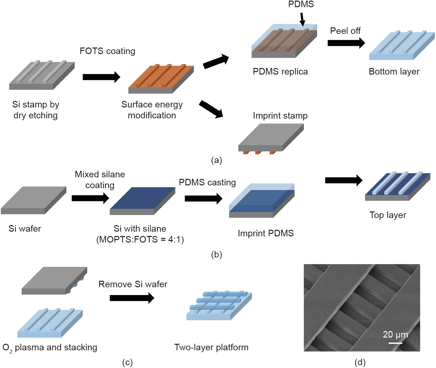
Fig. 1
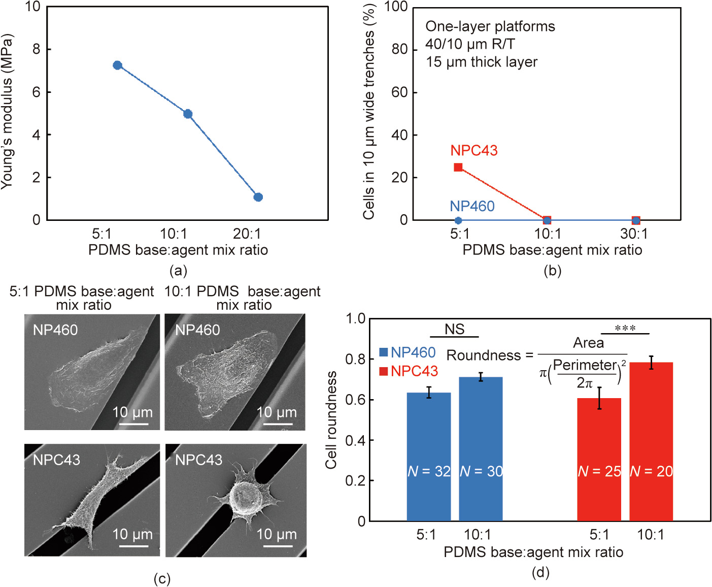
Fig. 2

Fig. 3

Fig. 4

Fig. 5

Fig. 6

Fig. 7

Fig. 8
References
[ 1 ] Gossett DR, Weaver WM, Mach AJ, Hur SC, Tse HTK, Lee W, et al. Label-free cell separation and sorting in microfluidic systems. Anal Bioanal Chem 2010;397 (8):3249–67. link1
[ 2 ] Fiddler M. Fetal cell based prenatal diagnosis: perspectives on the present and future. J Clin Med 2014;3(3):972–85. link1
[ 3 ] Blainey PC, Quake SR. Dissecting genomic diversity, one cell at a time. Nat Methods 2014;11(1):19–21. link1
[ 4 ] Schor SL, Schor AM. Phenotypic and genetic alterations in mammary stroma: implications for tumour progression. Breast Cancer Res 2001;3(6):373–9. link1
[ 5 ] Guo KT, SchÄfer R, Paul A, Gerber A, Ziemer G, Wendel HP. A new technique for the isolation and surface immobilization of mesenchymal stem cells from whole bone marrow using high-specific DNA aptamers. Stem Cells 2006;24 (10):2220–31. link1
[ 6 ] Cho SH, Chen CH, Tsai FS, Godin JM, Lo YH. Human mammalian cell sorting using a highly integrated micro-fabricated fluorescence-activated cell sorter (lFACS). Lab Chip 2010;10(12):1567–73. link1
[ 7 ] Pasut A, Oleynik P, Rudnicki MA. Isolation of muscle stem cells by fluorescence activated cell sorting cytometry. Methods Mol Biol 2012;798:53–64. link1
[ 8 ] Schulz KR, Danna EA, Krutzik PO, Nolan GP. Single-cell phospho-protein analysis by flow cytometry. Curr Protoc Immunol 2012;96(1):8.17.1-20. link1
[ 9 ] Wu M, Singh AK. Single-cell protein analysis. Curr Opin Biotechnol 2012;23 (1):83–8. link1
[10] Miltenyi S, Müller W, Weichel W, Radbruch A. High gradient magnetic cell separation with MACS. Cytometry 1990;11(2):231–8. link1
[11] Allan AL, Vantyghem SA, Tuck AB, Chambers AF, Chin-Yee IH, Keeney M. Detection and quantification of circulating tumor cells in mouse models of human breast cancer using immunomagnetic enrichment and multiparameter flow cytometry. Cytometry A 2005;65(1):4–14. link1
[12] Hejazian M, Li W, Nguyen NT. Lab on a chip for continuous-flow magnetic cell separation. Lab Chip 2015;15(4):959–70. link1
[13] Holt LM, Olsen ML. Novel applications of magnetic cell sorting to analyze celltype specific gene and protein expression in the central nervous system. PLoS ONE 2016;11(2):e0150290. link1
[14] Citri A, Pang ZP, Südhof TC, Wernig M, Malenka RC. Comprehensive qPCR profiling of gene expression in single neuronal cells. Nat Protoc 2011;7 (1):118–27. link1
[15] Paiè P, Zandrini T, Vázquez RM, Osellame R, Bragheri F. Particle manipulation by optical forces in microfluidic devices. Micromachines 2018;9(5):200. link1
[16] Eberwine J, Yeh H, Miyashiro K, Cao Y, Nair S, Finnell R, et al. Analysis of gene expression in single live neurons. Proc Natl Acad Sci USA 1992;89 (7):3010–4. link1
[17] Yousuff CM, Ho ETW, Hussain KI, Hamid NHB. Microfluidic platform for cell isolation and manipulation based on cell properties. Micromachines 2017;8 (1):15. link1
[18] Tai CH, Hsiung SK, Chen CY, Tsai ML, Lee GB. Automatic microfluidic platform for cell separation and nucleus collection. Biomed Microdevices 2007;9 (4):533–43. link1
[19] Yun H, Kim K, Lee WG. Cell manipulation in microfluidics. Biofabrication 2013;5(2):022001. link1
[20] Shields IV CW, Reyes CD, López GP. Microfluidic cell sorting: a review of the advances in the separation of cells from debulking to rare cell isolation. Lab Chip 2015;15(5):1230–49. link1
[21] Ji HM, Samper V, Chen Y, Heng CK, Lim TM, Yobas L. Silicon-based microfilters for whole blood cell separation. Biomed Microdevices 2008;10(2):251–7. link1
[22] Huang LR, Cox EC, Austin RH, Sturm JC. Continuous particle separation through deterministic lateral displacement. Science 2004;304(5673):987–90. link1
[23] Kuntaegowdanahalli SS, Bhagat AAS, Kumar G, Papautsky I. Inertial microfluidics for continuous particle separation in spiral microchannels. Lab Chip 2009;9(20):2973–80. link1
[24] Zheng S, Lin H, Liu JQ, Balic M, Datar R, Cote RJ, et al. Membrane microfilter device for selective capture, electrolysis and genomic analysis of human circulating tumor cells. J Chromatogr A 2007;1162(2):154–61. link1
[25] Takagi J, Yamada M, Yasuda M, Seki M. Continuous particle separation in a microchannel having asymmetrically arranged multiple branches. Lab Chip 2005;5(7):778–84. link1
[26] Yamada M, Kano K, Tsuda Y, Kobayashi J, Yamato M, Seki M, et al. Microfluidic devices for size-dependent separation of liver cells. Biomed Microdevices 2007;9(5):637–45. link1
[27] Kuo JS, Zhao Y, Schiro PG, Ng L, Lim DSW, Shelby JP, et al. Deformability considerations in filtration of biological cells. Lab Chip 2010;10(7):837–42. link1
[28] Mohamed H, Turner JN, Caggana M. Biochip for separating fetal cells from maternal circulation. J Chromatogr A 2007;1162(2):187–92. link1
[29] Hsu CH, Di Carlo D, Chen C, Irimia D, Toner M. Microvortex for focusing, guiding and sorting of particles. Lab Chip 2008;8(12):2128–34. link1
[30] Chen DF, Du H, Li WH. A 3D paired microelectrode array for accumulation and separation of microparticles. J Micromech Microeng 2006;16(7):1162. link1
[31] Cui HH, Voldman J, He XF, Lim KM. Separation of particles by pulsed dielectrophoresis. Lab Chip 2009;9(16):2306–12. link1
[32] MacDonald MP, Spalding GC, Dholakia K. Microfluidic sorting in an optical lattice. Nature 2003;426(6965):421–4. link1
[33] Milne G, Rhodes D, MacDonald M, Dholakia K. Fractionation of polydisperse colloid with acousto-optically generated potential energy landscapes. Opt Lett 2007;32(9):1144–6. link1
[34] McFaul SM, Lin BK, Ma H. Cell separation based on size and deformability using microfluidic funnel ratchets. Lab Chip 2012;12(13):2369–76. link1
[35] Preira P, Grandné V, Forel JM, Gabriele S, Camara M, Theodoly O. Passive circulating cell sorting by deformability using a microfluidic gradual filter. Lab Chip 2013;13(1):161–70. link1
[36] Lu X, Martin A, Soto F, Angsantikul P, Li J, Chen C, et al. Parallel label-free isolation of cancer cells using arrays of acoustic microstreaming traps. Adv Mater Technol 2019;4(2):1800374. link1
[37] Tang QY, Tong WY, Shi J, Shi P, Lam YW, Pang SW. Influence of engineered surface on cell directionality and motility. Biofabrication 2014;6(1):015011. link1
[38] Zhou SF, Gopalakrishnan S, Xu YH, Yang J, Lam YW, Pang SW. A unidirectional cell switching gate by engineering grating length and bending angle. PLoS ONE 2016;11(1):e0147801. link1
[39] Wei WI, Sham JST. Nasopharyngeal carcinoma. Lancet 2005;365 (9476):2041–54. link1
[40] Chan KCA, Hung ECW, Woo JKS, Chan PKS, Leung SF, Lai FPT, et al. Early detection of nasopharyngeal carcinoma by plasma Epstein–Barr virus DNA analysis in a surveillance program. Cancer 2013;119(10):1838–44. link1
[41] Siva Sankar P, Mat MFC, Muniandy K, Xiang BLS, Ling PS, Hoe SLL, et al. Modeling nasopharyngeal carcinoma in three dimensions. Oncol Lett 2017;13 (4):2034–44. link1
[42] Yip YL, Lin W, Deng W, Jia L, Lo KW, Busson P, et al. Establishment of a nasopharyngeal carcinoma cell line capable of undergoing lytic Epstein–Barr virus reactivation. Lab Invest 2018;98(8):1093–104. link1
[43] Dittmer DP, Hilscher CJ, Gulley ML, Yang EV, Chen M, Glaser R. Multiple pathways for Epstein–Barr virus episome loss from nasopharyngeal carcinoma. Int J Cancer 2008;123(9):2105–12. link1
[44] Lin W, Yip YL, Jia L, Deng W, Zheng H, Dai W, et al. Establishment and characterization of new tumor xenografts and cancer cell lines from EBVpositive nasopharyngeal carcinoma. Nat Commun 2018;9(1):4663. link1
[45] Wang Y, Wang G, Luo X, Qiu J, Tang C. Substrate stiffness regulates the proliferation, migration, and differentiation of epidermal cells. Burns 2012;38 (3):414–20. link1
[46] Bangasser BL, Shamsan GA, Chan CE, Opoku KN, Tüzel E, Schlichtmann BW, et al. Shifting the optimal stiffness for cell migration. Nat Commun 2017;8 (1):15313. link1
[47] Pathak A, Kumar S. Independent regulation of tumor cell migration by matrix stiffness and confinement. Proc Natl Acad Sci USA 2012;109(26):10334–9. link1
[48] Zhong Y, Ji B. Impact of cell shape on cell migration behavior on elastic substrate. Biofabrication 2013;5(1):015011. link1
[49] Huang L, Chua MLK. Surgery as an alternative to radiotherapy in early-stage nasopharyngeal carcinoma: innovation at the expense of uncertainty. Cancer Commun 2020;40(2–3):119–21. link1
[50] Zhao L, Lu YT, Li F, Wu K, Hou S, Yu J, et al. High-purity prostate circulating tumor cell isolation by a polymer nanofiber-embedded microchip for whole exome sequencing. Adv Mater 2013;25(21):2897–902. link1
[51] van Zijl F, Krupitza G, Mikulits W. Initial steps of metastasis: cell invasion and endothelial transmigration. Mutat Res 2011;728(1–2):23–34. link1
[52] Bange J, Prechtl D, Cheburkin Y, Specht K, Harbeck N, Schmitt M, et al. Cancer progression and tumor cell motility are associated with the FGFR4 Arg388 allele. Cancer Res 2002;62(3):840–7. link1
[53] Olson MF, Sahai E. The actin cytoskeleton in cancer cell motility. Clin Exp Metastasis 2009;26(4):273–87. link1
[54] Luga V, Zhang L, Viloria-Petit AM, Ogunjimi AA, Inanlou MR, Chiu E, et al. Exosomes mediate stromal mobilization of autocrine Wnt–PCP signaling in breast cancer cell migration. Cell 2012;151(7):1542–56. link1
[55] Zhang L, Luga V, Armitage SK, Musiol M, Won A, Yip CM, et al. A lateral signalling pathway coordinates shape volatility during cell migration. Nat Commun 2016;7(1):11714. link1
[56] Zhou SF, Gopalakrishnan S, Xu YH, To SKY, Wong AST, Pang SW, et al. Substrates with patterned topography reveal metastasis of human cancer cells. Biomed Mater 2017;12(5):055001. link1


 京公网安备 11010502051620号


