
2023, Volume 26, Issue 7
Engineering >> 2023, Volume 26, Issue 7 doi: 10.1016/j.eng.2022.08.016
Profound Diversity of the N-Glycome from Microdissected Regions of Colorectal Cancer, Stroma, and Normal Colon Mucosa
a Center for Proteomics and Metabolomics, Leiden University Medical Center, Leiden 2300 RC, the Netherlands
b Copenhagen Center for Glycomics, Department of Cellular and Molecular Medicine, University of Copenhagen, Copenhagen 2200, Denmark
Next Previous
Abstract
Aberrant glycosylation is considered to be a hallmark of colorectal cancer (CRC), as demonstrated by various studies. While the N-glycosylation of cell lines and serum has been widely examined, the analysis of cancer-associated N-glycans from tissues has been hampered by the heterogeneity of tumors and the complexity of N-glycan structures. To overcome these obstacles, we present a study using laser capture microdissection that makes it possible to largely deconvolute distinct N-glycomic signatures originating from different regions of heterogeneous tissues including cancerous, stromal, and healthy mucosa cells. N-glycan alditols were analyzed by means of porous graphitized carbon liquid chromatography-electrospray ionization tandem mass spectrometry, enabling the differentiation and structural characterization of isomeric species. In total, 116 N-glycans were identified that showed profound differences in expression among cancer, stroma, and normal mucosa. In comparison with healthy mucosa, the cancer cells showed an increase in α2-6 sialylation and monoantennary N-glycans, as well as a decrease in bisected N-glycans. Moreover, specific sialylated and (sialyl-)LewisA/X antigen-carrying N-glycans were exclusively expressed in cancers. In comparison with cancer, the stroma showed lower levels of oligomannosidic and monoantennary N-glycans, LewisA/X epitopes, and sulfation, as well as increased expression of (core-)fucosylation and α2-3 sialylation. Our study reveals the distinct N-glycomic profiles of different cell types in CRC tumor and control tissues, proving the necessity of their separate analysis for the discovery of cancer-associated glycans.
Keywords
Colorectal cancer ; Tumor ; Porous graphitized carbon liquid chromatography mass spectrometry ; N-glycomics ; Antibody response
SupplementaryMaterials
Figures
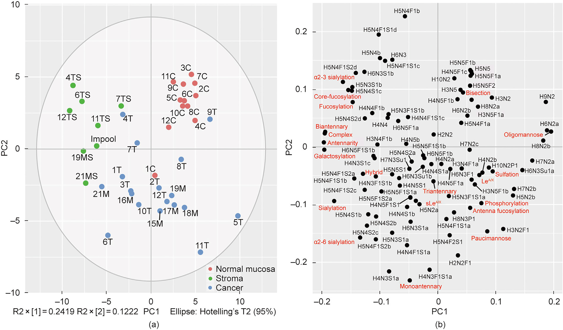
Fig. 1
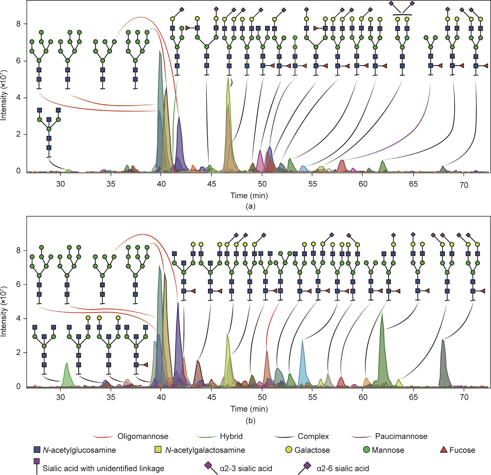
Fig. 2
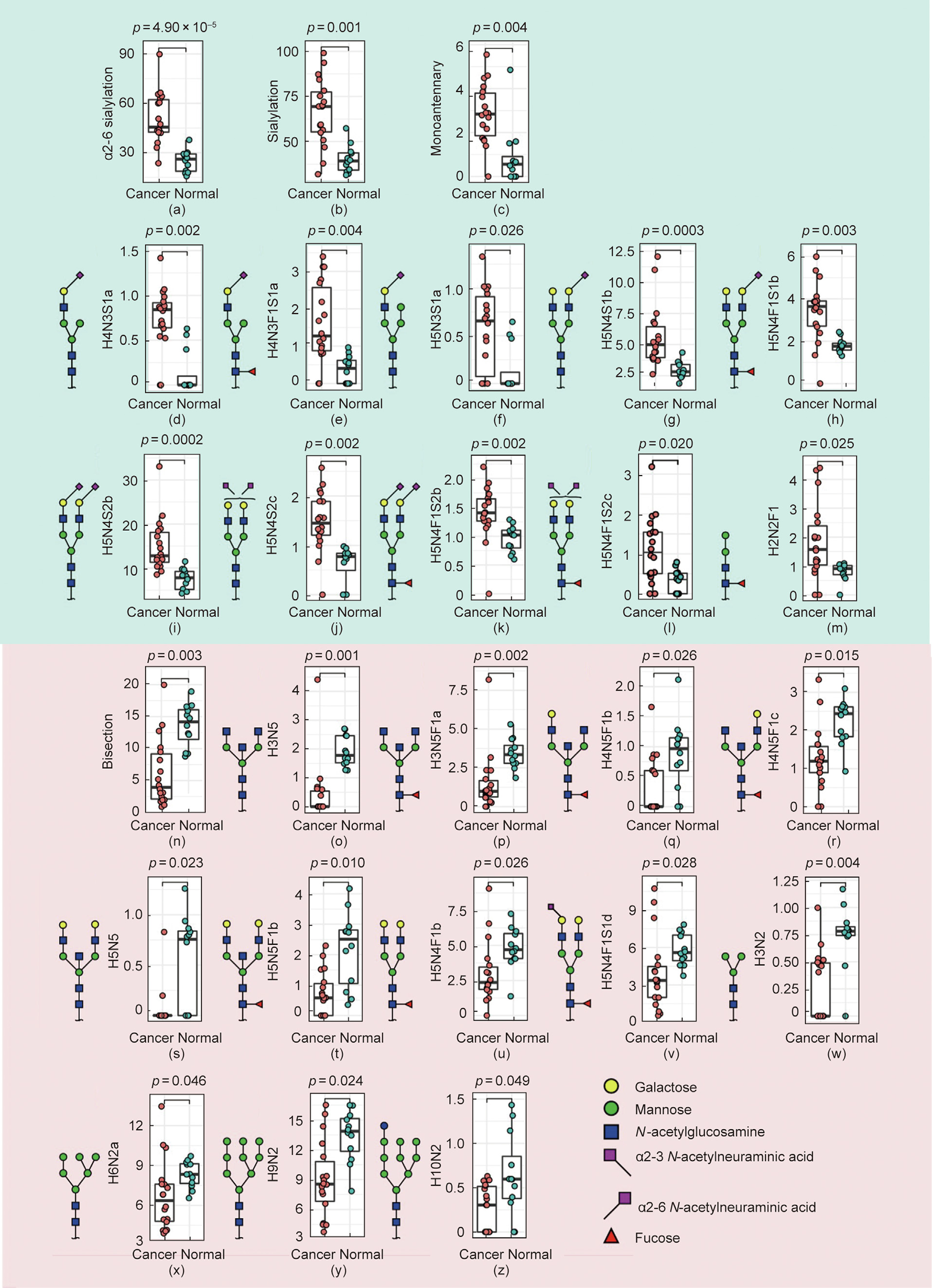
Fig. 3
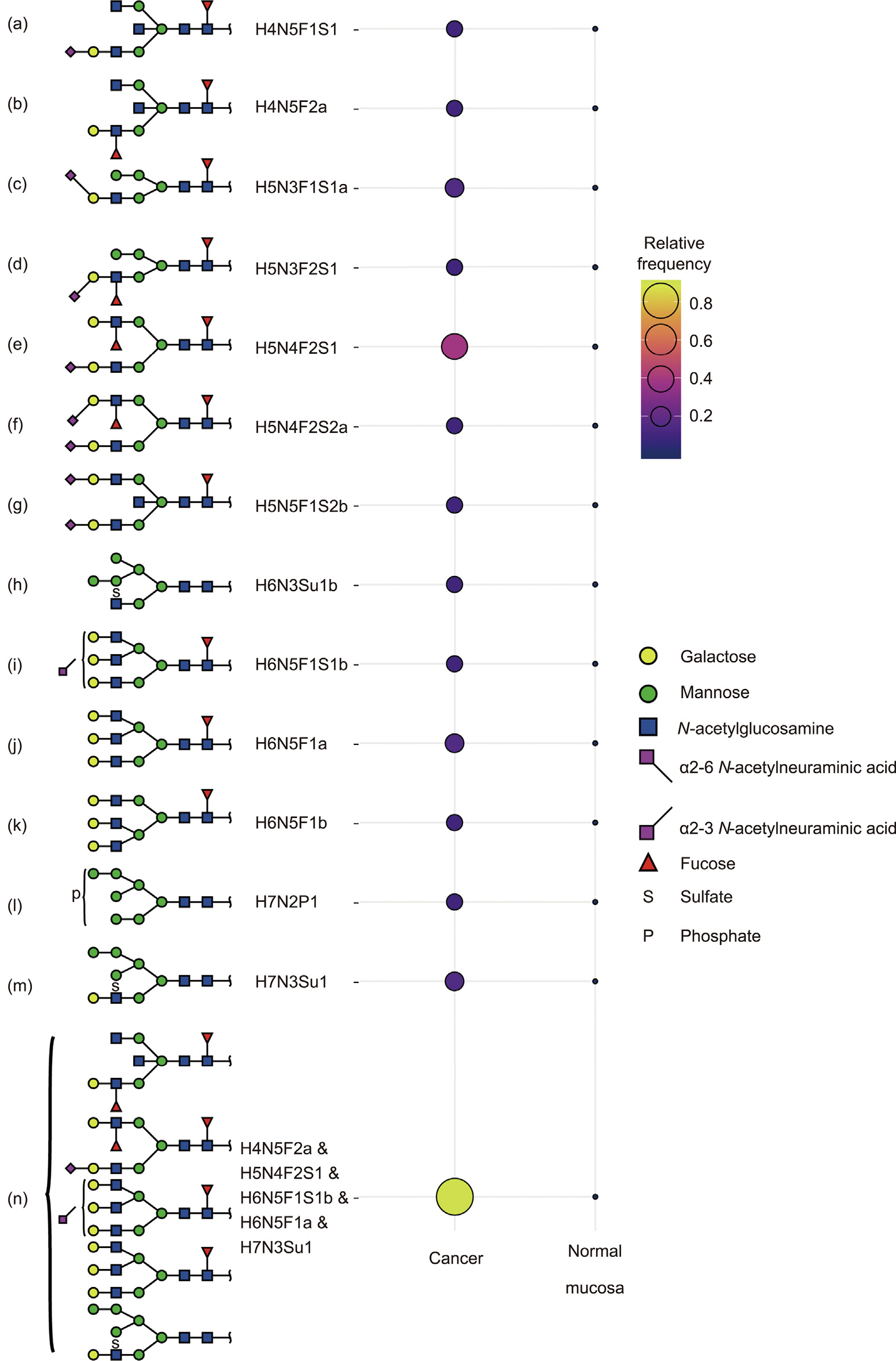
Fig. 4
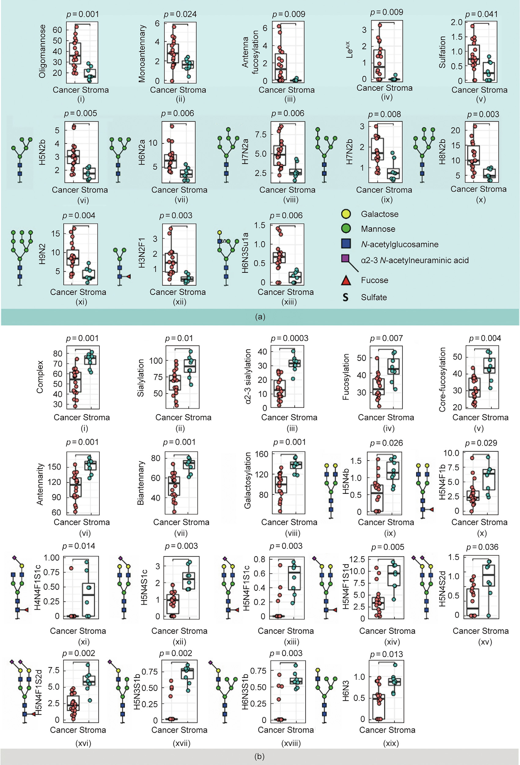
Fig. 5
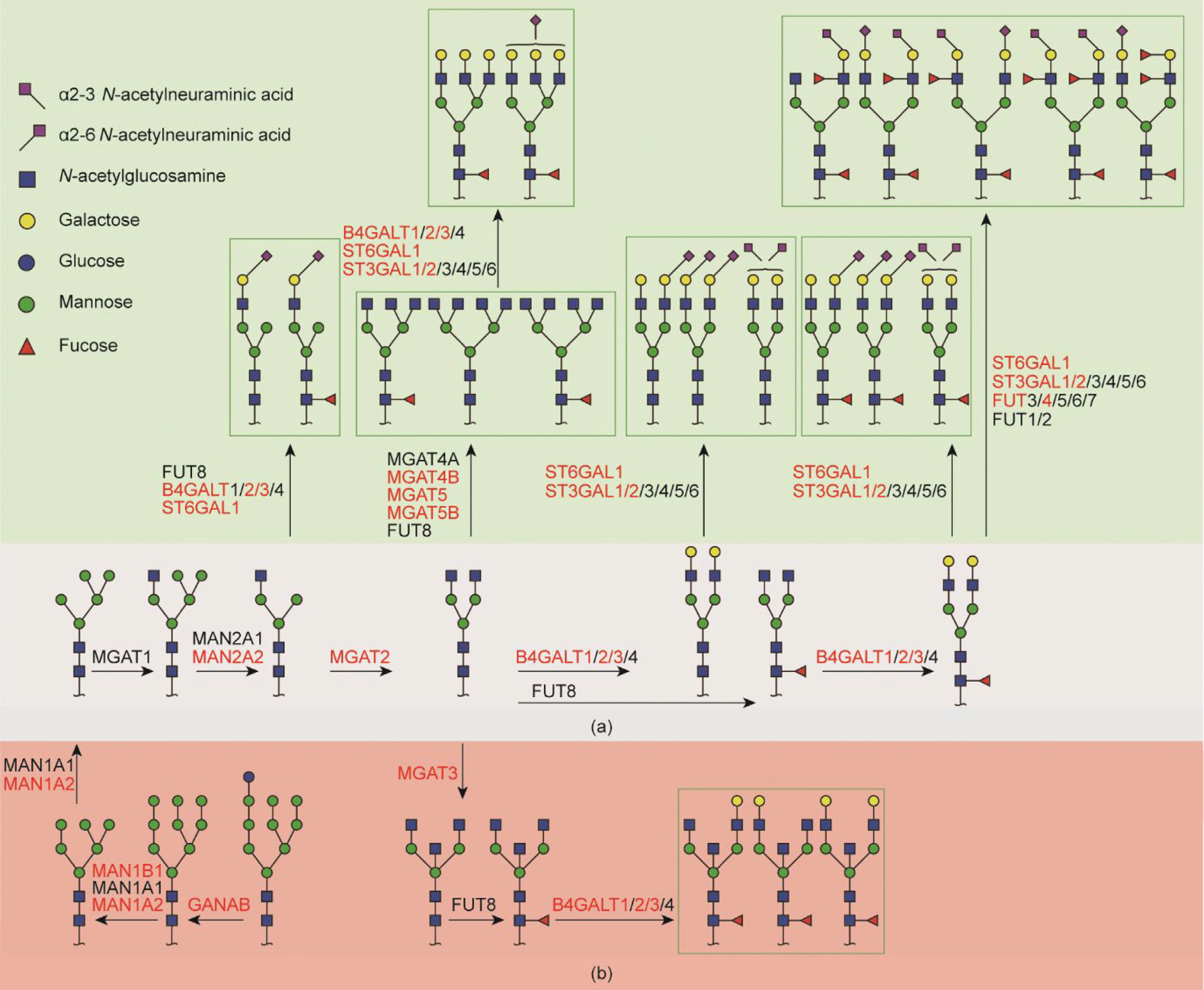
Fig. 6
References
[ 1 ] Bray F, Ferlay J, Soerjomataram I, Siegel RL, Torre LA, Jemal A. Global cancer statistics 2018: GLOBOCAN estimates of incidence and mortality worldwide for 36 cancers in 185 countries. CA Cancer J Clin 2018;68(6):394–424. link1
[ 2 ] Boyaval F, van Zeijl R, Dalebout H, Holst S, van Pelt G, Fariña-Sarasqueta A, et al. N-glycomic signature of stage II colorectal cancer and its association with the tumor microenvironment. Mol Cell Proteomics 2021;20:100057. link1
[ 3 ] Arnold M, Sierra MS, Laversanne M, Soerjomataram I, Jemal A, Bray F. Global patterns and trends in colorectal cancer incidence and mortality. Gut 2017;66 (4):683–91. link1
[ 4 ] Costa AF, Campos D, Reis CA, Gomes C. Targeting glycosylation: a new road for cancer drug discovery. Trends Cancer 2020;6(9):757–66. link1
[ 5 ] Balog CIA, Stavenhagen K, Fung WLJ, Koeleman CA, McDonnell LA, Verhoeven A, et al. N-glycosylation of colorectal cancer tissues: a liquid chromatography and mass spectrometry-based investigation. Mol Cell Proteomics 2012;11 (9):571–85. link1
[ 6 ] Davies RJ, Miller R, Coleman N. Colorectal cancer screening: prospects for molecular stool analysis. Nat Rev Cancer 2005;5(3):199–209. link1
[ 7 ] Pinho SS, Reis CA. Glycosylation in cancer: mechanisms and clinical implications. Nat Rev Cancer 2015;15(9):540–55. link1
[ 8 ] Munkley J, Elliott DJ. Hallmarks of glycosylation in cancer. Oncotarget 2016;7 (23):35478–89. link1
[ 9 ] Magalhães A, Duarte HO, Reis CA. Aberrant glycosylation in cancer: a novel molecular mechanism controlling metastasis. Cancer Cell 2017;31(6):733–5. link1
[10] Qiu Y, Patwa TH, Xu L, Shedden K, Misek DE, Tuck M, et al. Plasma glycoprotein profiling for colorectal cancer biomarker identification by lectin glycoarray and lectin blot. J Proteome Res 2008;7(4):1693–703. link1
[11] Peixoto A, Relvas-Santos M, Azevedo R, Santos LL, Ferreira JA. Protein glycosylation and tumor microenvironment alterations driving cancer hallmarks. Front Oncol 2019;9:380. link1
[12] Zhao YY, Takahashi M, Gu JG, Miyoshi E, Matsumoto A, Kitazume S, et al. Functional roles of N-glycans in cell signaling and cell adhesion in cancer. Cancer Sci 2008;99(7):1304–10. link1
[13] Gu J, Nishikawa A, Tsuruoka N, Ohno M, Yamaguchi N, Kangawa K, et al. Purification and characterization of UDP-N-acetylglucosamine: a-6-D-mannoside b 1–6N-acetylglucosaminyltransferase (N-acetylglucosaminyltransferase V) from a human lung cancer cell line. J Biochem 1993;113(5):614–9. link1
[14] Park JJ, Lee M. Increasing the a2, 6 sialylation of glycoproteins may contribute to metastatic spread and therapeutic resistance in colorectal cancer. Gut Liver 2013;7(6):629–41. link1
[15] Konno A, Hoshino Y, Terashima S, Motoki R, Kawaguchi T. Carbohydrate expression profile of colorectal cancer cells is relevant to metastatic pattern and prognosis. Clin Exp Metastasis 2002;19(1):61–70. link1
[16] Sethi MK, Kim H, Park CK, Baker MS, Paik YK, Packer NH, et al. In-depth Nglycome profiling of paired colorectal cancer and non-tumorigenic tissues reveals cancer-, stage- and EGFR-specific protein N-glycosylation. Glycobiology 2015;25(10):1064–78. link1
[17] Coura MMA, Barbosa EA, Brand GD, Bloch Jr C, de Sousa JB. Identification of differential N-glycan compositions in the serum and tissue of colon cancer patients by mass spectrometry. Biology 2021;10(4):343. link1
[18] Van Pelt GW, Sandberg TP, Morreau H, Gelderblom H, van Krieken JHJM, Tollenaar RAEM, et al. The tumour–stroma ratio in colon cancer: the biological role and its prognostic impact. Histopathology 2018;73(2):197–206. link1
[19] Huijbers A, Tollenaar RAEM, v Pelt GW, Zeestraten ECM, Dutton S, McConkey CC, et al. The proportion of tumor–stroma as a strong prognosticator for stage II and III colon cancer patients: validation in the VICTOR trial. Ann Oncol 2013;24(1):179–85. link1
[20] Hinneburg H, Korac´ P, Schirmeister F, Gasparov S, Seeberger PH, Zoldoš V, et al. Unlocking cancer glycomes from histopathological formalin-fixed and paraffin-embedded (FFPE) tissue microdissections. Mol Cell Proteomics 2017;16(4):524–36. link1
[21] Madunic´ K, Mayboroda OA, Zhang T, Weber J, Boons GJ, Morreau H, et al. Specific (sialyl-)Lewis core 2 O-glycans differentiate colorectal cancer from healthy colon epithelium. Theranostics 2022;12(10):4498–512. link1
[22] Zhang T, Madunic´ K, Holst S, Zhang J, Jin C, ten Dijke P, et al. Development of a 96-well plate sample preparation method for integrated N- and O-glycomics using porous graphitized carbon liquid chromatography-mass spectrometry. Mol Omics 2020;16(4):355–63. link1
[23] Jensen PH, Karlsson NG, Kolarich D, Packer NH. Structural analysis of N- and Oglycans released from glycoproteins. Nat Protoc 2012;7(7):1299–310. link1
[24] Madunic´ K, Wagt S, Zhang T, Wuhrer M, Lageveen-Kammeijer GSM. Dopantenriched nitrogen gas for enhanced electrospray ionization of released glycans in negative ion mode. Anal Chem 2021;93(18):6919–23. link1
[25] Kogo R, Shimamura T, Mimori K, Kawahara K, Imoto S, Sudo T, et al. Long noncoding RNA HOTAIR regulates polycomb-dependent chromatin modification and is associated with poor prognosis in colorectal cancers. Cancer Res 2011;71(20):6320–6. link1
[26] Fernández-Rodríguez J, Feijoo-Carnero C, Merino-Trigo A, Páez de la Cadena M, Rodríguez-Berrocal FJ, de Carlos A, et al. Immunohistochemical analysis of sialic acid and fucose composition in human colorectal adenocarcinoma. Tumour Biol 2000;21(3):153–64. link1
[27] Taniguchi N, Kizuka Y. Glycans and cancer: role of N-glycans in cancer biomarker, progression and metastasis, and therapeutics. Adv Cancer Res 2015;126:11–51. link1
[28] Gessner P, Riedl S, Quentmaier A, Kemmner W. Enhanced activity of CMPneuAc:Galb1-4GlcNAc:a2,6-sialyltransferase in metastasizing human colorectal tumor tissue and serum of tumor patients. Cancer Lett 1993;75 (3):143–9. link1
[29] Dall’Olio F, Malagolini N, di Stefano G, Minni F, Marrano D, Serafini-Cessi F. Increased CMP-NeuAc: Galb1,4GlcNAc-R a2,6 sialyltransferase activity in human colorectal cancer tissues. Int J Cancer 1989;44(3):434–9. link1
[30] Petretti T, Kemmner W, Schulze B, Schlag PM. Altered mRNA expression of glycosyltransferases in human colorectal carcinomas and liver metastases. Gut 2000;46(3):359–66. link1
[31] Seales EC, Jurado GA, Brunson BA, Wakefield JK, Frost AR, Bellis SL. Hypersialylation of b1 integrins, observed in colon adenocarcinoma, may contribute to cancer progression by up-regulating cell motility. Cancer Res 2005;65(11):4645–52. link1
[32] Lin S, Kemmner W, Grigull S, Schlag PM. Cell surface a2,6 sialylation affects adhesion of breast carcinoma cells. Exp Cell Res 2002;276(1):101–10. link1
[33] Vierbuchen MJ, Fruechtnicht W, Brackrock S, Krause KT, Zienkiewicz TJ. Quantitative lectin-histochemical and immunohistochemical studies on the occurrence of a(2,3)- and a(2,6)-linked sialic acid residues in colorectal carcinomas. Relation to clinicopathologic features. Cancer 1995;76(5):727–35. link1
[34] Kaprio T, Satomaa T, Heiskanen A, Hokke CH, Deelder AM, Mustonen H, et al. N-glycomic profiling as a tool to separate rectal adenomas from carcinomas. Mol Cell Proteomics 2015;14(2):277–88. link1
[35] Chatterjee S, Ugonotti J, Lee LY, Everest-Dass A, Kawahara R, ThaysenAndersen M. Trends in oligomannosylation and a1,2-mannosidase expression in human cancers. Oncotarget 2021;12(21):2188–205. link1
[36] Deˇdová T, Braicu EI, Sehouli J, Blanchard V. Sialic acid linkage analysis refines the diagnosis of ovarian cancer. Front Oncol 2019;9:261. link1
[37] Zhang D, Xie Q, Wang Q, Wang Y, Miao J, Li L, et al. Mass spectrometry analysis reveals aberrant N-glycans in colorectal cancer tissues. Glycobiology 2019;29 (5):372–84. link1
[38] Boyaval F, Dalebout H, Van Zeijl R, Wang W, Fariña-Sarasqueta A, LageveenKammeijer GSM, et al. High-mannose N-glycans as malignant progression markers in early-stage colorectal cancer. Cancers 2022;14(6):1552. link1
[39] Tjondro HC, Loke I, Chatterjee S, Thaysen-Andersen M. Human protein paucimannosylation: cues from the eukaryotic kingdoms. Biol Rev Camb Philos Soc 2019;94(6):2068–100. link1
[40] Holm M, Nummela P, Heiskanen A, Satomaa T, Kaprio T, Mustonen H, et al. Nglycomic profiling of colorectal cancer according to tumor stage and location. PLoS One 2020;15(6):e0234989. link1
[41] Holst S, Deuss AJM, van Pelt GW, van Vliet SJ, Garcia-Vallejo JJ, Koeleman CAM, et al. N-glycosylation profiling of colorectal cancer cell lines reveals association of fucosylation with differentiation and caudal type homebox 1 (CDX1)/villin mRNA expression. Mol Cell Proteomics 2016;15(1):124–40. link1
[42] Chen H, Deng Z, Huang C, Wu H, Zhao X, Li Y. Mass spectrometric profiling reveals association of N-glycan patterns with epithelial ovarian cancer progression. Tumour Biol 2017;39(7):1010428317716249. link1
[43] Wang X, Deng Z, Huang C, Zhu T, Lou J, Wang L, et al. Differential N-glycan patterns identified in lung adenocarcinoma by N-glycan profiling of formalin-fixed paraffin-embedded (FFPE) tissue sections. J Proteomics 2018;172:1–10. link1
[44] Miyoshi E, Moriwaki K, Nakagawa T. Biological function of fucosylation in cancer biology. J Biochem 2008;143(6):725–9. link1
[45] Paschos KA, Canovas D, Bird NC. The engagement of selectins and their ligands in colorectal cancer liver metastases. J Cell Mol Med 2010;14(1–2):165–74. link1
[46] Lu HH, Lin SY, Weng RR, Juan YH, Chen YW, Hou HH, et al. Fucosyltransferase 4 shapes oncogenic glycoproteome to drive metastasis of lung adenocarcinoma. EBioMedicine 2020;57:102846. link1
[47] Buckhaults P, Chen L, Fregien N, Pierce M. Transcriptional regulation of Nacetylglucosaminyltransferase V by the src oncogene. J Biol Chem 1997;272 (31):19575–81. link1
[48] Nagae M, Kizuka Y, Mihara E, Kitago Yu, Hanashima S, Ito Y, et al. Structure and mechanism of cancer-associated N-acetylglucosaminyltransferase-V. Nat Commun 2018;9(1):3380. link1
[49] Zhao Y, Sato Y, Isaji T, Fukuda T, Matsumoto A, Miyoshi E, et al. Branched Nglycans regulate the biological functions of integrins and cadherins. FEBS J 2008;275(9):1939–48. link1
[50] Murata K, Miyoshi E, Kameyama M, Ishikawa O, Kabuto T, Sasaki Y, et al. Expression of N-acetylglucosaminyltransferase V in colorectal cancer correlates with metastasis and poor prognosis. Clin Cancer Res 2000;6 (5):1772–7. link1
[51] Brockhausen I, Narasimhan S, Schachter H. The biosynthesis of highly branched N-glycans: studies on the sequential pathway and functional role of N-acetylglucosaminyltransferases I, II, III, IV, V and VI. Biochimie 1988;70 (11):1521–33. link1
[52] Raffaghello L, Dazzi F. Classification and biology of tumour associated stromal cells. Immunol Lett 2015;168(2):175–82. link1


 京公网安备 11010502051620号


