
2019, Volume 5, Issue 3
Engineering >> 2019, Volume 5, Issue 3 doi: 10.1016/j.eng.2019.01.006
Applications for Nanoscale X-Ray Imaging at High Pressure
a Department of Geological Sciences, Stanford University, Stanford, CA 94305, USA
b Stanford Institute for Materials and Energy Sciences, SLAC National Accelerator Laboratory, Menlo Park, CA 94025, USA
c Stanford Synchrotron Radiation Lightsource, SLAC National Accelerator Laboratory, Menlo Park, CA 94025, USA.
Next Previous
Abstract
Coupling nanoscale X-ray transmission microscopy (nanoTXM) with a diamond anvil cell (DAC) has exciting potential as a powerful three-dimensional probe for non-destructive imaging at high spatial resolution of materials under extreme conditions. In this article, we discuss current developments in high-resolution X-ray imaging and its application in highpressure nanoTXM experiments in a DAC with third-generation synchrotron X-ray sources, including technical considerations for preparing successful measurements. We then present results from a number of recent in situ highpressure measurements investigating equations of state (EOS) in amorphous or poorly crystalline materials and in pressure-induced phase transitions and electronic changes. These results illustrate the potential this technique holds for
addressing a wide range of research areas, ranging from condensed matter physics and solid-state chemistry to materials science and planetary interiors. Future directions for this exciting technique and opportunities to improve its capabilities for broader application in high-pressure science are discussed.
Keywords
Figures

Fig. 1

Fig. 2

Fig. 3
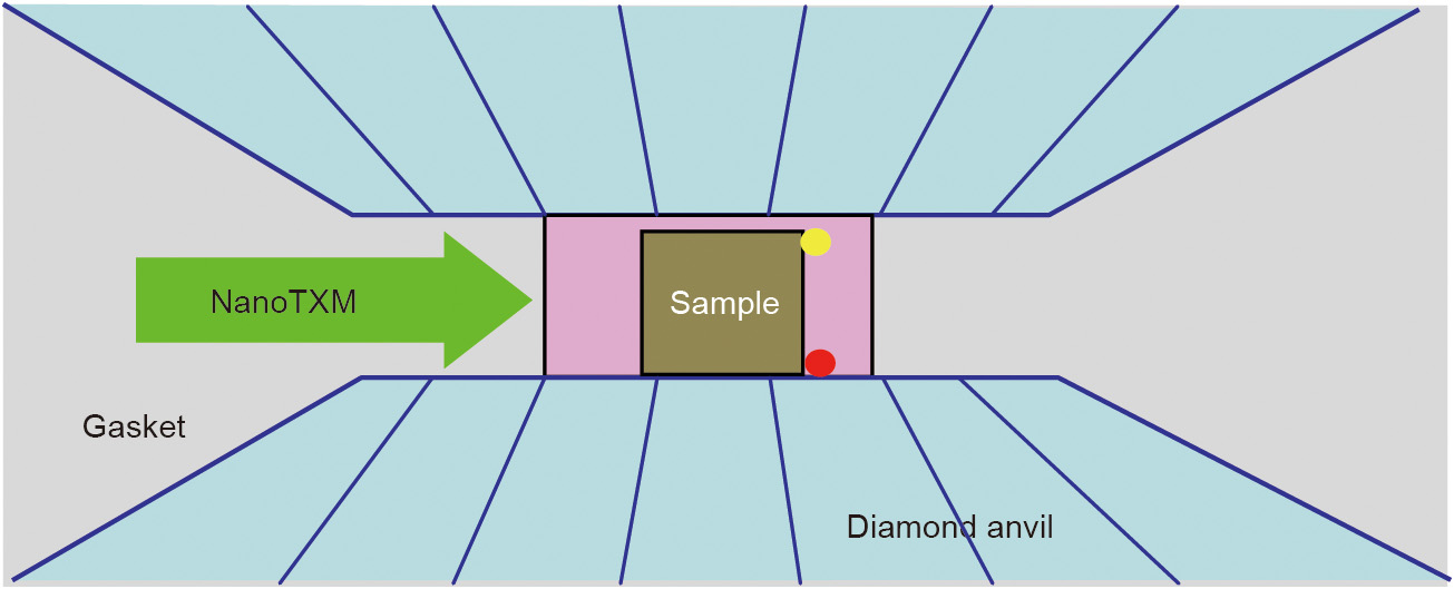
Fig. 4

Fig. 5
Fig. 6
Fig. 7
Fig. 8
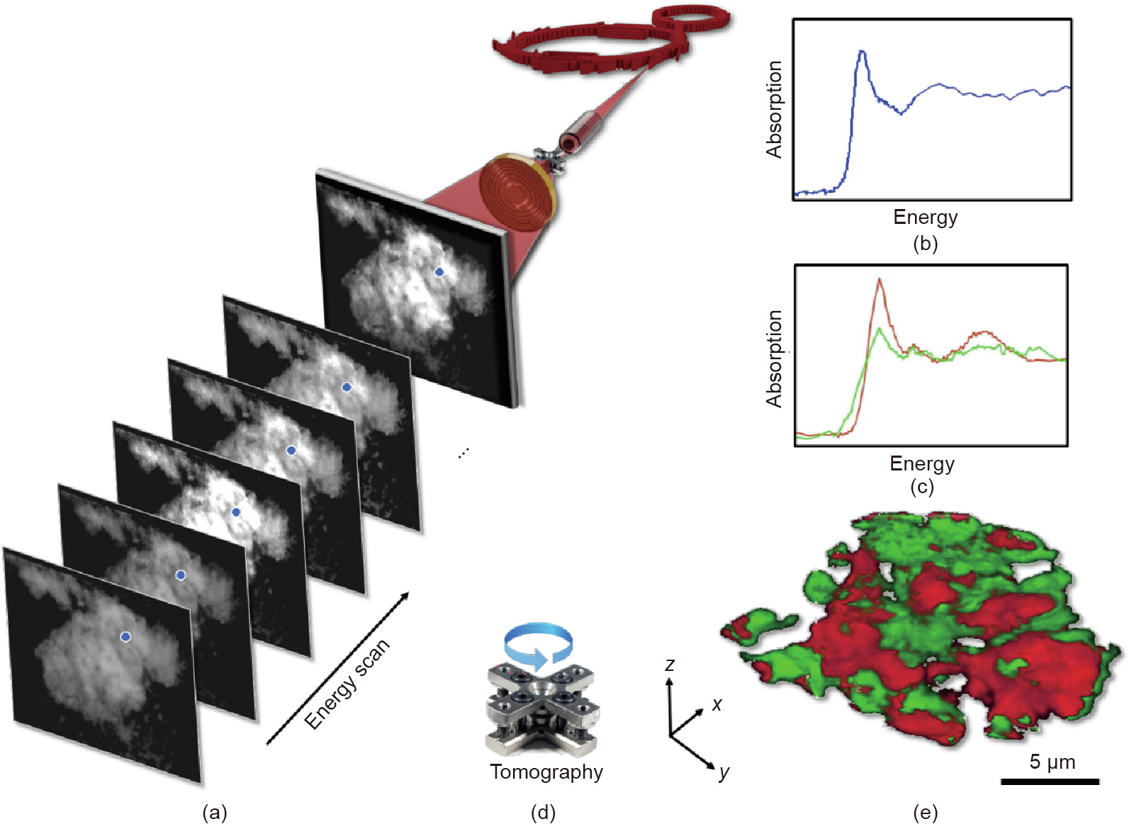
Fig. 9

Fig. 10

Fig. 11
References
[ 1 ] Dubrovinsky L, Dubrovinskaia N, Bykova E, Bykov M, Prakapenka V, Prescher C, et al. The most incompressible metal osmium at static pressures above 750 gigapascals. Nature 2015;525(7568):226–9. link1
[ 2 ] Mao HK, Mao WL. Theory and practice—diamond-anvil cells and probes for high P-T mineral physics studies. In: Price GD, editor. Treatise on geophysics: mineral physics. Amsterdam: Elsevier; 2007. p. 231–68. link1
[ 3 ] Frankel RI. Centennial of Röntgen’s discovery of X-rays. West J Med 1996;164 (6):497–501. link1
[ 4 ] Du Plessis A, Le Roux SG, Guelpa A. Comparison of medical and industrial X-ray computed tomography for non-destructive testing. Case Stud Nondestr Test Eval 2016;6:17–25. link1
[ 5 ] Landis EN, Keane DT. X-ray microtomography. Mater Charact 2010;61 (12):1305–16. link1
[ 6 ] Liu Y, Kiss AM, Larsson DH, Yang F, Pianetta P. To get the most out of high resolution X-ray tomography: a review of the post-reconstruction analysis. Spectrochim Acta B At Spectrosc 2016;117:29–41. link1
[ 7 ] Bautz W, Kalender W, Godfrey N. Hounsfield and his effect on radiology. Radiologe 2005;45(4):350–5. German. link1
[ 8 ] Sakdinawat A, Attwood D. Nanoscale X-ray imaging. Nat Photonics 2010;4 (12):840–8. link1
[ 9 ] Chang C, Sakdinawat A. Ultra-high aspect ratio high-resolution nanofabrication for hard X-ray diffractive optics. Nat Commun 2014;5(1):4243. link1
[10] Shi CY, Zhang L, Yang W, Liu Y, Wang J, Meng Y, et al. Formation of an interconnected network of iron melt at Earth’s lower mantle conditions. Nat Geosci 2013;6(11):971–5. link1
[11] Larabell CA, Nugent KA. Imaging cellular architecture with X-rays. Curr Opin Struct Biol 2010;20(5):623–31. link1
[12] Wei C, Xia S, Huang H, Mao Y, Pianetta P, Liu Y. Mesoscale battery science: the behavior of electrode particles caught on a multispectral X-ray camera. Acc Chem Res 2018;51(10):2484–92. link1
[13] Andrews JC, Weckhuysen BM. Hard X-ray spectroscopic nano-imaging of hierarchical functional materials at work. Chem Phys Chem 2013;14 (16):3655–66. link1
[14] Meirer F, Cabana J, Liu Y, Mehta A, Andrews JC, Pianetta P. Three-dimensional imaging of chemical phase transformations at the nanoscale with full-field transmission X-ray microscopy. J Synchrotron Radiat 2011;18:773–81. link1
[15] Liu Y, Meirer F, Wang J, Requena G, Williams P, Nelson J, et al. 3D elemental sensitive imaging using transmission X-ray microscopy. Anal Bioanal Chem 2012;404(5):1297–301. link1
[16] Andrews JC, Almeida E, Van der Meulen MC, Alwood JS, Lee C, Liu Y, et al. Nanoscale X-ray microscopic imaging of mammalian mineralized tissue. Microsc Microanal 2010;16(3):327–36. link1
[17] Liu Y, Meirer F, Williams PA, Wang J, Andrews JC, Pianetta P. TXM-Wizard: a program for advanced data collection and evaluation in full-field transmission X-ray microscopy. J Synchrotron Radiat 2012;19(Pt 2):281–7. link1
[18] Gürsoy D, De Carlo F, Xiao X, Jacobsen C. TomoPy: a framework for the analysis of synchrotron tomographic data. J Synchrotron Radiat 2014;21:1188–93. link1
[19] Yang X, De Carlo F, Phatak C, Gürsoy D. A convolutional neural network approach to calibrating the rotation axis for X-ray computed tomography. J Synchrotron Radiat 2017;24:469–75. link1
[20] Yang Y, Yang F, Hingerl FF, Xiao X, Liu Y, Wu Z, et al. Registration of the rotation axis in X-ray tomography. J Synchrotron Radiat 2015;22(2):452–7. link1
[21] Guizar-Sicairos M, Boon JJ, Mader K, Diaz A, Menzel A, Bunk O. Quantitative interior X-ray nanotomography by a hybrid imaging technique. Optica 2015;2 (3):259–66. link1
[22] Gürsoy D, Hong YP, He K, Hujsak K, Yoo S, Chen S, et al. Rapid alignment of nanotomography data using joint iterative reconstruction and reprojection. Sci Rep 2017;7(1):11818. link1
[23] Yu H, Xia S, Wei C, Mao Y, Larsson D, Xiao X, et al. Automatic projection image registration for nanoscale X-ray tomographic reconstruction. J Synchrotron Radiat 2018;25:1819–26. link1
[24] Liu Y, Wang J, Azuma M, Mao WL, Yang W. Five-dimensional visualization of phase transition in BiNiO3 under high pressure. Appl Phys Lett 2014;104 (4):043108. link1
[25] Wang JY, Yang W, Wang S, Xiao X, De Carlo F, Liu Y, et al. High pressure nanotomography using an iterative method. J Appl Phys 2012;111(11):112626. link1
[26] Duan X, Yang F, Antono E, Yang W, Pianetta P, Ermon S, et al. Unsupervised data mining in nanoscale X-ray spectro-microscopic study of NdFeB magnet. Sci Rep 2016;6(1):34406. link1
[27] Xu Y, Hu E, Zhang K, Wang X, Borzenets V, Sun Z, et al. In situ visualization of state-of-charge heterogeneity within a LiCoO2 particle that evolves upon cycling at different rates. ACS Energy Lett 2017;2(5):1240–5. link1
[28] Lin Y, Zeng Q, Yang W, Mao WL. Pressure-induced densification in GeO2 glass: a transmission X-ray microscopy study. Appl Phys Lett 2013;103(26):261909. link1
[29] Zeng Q, Kono Y, Lin Y, Zeng Z, Wang J, Sinogeikin SV, et al. Universal fractional noncubic power law for density of metallic glasses. Phys Rev Lett 2014;112 (18):185502. link1
[30] Liu H, Wang L, Xiao X, De Carlo F, Feng J, Mao HK, et al. Anomalous highpressure behavior of amorphous selenium from synchrotron X-ray diffraction and microtomography. Proc Natl Acad Sci USA 2008;105(36):13229–34. link1
[31] Zeng Q, Lin Y, Liu Y, Zeng Z, Shi CY, Zhang B, et al. General 2.5 power law of metallic glasses. Proc Natl Acad Sci USA 2016;113(7):1714–8. link1
[32] Chen DZ, Shi CY, An Q, Zeng Q, Mao WL, Goddard 3rd WA, et al. Fractal atomiclevel percolation in metallic glasses. Science 2015;349(6254):1306–10. link1
[33] Lin Y, Zhang L, Mao HK, Chow P, Xiao Y, Baldini M, et al. Amorphous diamond: a high-pressure superhard carbon allotrope. Phys Rev Lett 2011;107 (17):175504. link1
[34] Lee SK, Lin JF, Cai YQ, Hiraoka N, Eng PJ, Okuchi T, et al. X-ray Raman scattering study of MgSiO3 glass at high pressure: implication for triclustered MgSiO3 melt in Earth’s mantle. Proc Natl Acad Sci USA 2008;105(23):7925–9. link1
[35] Murakami M, Goncharov AF, Hirao N, Masuda R, Mitsui T, Thomas SM, et al. High-pressure radiative conductivity of dense silicate glasses with potential implications for dark magmas. Nat Commun 2014;5(1):5428. link1
[36] Petitgirard S, Malfait WJ, Sinmyo R, Kupenko I, Hennet L, Harries D, et al. Fate of MgSiO3 melts at core-mantle boundary conditions. Proc Natl Acad Sci USA 2015;112(46):14186–90. link1
[37] Sato T, Funamori N. High-pressure structural transformation of SiO2 glass up to 100 GPa. Phys Rev B Condens Matter Mater Phys 2010;82(18):184102. link1
[38] Wu M, Liang Y, Jiang JZ, Tse JS. Structure and properties of dense silica glass. Sci Rep 2012;2(1):398. link1
[39] Zha C, Hemley RJ, Mao H, Duffy TS, Meade C. Acoustic velocities and refractive index of SiO2 glass to 57.5 GPa by Brillouin scattering. Phys Rev B Condens Matter 1994;50(18):13105–12. link1
[40] Sato T, Funamori N. Sixfold-coordinated amorphous polymorph of SiO2 under high pressure. Phys Rev Lett 2008;101(25):255502. link1
[41] Murakami M, Bass JD. Spectroscopic evidence for ultrahigh-pressure polymorphism in SiO2 glass. Phys Rev Lett 2010;104(2):025504. link1
[42] Williams Q, Jeanloz R. Spectroscopic evidence for pressure-induced coordination changes in silicate glasses and melts. Science 1988;239 (4842):902–5. link1
[43] Stixrude L, Karki B. Structure and freezing of MgSiO3 liquid in Earth’s lower mantle. Science 2005;310(5746):297–9. link1
[44] Shen G, Mei Q, Prakapenka VB, Lazor P, Sinogeikin S, Meng Y, et al. Effect of helium on structure and compression behavior of SiO2 glass. Proc Natl Acad Sci USA 2011;108(15):6004–7. link1
[45] Clark AN, Lesher CE, Jacobsen SD, Wang Y. Anomalous density and elastic properties of basalt at high pressure: reevaluating of the effect of melt fraction on seismic velocity in the Earth’s crust and upper mantle. J Geophys Res Solid Earth 2016;121(6):4232–48. link1
[46] Ghosh DB, Karki BB, Stixrude L. First-principles molecular dynamics simulations of MgSiO3 glass: structure, density, and elasticity at high pressure. Am Mineral 2014;99(7):1304–14. link1
[47] Jiang H, Xu R, Chen CC, Yang W, Fan J, Tao X, et al. Three-dimensional coherent X-ray diffraction imaging of molten iron in mantle olivine at nanoscale resolution. Phys Rev Lett 2013;110(20):205501. link1
[48] Miao J, Ishikawa T, Robinson IK, Murnane MM. Beyond crystallography: diffractive imaging using coherent X-ray light sources. Science 2015;348 (6234):530–5. link1
[49] Yang W, Huang X, Harder R, Clark JN, Robinson IK, Mao HK. Coherent diffraction imaging of nanoscale strain evolution in a single crystal under high pressure. Nat Commun 2013;4(1):1680. link1
[50] Eriksson M, Van der Veen JF, Quitmann C. Diffraction-limited storage rings— a window to the science of tomorrow. J Synchrotron Radiat 2014;21: 837–42. link1
[51] McNeil BWJ, Thompson NR. X-ray free-electron lasers. Nat Photonics 2010;4 (12):814–21. link1


 京公网安备 11010502051620号


