2022, Volume 13, Issue 6
Engineering >> 2022, Volume 13, Issue 6 doi: 10.1016/j.eng.2021.01.010
Tailoring Resorption Rates and Osteogenic Response in Xeno-Hybrid Bone Grafts: The Effect of Added Gelatins
a Department of Orthopedic Surgery, Tongji Hospital, Tongji Medical College, Huazhong University of Science and Technology, Wuhan 430030, China
b Department of Biomaterials, Institute of Clinical Dentistry, University of Oslo, Oslo 0317, Norway
c Industrie Biomediche Insubri SA, Mezzovico-Vira 6805, Switzerland
d Ludwig Boltzmann Institute for Experimental and Clinical Traumatology, Donaueschingenstrasse 13, 1200 Vienna, Austria
e Faculty of Biomedical Sciences, University of Southern Switzerland, Lugano 6900, Switzerland
f Faculty of Veterinary, University of Santiago de Compostela, Lugo 27002, Spain
Next Previous
Abstract
Bone defects resulting from trauma, surgery, congenital malformations, and other factors are among the most common health problems nowadays. Although current strategies such as autografts and allografts are recognized as the most successful treatments for stimulating bone regeneration, limitations such as graft source and complications still exist. SmartBone® is a xeno-hybrid bone graft (made from bovine bone matrix, poly(L-lactic-co-ε-caprolactone), and gelatin) with a positive clinical record for bone regeneration. In this study, the formulation for designing xeno-hybrid bone grafts using gelatins from different sources (bovine- and porcine-derived gelatin, with bone grafts named SBN and SPK, respectively) was investigated, and the biological responses were evaluated in vitro and in vivo. The results demonstrate that gelatins from both bovine and porcine sources can be loaded onto SmartBone® successfully and safely, withstanding the aggressive manufacturing processes. Different bone cell responses were observed in vitro. SBN was found to enhance osteocalcin secretion while SPK was found to upregulate osteopontin from human osteoblasts. In vivo, both bone grafts promoted osteogenesis, but SPK degraded earlier than SBN. Our findings suggest that SBN and SPK provide different yet comparable solutions for optimizing the bone resorption and regeneration balance. These xeno-hybrid bone grafts possess ideal potential for bone defect repairing.
Keywords
Bone graft ; Xeno-hybrid ; Gelatin source ; Resorption ; Bone regeneration
SupplementaryMaterials
Figures

Fig. 1
Fig. 2

Fig. 3
Fig. 4
Fig. 5
Fig. 6
Fig. 7
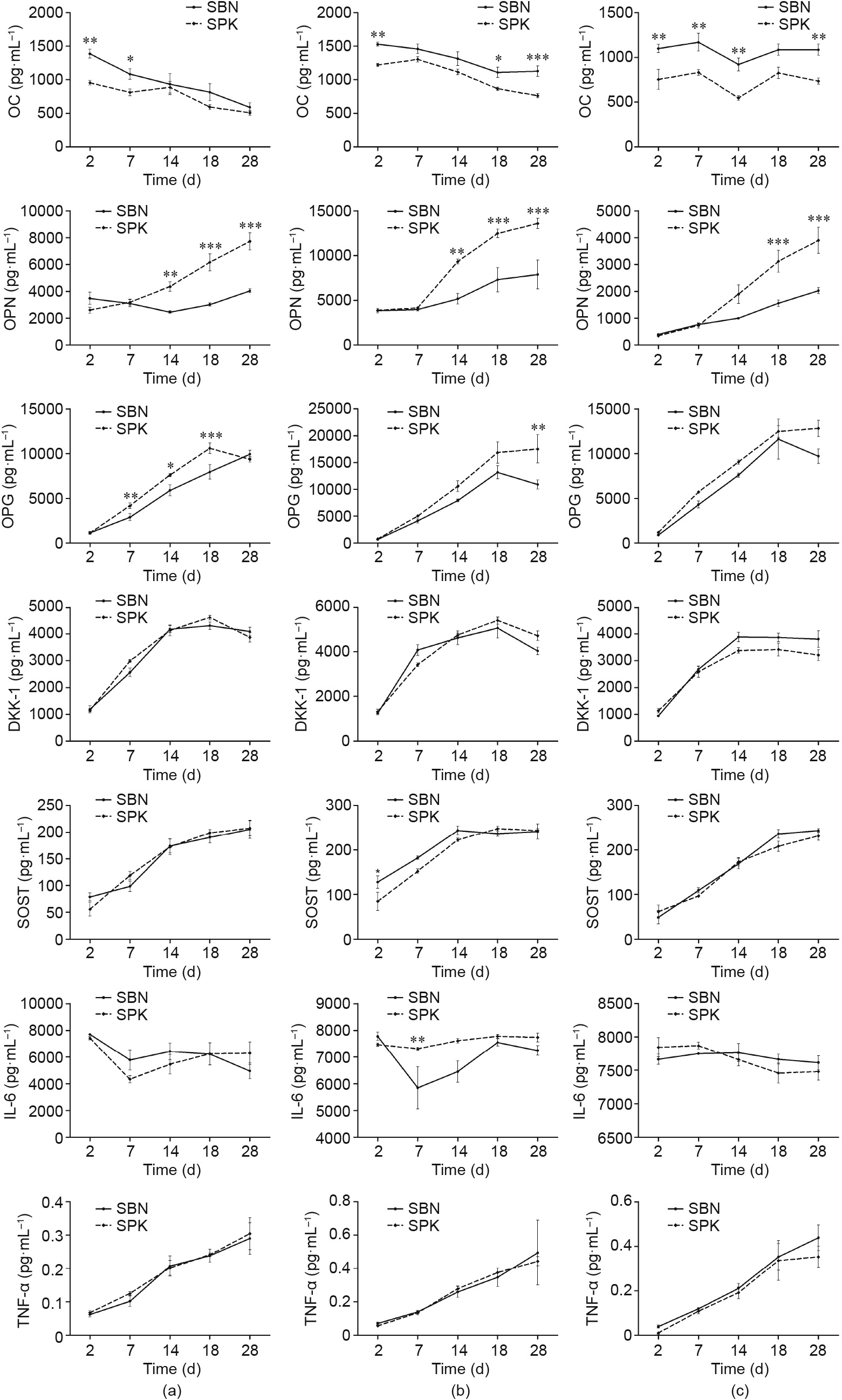
Fig. 8
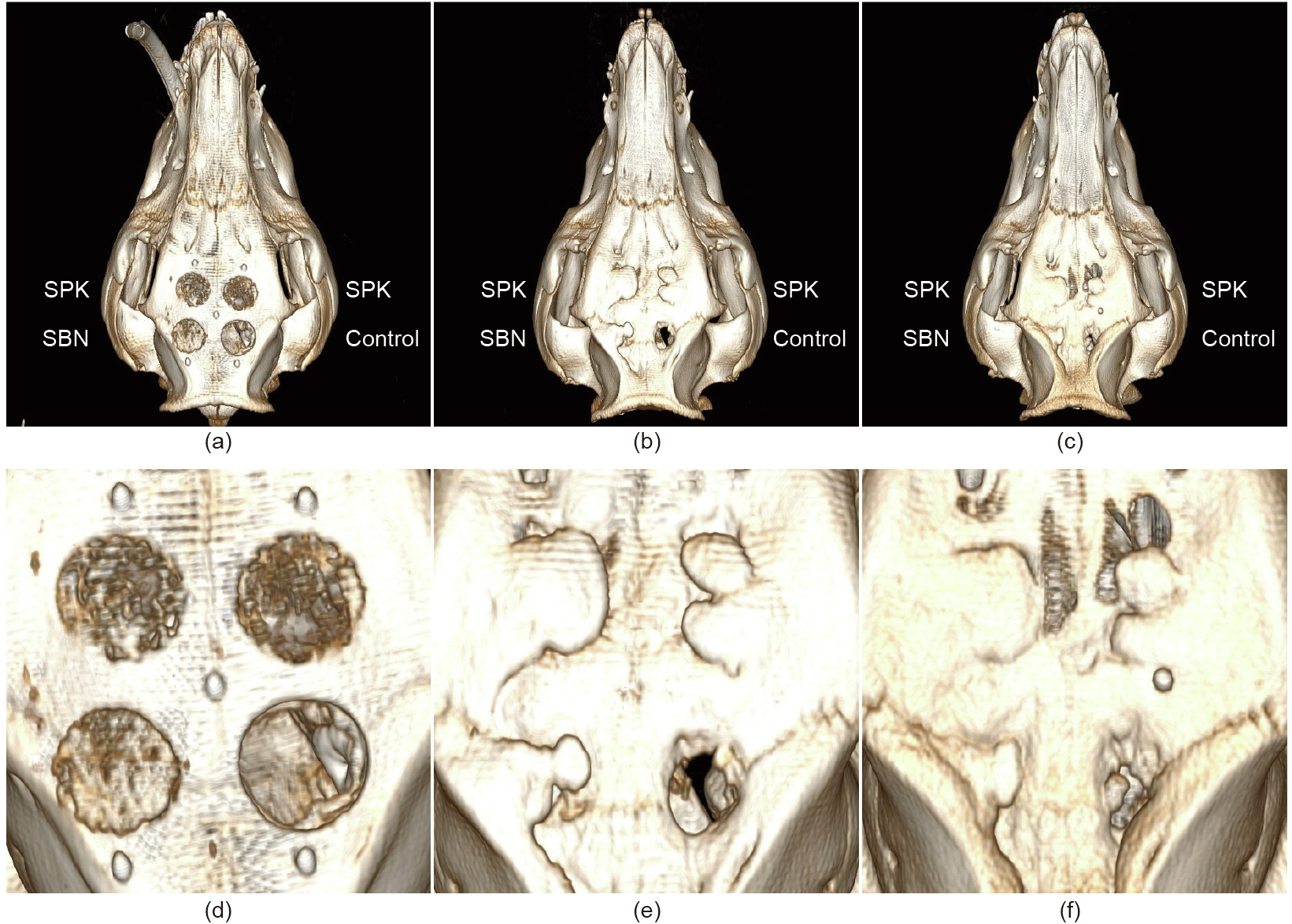
Fig. 9
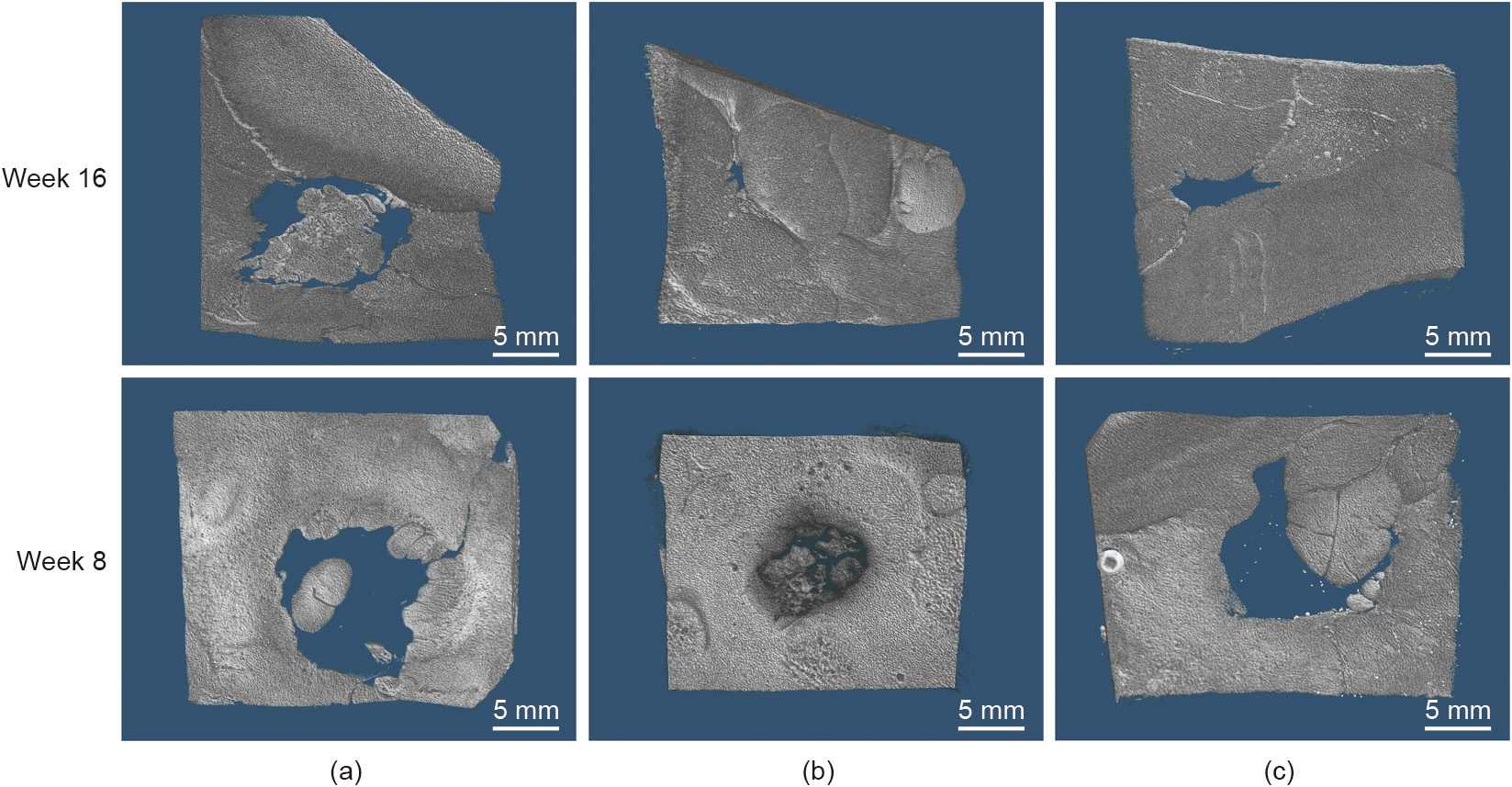
Fig. 10

Fig. 11
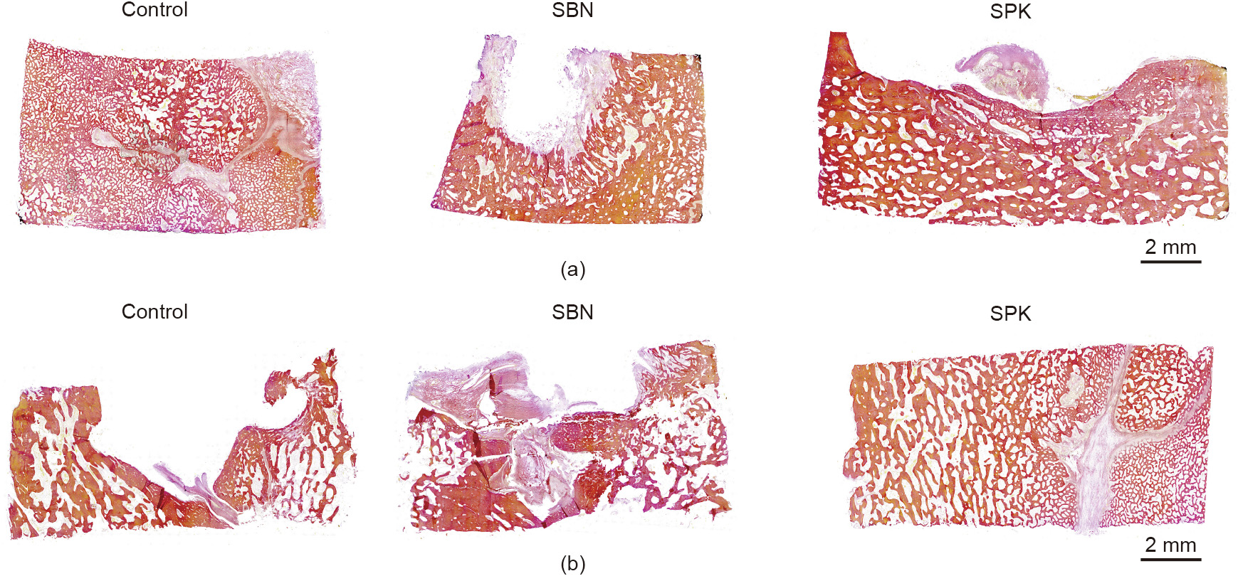
Fig. 12
References
[ 1 ] Wang W, Yeung KWK. Bone grafts and biomaterials substitutes for bone defect repair: a review. Bioact Mater 2017;2(4):224–47. link1
[ 2 ] Winkler T, Sass FA, Duda GN, Schmidt-Bleek K. A review of biomaterials in bone defect healing, remaining shortcomings and future opportunities for bone tissue engineering. Bone Joint Res 2018;7(3):232–43. link1
[ 3 ] Kaur M, Singh K. Review on titanium and titanium based alloys as biomaterials for orthopaedic applications. Mater Sci Eng C 2019;102:844–62. link1
[ 4 ] Pierannunzii L, Zagra L. Bone grafts, bone graft extenders, substitutes and enhancers for acetabular reconstruction in revision total hip arthroplasty. EFORT Open Rev 2016;1(12):431–9. link1
[ 5 ] Fillingham Y, Jacobs J. Bone grafts and their substitutes. Bone Joint J 2016;98-B (1 Suppl A):6–9. link1
[ 6 ] Sakkas A, Wilde F, Heufelder M, Winter K, Schramm A. Autogenous bone grafts in oral implantology—is it still a ‘‘gold standard”? A consecutive review of 279 patients with 456 clinical procedures. Int J Implant Dent 2017;3(1):23. link1
[ 7 ] Roseti L, Parisi V, Petretta M, Cavallo C, Desando G, Bartolotti I, et al. Scaffolds for bone tissue engineering: state of the art and new perspectives. Mater Sci Eng C 2017;78:1246–62. link1
[ 8 ] Sanz M, Dahlin C, Apatzidou D, Artzi Z, Bozic D, Calciolari E, et al. Biomaterials and regenerative technologies used in bone regeneration in the craniomaxillofacial region: consensus report of group 2 of the 15th European Workshop on Periodontology on Bone Regeneration. J Clin Periodontol 2019;46:82–91. link1
[ 9 ] Agarwal R, García AJ. Biomaterial strategies for engineering implants for enhanced osseointegration and bone repair. Adv Drug Deliv Rev 2015;94:53–62. link1
[10] Skaggs DL, Samuelson MA, Hale JM, Kay RM, Tolo VT. Complications of posterior iliac crest bone grafting in spine surgery in children. Spine 2000;25 (18):2400–2. link1
[11] Colo E, Rijnen WHC, Schreurs BW. The biological approach in acetabular revision surgery: impaction bone grafting and a cemented cup. Hip Int 2015;25(4):361–7. link1
[12] Issack PS, Nousiainen M, Beksac B, Helfet DL, Sculco TP, Buly RL. Acetabular component revision in total hip arthroplasty. Part II: management of major bone loss and pelvic discontinuity. Am J Orthop 2009;38(11):550–6. link1
[13] Greenwald AS, Boden SD, Goldberg VM, Khan Y, Laurencin CT, Rosier RN; American Academy of Orthopaedic Surgeons, The Committee on Biological Implants. Bone-graft substitutes: facts, fictions, and applications. J Bone Joint Surg Am 2001;83-A(Suppl 2 Pt 2):98–103.
[14] Sang T, Li S, Ting HK, Stevens MM, Becer CR, Jones JR. Hybrids of silica/poly (caprolactone coglycidoxypropyl trimethoxysilane) as biomaterials. Chem Mater 2018;30(11):3743–51. link1
[15] Saigo L, Kumar V, Liu Y, Lim J, Teoh SH, Goh BT. A pilot study: clinical efficacy of novel polycaprolactone–tricalcium phosphate membrane for guided bone regeneration in rabbit calvarial defect model. J Oral Max Surg Med 2018;30 (3):212–9. link1
[16] Nasajpour A, Ansari S, Rinoldi C, Rad AS, Aghaloo T, Shin SR, et al. A multifunctional polymeric periodontal membrane with osteogenic and antibacterial characteristics. Adv Funct Mater 2018;28(3):1703437. link1
[17] Huang B, Caetano G, Vyas C, Blaker JJ, Diver C, Bártolo P. Polymer-ceramic composite scaffolds: the effect of hydroxyapatite and b-tri-calcium phosphate. Materials 2018;11(1):129. link1
[18] Cristofaro F, Gigli M, Bloise N, Chen H, Bruni G, Munari A, et al. Influence of the nanofiber chemistry and orientation of biodegradable poly(butylene succinate)-based scaffolds on osteoblast differentiation for bone tissue regeneration. Nanoscale 2018;10(18):8689–703. link1
[19] Domingos M, Gloria A, Coelho J, Bartolo P, Ciurana J. Three-dimensional printed bone scaffolds: the role of nano-/micro-hydroxyapatite particles on the adhesion and differentiation of human mesenchymal stem cells. Proc Inst Mech Eng H 2017;231(6):555–64. link1
[20] Haugen HJ, Lyngstadaas SP, Rossi F, Perale G. Bone grafts: which is the ideal biomaterial? J Clin Periodontol 2019;46(Suppl 21):92–102. link1
[21] Amini AR, Laurencin CT, Nukavarapu SP. Bone tissue engineering: recent advances and challenges. Crit Rev Biomed Eng 2012;40(5):363–408. link1
[22] Athanasiou VT, Papachristou DJ, Panagopoulos A, Saridis A, Scopa CD, Megas P. Histological comparison of autograft, allograft-DBM, xenograft, and synthetic grafts in a trabecular bone defect: an experimental study in rabbits. Med Sci Monit 2010;16(1):BR24–31. link1
[23] Meloni SM, Jovanovic SA, Pisano M, Xhanari E, De Riu G, Tullio A, et al. Sinus lift grafting with anorganic bovine bone vs 50% autologous bone mixed with 50% anorganic bovine bone: 2 years after loading results from a randomised controlled trial. Eur J Oral Implantol 2017;10(4):425–32. link1
[24] Cheng L, Wang Yi, Sun G, Wen S, Deng L, Zhang H, et al. Hydration-enhanced lubricating electrospun nanofibrous membranes prevent tissue adhesion. Research 2020;2020:1–12. link1
[25] Chen W, Tian X, He W, Li J, Feng Y, Pan G. Emerging functional materials based on chemically designed molecular recognition. BMC Materials 2020;2(1):1. link1
[26] Qian Y, Shen Y, Deng S, Liu T, Qi F, Lu Z, et al. Dual functional b-peptide polymer-modified resin beads for bacterial killing and endotoxin adsorption. BMC Materials 2019;1(1):5. link1
[27] D’Alessandro D, Perale G, Milazzo M, Moscato S, Stefanini C, Pertici G, et al. Bovine bone matrix/poly(L-lactic-co-e-caprolactone)/gelatin hybrid scaffold (SmartBone) for maxillary sinus augmentation: a histologic study on bone regeneration. Int J Pharm 2017;523(2):534–44. link1
[28] Facciuto E, Grottoli CF, Mattarocci M, Illiano F, Compagno M, Ferracini R, et al. Three-dimensional craniofacial bone reconstruction with SmartBone on demand. J Craniofac Surg 2019;30(3):739–41. link1
[29] Grottoli CF, Cingolani A, Zambon F, Ferracini R, Villa T, Perale G. Simulated performance of a xenohybrid bone graft (SmartBone) in the treatment of acetabular prosthetic reconstruction. J Funct Biomater 2019;10(4):53. link1
[30] Cingolani A, Grottoli CF, Esposito R, Villa T, Rossi F, Perale G. Improving bovine bone mechanical characteristics for the development of xenohybrid bone grafts. Curr Pharm Biotechnol 2019;19(12):1005–13. link1
[31] van den Bosch E, Gielens C. Gelatin degradation at elevated temperature. Int J Biol Macromol 2003;32(3-5):129–38. link1
[32] Esposito M, Grusovin MG, Papanikolaou N, Coulthard P, Worthington HV. Enamel matrix derivative (Emdogain) for periodontal tissue regeneration in intrabony defects. Eur J Oral Implantol 2009;2(4):247–66. link1
[33] Leijten J, Chai YC, Papantoniou I, Geris L, Schrooten J, Luyten FP. Cell based advanced therapeutic medicinal products for bone repair: keep it simple? Adv Drug Deliv Rev 2015;84:30–44. link1
[34] Gallop PM, Lian JB, Hauschka PV. Carboxylated calcium-binding proteins and vitamin K. N Engl J Med 1980;302(26):1460–6. link1
[35] McKee MD, Cole WG. Bone matrix and mineralization. In: Glorieux FH, Pettifor JM, Jüppner H, editors. Pediatric bone. San Diego: Academic Press; 2012. p. 9–37. link1
[36] Price PA, Otsuka AA, Poser JW, Kristaponis J, Raman N. Characterization of a ccarboxyglutamic acid-containing protein from bone. Proc Natl Acad Sci USA 1976;73(5):1447–51. link1
[37] Eastell R, Hannon RA. Biochemical markers of bone turnover. In: Lobo RA, editor. Treatment of the postmenopausal woman. St. Louis: Academic Press; 2007. p. 337–49. link1
[38] Hall BK, Part I. Vertebrate skeletal tissues. In: Hall BK, editor. Bones and cartilage. San Diego: Academic Press; 2015. p. 1. link1
[39] Maeda H, Wada N, Tomokiyo A, Monnouchi S, Akamine A. Prospective potency of TGF-a1 on maintenance and regeneration of periodontal tissue. In: Jeon KW, editor. International review of cell and molecular biology. New York: Academic Press; 2013. p. 283–367. link1
[40] Butler WT. The nature and significance of osteopontin. Connect Tissue Res 1989;23(2–3):123–36. link1
[41] Sodek J, Ganss B, McKee MD. Osteopontin. Crit Rev Oral Biol Med 2000;11 (3):279–303. link1
[42] Chabas D. L’ostéopontine, une molécule aux multiples facettes. Med Sci 2005;21(10):832–8. French. link1
[43] Giachelli CM, Steitz S. Osteopontin: a versatile regulator of inflammation and biomineralization. Matrix Biol 2000;19(7):615–22. link1
[44] Nikel O, Laurencin D, McCallum SA, Gundberg CM, Vashishth D. NMR investigation of the role of osteocalcin and osteopontin at the organic– inorganic interface in bone. Langmuir 2013;29(45):13873–82. link1
[45] Dashdulam D, Kim ID, Lee H, Lee HK, Kim SW, Lee JK. Osteopontin heptamer peptide containing the RGD motif enhances the phagocytic function of microglia. Biochem Biophys Res Commun 2020;524(2):371–7. link1
[46] Denhardt DT, Guo X. Osteopontin: a protein with diverse functions. FASEB J 1993;7(15):1475–82. link1
[47] Kaminska B. Role of osteopontin–integrin signalling in glioma–microglia crosstalk. FEBS Open Bio 2019;9:33. link1
[48] Schett G, Redlich K, Smolen JS. The role of osteoprotegerin in arthritis. Arthritis Res Ther 2003;5(5):239–45. link1
[49] Lories RJU, Luyten FP. Osteoprotegerin and osteoprotegerin-ligand balance: a new paradigm in bone metabolism providing new therapeutic targets. Clin Rheumatol 2001;20(1):3–9. link1
[50] Yeung RSM. The osteoprotegerin/osteoprotegerin ligand family: role in inflammation and bone loss. J Rheumatol 2004;31(5):844–6. link1
[51] Kurban S, Mehmetoglu I. Osteoprotegerin, RANK and RANK ligand. Turk J Biochem 2007;32(4):178–84. link1
[52] Martin TJ, Romas E, Gillespie MT. Interleukins in the control of osteoclast differentiation. Crit Rev Eukaryot Gene Expr 1998;8(2):107–23. link1
[53] Wang T, He C. TNF-a and IL-6: the link between immune and bone system. Curr Drug Targets 2020;21(3):213–27. link1
[54] Williams DF. On the mechanisms of biocompatibility. Biomaterials 2008;29 (20):2941–53. link1
[55] Murphy CM, Haugh MG, O’Brien FJ. The effect of mean pore size on cell attachment, proliferation and migration in collagen–glycosaminoglycan scaffolds for bone tissue engineering. Biomaterials 2010;31(3):461–6. link1


 京公网安备 11010502051620号


