2022, Volume 15, Issue 8
Engineering >> 2022, Volume 15, Issue 8 doi: 10.1016/j.eng.2021.03.013
Multi-Omics Analysis Provides Insight into the Possible Molecular Mechanism of Hay Fever Based on Gut Microbiota
a State Key Laboratory of Bioactive Substance and Function of Natural Medicines, Institute of Materia Medica, Chinese Academy of Medical Sciences & Peking Union Medical College, Beijing 100050, China
b Department of Allergy, National Clinical Research Center for Dematologic and Immunologic Diseases, Beijing Key Laboratory of Precision Medicine for Diagnosis and Treatment on Allergic Diseases, Peking Union Medical College Hospital, Chinese Academy of Medical Sciences & Peking Union Medical College, Beijing 100730, China
c Department of Allergy & Department of Otorhinolaryngology Head and Neck Surgery & Key Laboratory of Otolaryngology Head and Neck Surgery, Ministry of Education, Beijing Tongren Hospital, Capital Medical University, Beijing 100005, China
d Department of Allergy, Beijing Shijitan Hospital, Capital Medical University, Beijing 100038, China
e Beijing Key Laboratory of Nasal diseases, Beijing Institute of Otolaryngology, Beijing Tongren Hospital, Capital Medical University, Beijing 100005, China
Next Previous
Abstract
Due to the worldwide epidemic of allergic disease and a cure nowhere in sight, there is a crucial need to explore its pathophysiological mechanisms. As allergic disease has been associated with gut dysbiosis, we searched for a possible mechanism from the perspective of the molecular interface between host and microbiota with concurrent metabolomics and microbiome composition analysis. Sprague-Dawley rats were injected with Artemisia pollen extract to stimulate a hyper reaction to pollen. This hyper reaction decreased the circulation of valine, isoleucine, aspartate, glutamate, glutamine, indole-propionate (IPA), and myo-inositol, and reduced short-chain fatty acids (SCFAs) in feces. Several beneficial genera belonging to Ruminococcaceae, Lachnospiraceae, and Clostridiales declined in the model group, whereas Helicobacter and Akkermansia were only expressed in the model group. Furthermore, the expression of intestinal claudin-3 and liver fatty acid binding protein was downregulated in the model group and associated with metabolic changes and bacteria. Our results suggest that alterations in amino acids as well as their derivatives (especially valine, and IPA which is the reductive product of tryptophan) , SCFAs, and the gut microbiome (specifically Akkermansia and Helicobacter) may disrupt the intestinal barrier function by inhibiting the expression of claudin proteins and affecting the mucus layer, which further results in hay fever.
Keywords
Metabolome ; Gut microbiota ; Hay fever ; Allergic diseases ; Intestinal barrier dysfunction
SupplementaryMaterials
Figures

Fig. 1

Fig. 2

Fig. 3

Fig. 4
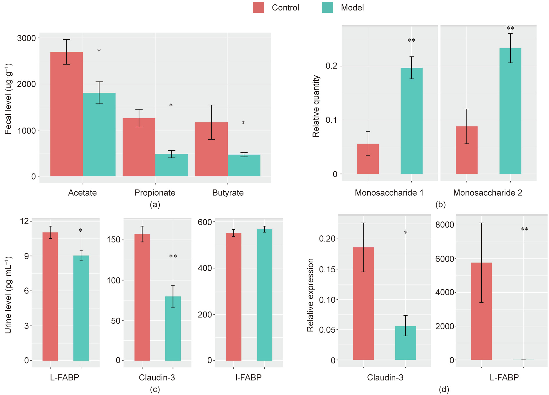
Fig. 5
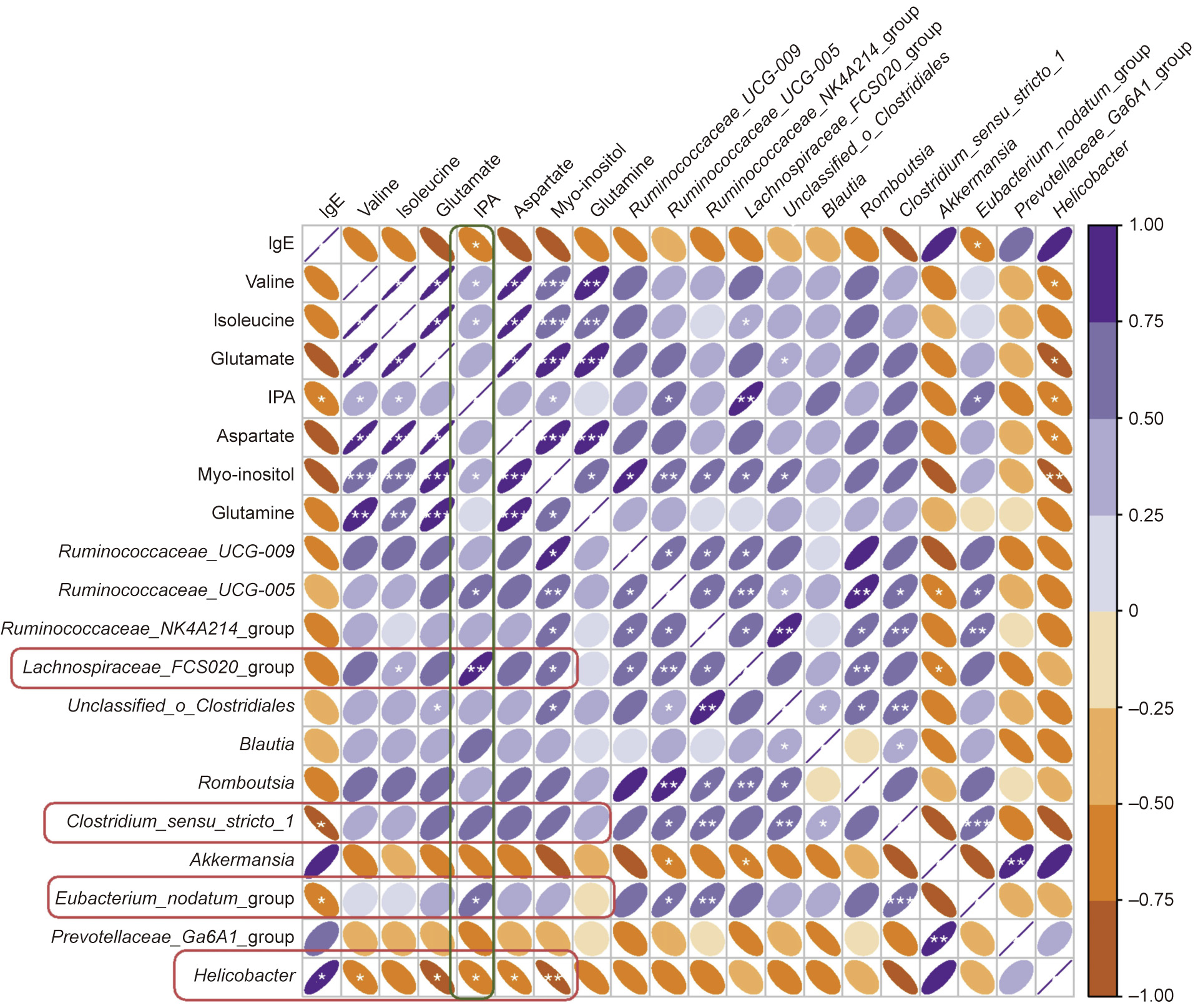
Fig. 6

Fig. 7
References
[ 1 ] B W. Hay fever. Nature 1923;111(2798):812–4.
[ 2 ] Victorio Puche L, Somoza ML, López-Sánchez JD, Garrido-Arandia M, DíazPerales A, Blanca M. Peach tree pollen and prunus persica 9 sensitisation and allergy in children and adolescents. Int Arch Allergy Immunol 2019;180 (3):212–20. link1
[ 3 ] Voukantsis D, Berger U, Tzima F, Karatzas K, Jaeger S, Bergmann KC. Personalized symptoms forecasting for pollen-induced allergic rhinitis sufferers. Int J Biometeorol 2015;59(7):889–97. link1
[ 4 ] Pablos I, Wildner S, Asam C, Wallner M, Gadermaier G. Pollen allergens for molecular diagnosis. Curr Allergy Asthma Rep 2016;16(4):31. link1
[ 5 ] Wang XY, Ma TT, Wang XY, Zhuang Y, Wang XD, Ning HY, et al. Prevalence of pollen-induced allergic rhinitis with high pollen exposure in grasslands of northern China. Allergy 2018;73(6):1232–43. link1
[ 6 ] Xie ZJ, Guan K, Yin J. Advances in the clinical and mechanism research of pollen induced seasonal allergic asthma. Am J Clin Exp Immunol 2019;8(1):1–8. link1
[ 7 ] Wambre E, Bajzik V, DeLong JH, O’Brien K, Nguyen QA, Speake C, et al. A phenotypically and functionally distinct human TH2 cell subpopulation is associated with allergic disorders. Sci Transl Med 2017;9(401):eaam9171. link1
[ 8 ] Wahn U. Considering 25 years of research on allergy prevention—have we let ourselves down? Pediatr Allergy Immunol 2013;24(4):308–10. link1
[ 9 ] Bernard A, Nickmilder M, Dumont X. Airway epithelium defects and risks of allergic diseases: multiple associations revealed by a biomarker study among adolescents. Am J Respir Crit Care Med 2015;191(6):714–7. link1
[10] Åsa J, Mathias RA, Torgny K, Ek WE. Genome-wide association analysis of 350 000 Caucasians from the UK Biobank identifies novel loci for asthma, hay fever and eczema. Hum Mol Genet 2019;28(23):4022–41. link1
[11] Ferreira MA, Matheson MC, Tang CS, Granell R, Ang W, Hui J, et al.; Australian Asthma Genetics Consortium Collaborators. Genome-wide association analysis identifies 11 risk variants associated with the asthma with hay fever phenotype. J Allergy Clin Immunol 2014;133(6):1564–71. link1
[12] Lambrecht BN, Hammad H. The immunology of the allergy epidemic and the hygiene hypothesis. Nat Immunol 2017;18(10):1076–83. link1
[13] Crestani E, Harb H, Charbonnier LM, Leirer J, Motsinger-Reif A, Rachid R, et al. Untargeted metabolomic profiling identifies disease-specific signatures in food allergy and asthma. J Allergy Clin Immunol 2020;145(3):897–906. link1
[14] Ursell LK, Metcalf JL, Parfrey LW, Knight R. Defining the human microbiome. Nutr Rev 2012;70(Suppl 1):S38–44. link1
[15] Wikoff WR, Anfora AT, Liu J, Schultz PG, Lesley SA, Peters EC, et al. Metabolomics analysis reveals large effects of gut microflora on mammalian blood metabolites. Proc Natl Acad Sci USA 2009;106(10):3698–703. link1
[16] Marcobal A, Kashyap PC, Nelson TA, Aronov PA, Donia MS, Spormann A, et al. A metabolomic view of how the human gut microbiota impacts the host metabolome using humanized and gnotobiotic mice. ISME J 2013;7 (10):1933–43. link1
[17] Vernocchi P, Del Chierico F, Putignani L. Gut microbiota profiling: metabolomics based approach to unravel compounds affecting human health. Front Microbiol 2016;7:1144. link1
[18] Fiehn O. Combining genomics, metabolome analysis, and biochemical modelling to understand metabolic networks. Comp Funct Genomics 2001;2 (3):155–68. link1
[19] Bundy JG, Davey MP, Viant MR. Environmental metabolomics: a critical review and future perspectives. Metabolomics 2008;5(1):3–21. link1
[20] Wang F, Daugherty B, Keise LL, Wei Z, Foley JP, Savani RC, et al. Heterogeneity of claudin expression by alveolar epithelial cells. Am J Respir Cell Mol Biol 2003;29(1):62–70. link1
[21] Guthmann F, Hohoff C, Fechner H, Humbert P, Börchers T, Spener F, et al. Expression of fatty-acid-binding proteins in cells involved in lung-specific lipid metabolism. Eur J Biochem 1998;253(2):430–6. link1
[22] Feng R, Zhao ZX, Ma SR, Guo F, Wang Y, Jiang JD. Gut microbiota-regulated pharmacokinetics of berberine and active metabolites in beagle dogs after oral administration. Front Pharmacol 2018;9:214. link1
[23] Pan L, Han P, Ma S, Peng R, Wang C, Kong W, et al. Abnormal metabolism of gut microbiota reveals the possible molecular mechanism of nephropathy induced by hyperuricemia. Acta Pharm Sin B 2020;10(2):249–61. link1
[24] Zhou YJ, Li LS, Sun JL, Guan K, Wei JF. 1 H NMR-based metabolomic study of metabolic profiling for pollinosis. World Allergy Organ J 2019;12(1):100005. link1
[25] Jennis M, Cavanaugh CR, Leo GC, Mabus JR, Lenhard J, Hornby PJ. Microbiotaderived tryptophan indoles increase after gastric bypass surgery and reduce intestinal permeability in vitro and in vivo. Neurogastroenterol Motil 2018;30 (2):e13178. link1
[26] Drabin´ ska N, Krupa-Kozak U, Abramowicz P, Jarocka-Cyrta E. Beneficial effect of oligofructose-enriched inulin on vitamin D and E status in children with celiac disease on a long-term gluten-free diet: a preliminary randomized, placebo-controlled nutritional intervention study. Nutrients 2018;10 (11):1768. link1
[27] Menni C, Hernandez MM, Vital M, Mohney RP, Spector TD, Valdes AM. Circulating levels of the anti-oxidant indoleproprionic acid are associated with higher gut microbiome diversity. Gut Microbes 2019;10(6):688–95. link1
[28] Roager HM, Licht TR. Microbial tryptophan catabolites in health and disease. Nat Commun 2018;9(1):3294. link1
[29] Li N, Lewis P, Samuelson D, Liboni K, Neu J. Glutamine regulates Caco-2 cell tight junction proteins. Am J Physiol Gastrointest Liver Physiol 2004;287(3): G726–33. link1
[30] Wang B, Wu Z, Ji Y, Sun K, Dai Z, Wu G. L-Glutamine enhances tight junction integrity by activating CaMK kinase 2-AMP-activated protein kinase signaling in intestinal porcine epithelial cells. J Nutr 2016;146(3):501–8. link1
[31] Bertrand J, Ghouzali I, Guérin C, Bôle-Feysot C, Gouteux M, Déchelotte P, et al. Glutamine restores tight junction protein claudin-1 expression in colonic mucosa of patients with diarrhea-predominant irritable bowel syndrome. JPEN J Parenter Enteral Nutr 2016;40(8):1170–6. link1
[32] Powell JD, Pollizzi KN, Heikamp EB, Horton MR. Regulation of immune responses by mTOR. Annu Rev Immunol 2012;30(1):39–68. link1
[33] Ma N, Guo P, Zhang J, He T, Kim SW, Zhang G, et al. Nutrients mediate intestinal bacteria–mucosal immune crosstalk. Front Immunol 2018;9:5. link1
[34] Mao X, Qi S, Yu B, He J, Yu J, Chen D. Zn2+ and L-isoleucine induce the expressions of porcine b-defensins in IPEC-J2 cells. Mol Biol Rep 2013;40 (2):1547–52. link1
[35] Beutheu S, Ghouzali I, Galas L, Déchelotte P, Coëffier M. Glutamine and arginine improve permeability and tight junction protein expression in methotrexate-treated Caco-2 cells. Clin Nutr 2013;32(5):863–9. link1
[36] Luo JB, Feng L, Jiang WD, Liu Y, Wu P, Jiang J, et al. The impaired intestinal mucosal immune system by valine deficiency for young grass carp (Ctenopharyngodon idella) is associated with decreasing immune status and regulating tight junction proteins transcript abundance in the intestine. Fish Shellfish Immunol 2014;40(1):197–207. link1
[37] Wang B, Wu G, Zhou Z, Dai Z, Sun Y, Ji Y, et al. Glutamine and intestinal barrier function. Amino Acids 2015;47(10):2143–54. link1
[38] Ree R, Hummelshøj L, Plantinga M, Poulsen LK, Swindle E. Allergic sensitization: host-immune factors. Clin Transl Allergy 2014;4(1):12. link1
[39] Chiu CY, Chan YL, Tsai MH, Wang CJ, Chiang MH, Chiu CC. Gut microbial dysbiosis is associated with allergen-specific IgE responses in young children with airway allergies. World Allergy Organ J 2019;12(3):100021. link1
[40] Bach Knudsen KE, Lærke HN, Hedemann MS, Nielsen TS, Ingerslev AK, Gundelund Nielsen DS, et al. Impact of diet-modulated butyrate production on intestinal barrier function and inflammation. Nutrients 2018;10(10):1499. link1
[41] Rowland I, Gibson G, Heinken A, Scott K, Swann J, Thiele I, et al. Gut microbiota functions: metabolism of nutrients and other food components. Eur J Nutr 2018;57(1):1–24. link1
[42] Fazlollahi M, Chun Y, Grishin A, Wood RA, Burks AW, Dawson P, et al. Early-life gut microbiome and egg allergy. Allergy 2018;73(7):1515–24. link1
[43] Johansson MEV, Hansson GC. Immunological aspects of intestinal mucus and mucins. Nat Rev Immunol 2016;16(10):639–49. link1
[44] Li Y, Faden HS, Zhu L. The response of the gut microbiota to dietary changes in the first two years of life. Front Pharmacol 2020;11:334. link1
[45] Parada Venegas D, De la Fuente MK, Landskron G, González MJ, Quera R, Dijkstra G, et al. Short chain fatty acids (SCFAs)-mediated gut epithelial and immune regulation and its relevance for inflammatory bowel diseases. Front Immunol 2019;10:277. link1
[46] Meng J, Banerjee S, Zhang L, Sindberg G, Moidunny S, Li B, et al. Opioids impair intestinal epithelial repair in HIV-infected humanized mice. Front Immunol 2020;10:2999. link1
[47] Feng Y, Wang Y, Wang P, Huang Y, Wang F. Short-chain fatty acids manifest stimulative and protective effects on intestinal barrier function through the inhibition of NLRP3 inflammasome and autophagy. Cell Physiol Biochem 2018;49(1):190–205. link1
[48] Jirsova Z, Heczkova M, Dankova H, Malinska H, Videnska P, Vespalcova H, et al. The effect of butyrate-supplemented parenteral nutrition on intestinal defence mechanisms and the parenteral nutrition-induced shift in the gut microbiota in the rat model. BioMed Res Int 2019;2019:7084734. link1
[49] Kelly CJ, Zheng L, Campbell EL, Saeedi B, Scholz CC, Bayless AJ, et al. Crosstalk between microbiota-derived short-chain fatty acids and intestinal epithelial HIF augments tissue barrier function. Cell Host Microbe 2015;17(5):662–71. link1
[50] Diao H, Jiao AR, Yu B, Mao XB, Chen DW. Gastric infusion of short-chain fatty acids can improve intestinal barrier function in weaned piglets. Genes Nutr 2019;14(1):4. link1
[51] Yang H, Meng L, Ai D, Hou N, Li H, Shuai X, et al. Acetic acid alleviates the inflammatory response and liver injury in septic mice by increasing the expression of TRIM40. Exp Ther Med 2019;17(4):2789–98. link1
[52] Stefka AT, Feehley T, Tripathi P, Qiu J, McCoy K, Mazmanian SK, et al. Commensal bacteria protect against food allergen sensitization. Proc Natl Acad Sci USA 2014;111(36):13145–50. link1
[53] Atarashi K, Tanoue T, Oshima K, Suda W, Nagano Y, Nishikawa H, et al. Treg induction by a rationally selected mixture of Clostridia strains from the human microbiota. Nature 2013;500(7461):232–6. link1
[54] Caron TJ, Scott KE, Fox JG, Hagen SJ. Tight junction disruption: Helicobacter pylori and dysregulation of the gastric mucosal barrier. World J Gastroenterol 2015;21(40):11411–27. link1
[55] Terrés AM, Pajares JM, Hopkins AM, Murphy A, Moran A, Baird AW, et al. Helicobacter pylori disrupts epithelial barrier function in a process inhibited by protein kinase C activators. Infect Immun 1998;66(6):2943–50. link1
[56] Solnick JV, Solnick JV. Clinical significance of Helicobacter species other than Helicobacter pylori. Clin Infect Dis 2003;36(3):349–54. link1
[57] O’Rourke JL, Grehan M, Lee A. Non-pylori Helicobacter species in humans. Gut 2001;49(5):601–6. link1
[58] Dautriche CN, Zaba LC, Kim R, Marmon S. A persistent dermal hypersensitivity reaction associated with Helicobacter pylori infection. JAAD Case Rep 2020;6 (2):156–8. link1
[59] Bodogai M, O’Connell J, Kim K, Kim Y, Moritoh K, Chen C, et al. Commensal bacteria contribute to insulin resistance in aging by activating innate B1a cells. Sci Transl Med 2018;10(467):eaat4271. link1
[60] Everard A, Belzer C, Geurts L, Ouwerkerk JP, Druart C, Bindels LB, et al. Crosstalk between Akkermansia muciniphila and intestinal epithelium controls dietinduced obesity. Proc Natl Acad Sci USA 2013;110(22):9066–71. link1
[61] Chelakkot C, Choi Y, Kim DK, Park HT, Ghim J, Kwon Y, et al. Akkermansia muciniphila-derived extracellular vesicles influence gut permeability through the regulation of tight junctions. Exp Mol Med 2018;50(2):e450. link1
[62] Nishiwaki H, Ito M, Ishida T, Hamaguchi T, Maeda T, Kashihara K, et al. Metaanalysis of gut dysbiosis in parkinson’s disease. Mov Disord 2020;35 (9):1626–35. link1
[63] Qin J, Li Y, Cai Z, Li S, Zhu J, Zhang F, et al. A metagenome-wide association study of gut microbiota in type 2 diabetes. Nature 2012;490(7418):55–60. link1
[64] Linden SK, Sutton P, Karlsson NG, Korolik V, McGuckin MA. Mucins in the mucosal barrier to infection. Mucosal Immunol 2008;1(3):183–97. link1
[65] Chang M, Alsaigh T, Kistler EB, Schmid-Schönbein GW. Breakdown of mucin as barrier to digestive enzymes in the ischemic rat small intestine. PLoS ONE 2012;7(6):e40087. link1
[66] Moen AEF, Lindstrøm JC, Tannæs TM, Vatn S, Ricanek P, Vatn MH, et al.; IBD-Character Consortium. The prevalence and transcriptional activity of the mucosal microbiota of ulcerative colitis patients. Sci Rep 2018;8 (1):17278.
[67] Elinav E, Strowig T, Kau AL, Henao-Mejia J, Thaiss CA, Booth CJ, et al. NLRP6 inflammasome regulates colonic microbial ecology and risk for colitis. Cell 2011;145(5):745–57. link1
[68] Iljazovic A, Roy U, Gálvez EJC, Lesker TR, Zhao B, Gronow A, et al. Perturbation of the gut microbiome by Prevotella spp. enhances host susceptibility to mucosal inflammation. Mucosal Immunol 2021;14(1):113–24. link1
[69] Nakamura S, Irie K, Tanaka H, Nishikawa K, Suzuki H, Saitoh Y, et al. Morphologic determinant of tight junctions revealed by claudin-3 structures. Nat Commun 2019;10(1):816. link1
[70] Sikora M, Chraba˛szcz M, Was´kiel-Burnat A, Rakowska A, Olszewska M, Rudnicka L. Claudin-3—a new intestinal integrity marker in patients with psoriasis: association with disease severity. J Eur Acad Dermatol Venereol 2019;33(10):1907–12. link1
[71] Jin HJ, Park HS. Claudin may be a potential biomarker for epithelial barrier dysfunction in asthma. Allergy Asthma Immunol Res 2018;10(1):4–5. link1
[72] Yamaga K, Murota H, Tamura A, Miyata H, Ohmi M, Kikuta J, et al. Claudin-3 loss causes leakage of sweat from the sweat gland to contribute to the pathogenesis of atopic dermatitis. J Invest Dermatol 2018;138(6):1279–87. link1
[73] Gonçalves FLL, Soares LMM, Figueira RL, Simões ALB, Gallindo RM, Sbragia L. Evaluation of the expression of I-FABP and L-FABP in a necrotizing enterocolitis model after the use of Lactobacillus acidophilus. J Pediatr Surg 2015;50(4):543–9. link1
[74] Derikx JP, Vreugdenhil AC, Van den Neucker AM, Grootjans J, van Bijnen AA, Damoiseaux JG, et al. A pilot study on the noninvasive evaluation of intestinal damage in celiac disease using I-FABP and L-FABP. J Clin Gastroenterol 2009;43(8):727–33. link1
[75] Zimmerman AW, Veerkamp JH. New insights into the structure and function of fatty acid-binding proteins. Cell Mol Life Sci 2002;59(7):1096–116. link1


 京公网安备 11010502051620号


