
2022, Volume 9, Issue 2
Engineering >> 2022, Volume 9, Issue 2 doi: 10.1016/j.eng.2021.06.015
Next-Generation Imaging: New Insights from Multicolor Microscopy in Liver Biology and Disease
Department of Hepatology and Gastroenterology, Charité Universitätsmedizin Berlin, Campus Virchow-Klinikum (CVK) and Campus Charite Mitte (CCM), Berlin 13353, Germany
Next Previous
Figures

Fig. 1
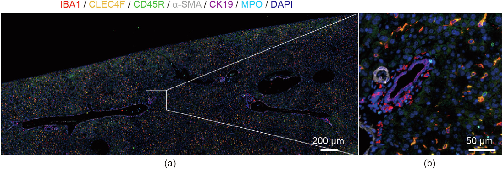
Fig. 2
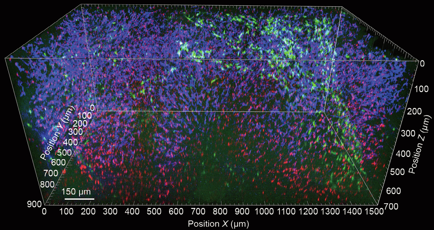
Fig. 3
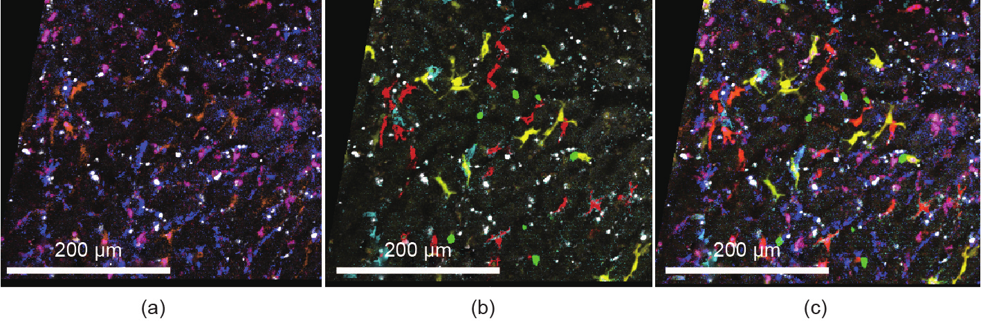
Fig. 4
References
[ 1 ] Faget L, Hnasko TS. Tyramide signal amplification for immunofluorescent enhancement. Methods Mol Biol 2015;1318:161–72. link1
[ 2 ] Guillot A, Kohlhepp MS, Bruneau A, Heymann F, Tacke F. Deciphering the immune microenvironment on a single archival formalin-fixed paraffinembedded tissue section by an immediately implementable multiplex fluorescence immunostaining protocol. Cancers 2020;12(9):12. link1
[ 3 ] Holzwarth K, Köhler R, Philipsen L, Tokoyoda K, Ladyhina V, Wählby C, et al. Multiplexed fluorescence microscopy reveals heterogeneity among stromal cells in mouse bone marrow sections. Cytometry A 2018;93 (9):876–88. link1
[ 4 ] Bonnardel J, T’Jonck W, Gaublomme D, Browaeys R, Scott CL, Martens L, et al. Stellate cells, hepatocytes, and endothelial cells imprint the Kupffer cell identity on monocytes colonizing the liver macrophage niche. Immunity 2019;51(4):638–54.e9. link1
[ 5 ] Richardson DS, Lichtman JW. Clarifying tissue clearing. Cell 2015;162 (2):246–57. link1
[ 6 ] Hägerling R, Drees D, Scherzinger A, Dierkes C, Martin-Almedina S, Butz S, et al. VIPAR, a quantitative approach to 3D histopathology applied to lymphatic malformations. JCI Insight 2017;2(16):2.
[ 7 ] Ishizawa K, Togami K, Tada H, Chono S. Evaluation of tissue-clearing techniques for intraorgan imaging of distribution of polymeric nanoparticles as drug carriers. Drug Dev Ind Pharm 2020;46(12):2061–9. link1
[ 8 ] Wassie AT, Zhao Y, Boyden ES. Expansion microscopy: principles and uses in biological research. Nat Methods 2019;16(1):33–41. link1
[ 9 ] Heymann F, Niemietz PM, Peusquens J, Ergen C, Kohlhepp M, Mossanen JC, et al. Long term intravital multiphoton microscopy imaging of immune cells in healthy and diseased liver using CXCR6.gfp reporter mice. J Vis Exp 2015; (97):52607.
[10] Surewaard BGJ, Kubes P. Measurement of bacterial capture and phagosome maturation of Kupffer cells by intravital microscopy. Methods 2017;128: 12–9. link1
[11] Marques PE, Antunes MM, David BA, Pereira RV, Teixeira MM, Menezes GB. Imaging liver biology in vivo using conventional confocal microscopy. Nat Protoc 2015;10(2):258–68. link1
[12] Thorling CA, Crawford D, Burczynski FJ, Liu X, Liau I, Roberts MS. Multiphoton microscopy in defining liver function. J Biomed Opt 2014;19 (9):090901. link1
[13] Ryan J, Morgan RE, Chen Y, Volak LP, Dunn RT, Dunn KW. Intravital multiphoton microscopy with fluorescent bile salts in rats as an in vivo biomarker for hepatobiliary transport inhibition. Drug Metab Dispos 2018;46 (5):704–18. link1
[14] Heymann F, Peusquens J, Ludwig-Portugall I, Kohlhepp M, Ergen C, Niemietz P, et al. Liver inflammation abrogates immunological tolerance induced by Kupffer cells. Hepatology 2015;62(1):279–91. link1
[15] Ritsma L, Steller EJA, Ellenbroek SIJ, Kranenburg O, Borel Rinkes IHM, van Rheenen J. Surgical implantation of an abdominal imaging window for intravital microscopy. Nat Protoc 2013;8(3):583–94. link1
[16] Rakhilin N, Garrett A, Eom CY, Chavez KR, Small DM, Daniel AR, et al. An intravital window to image the colon in real time. Nat Commun 2019;10 (1):5647. link1
[17] Stenzinger A, Alber M, Allgäuer M, Jurmeister P, Bockmayr M, Budczies J, et al. Artificial intelligence and pathology: from principles to practice and future applications in histomorphology and molecular profiling. Semin Cancer Biol. In press.
[18] Arganda-Carreras I, Kaynig V, Rueden C, Eliceiri KW, Schindelin J, Cardona A, et al. Trainable Weka Segmentation: a machine learning tool for microscopy pixel classification. Bioinformatics 2017;33(15):2424–6.
[19] Berg S, Kutra D, Kroeger T, Straehle CN, Kausler BX, Haubold C, et al. Ilastik: interactive machine learning for (bio)image analysis. Nat Methods 2019;16 (12):1226–32. link1


 京公网安备 11010502051620号


