
2021, Volume 7, Issue 12
Engineering >> 2021, Volume 7, Issue 12 doi: 10.1016/j.eng.2021.08.019
Mechanisms of Steatosis-Derived Hepatocarcinogenesis: Lessons from HCV Core Gene Transgenic Mice
a Department of Metabolic Regulation, Shinshu University School of Medicine, Matsumoto 390-8621, Japan
b State Key Laboratory for Diagnosis and Treatment of Infectious Diseases, National Clinical Research Center for Infectious Diseases, Collaborative Innovation Center for Diagnosis and Treatment of Infectious Diseases, Department of Infectious Diseases, The First Affiliated Hospital, College of Medicine, Zhejiang University, Hangzhou 310003, China
c Department of Gastroenterology, Lishui Hospital, Zhejiang University School of Medicine, Lishui 323000, China
d Department of Pathophysiology, Hebei Medical University, Shijiazhuang 050017, China
e Department of Gastroenterology, Shinshu University School of Medicine, Matsumoto 390-8621, Japan
f Molecular Signaling Section, Laboratory of Bioorganic Chemistry, National Institute of Diabetes and Digestive and Kidney Diseases, National Institutes of Health, Bethesda, Bethesda, MD, 20892, United States
g Department of Infection Control and Prevention, The University of Tokyo, Tokyo 113-0033, Japan
h Department of Gastroenterology, The University of Tokyo, Tokyo 113-0033, Japan
i International Relations Office, Shinshu University School of Medicine, Matsumoto 390-8621, Japan
j Research Center for Social Systems, Shinshu University, Matsumoto 390-8621, Japan
Next Previous
Abstract
Hepatitis C virus (HCV) is a major cause of chronic hepatitis, liver cirrhosis, and hepatocellular carcinoma (HCC) worldwide. Among the structural proteins of HCV, the HCV core protein has the ability to regulate gene transcription, lipid metabolism, cell proliferation, apoptosis, and autophagy, all of which are closely related to the development of HCC. Transgenic mice carrying the HCV core gene exhibited age-dependent insulin resistance, hepatic steatosis, and HCC that resembled the clinical characteristics of chronic hepatitis C patients. Several dietary modifications, including calorie restriction and diets rich in saturated fatty acids (SFAs), trans fatty acids (TFAs), or cholesterol, were found to influence hepatic steatogenesis and tumorigenesis in HCV core gene transgenic mice. These strategies modulated hepatocellular stress and proliferation, in addition to hepatic fibrotic processes and the microenvironment, thereby corroborating a close interconnection between dietary habits and steatosis-related hepatocarcinogenesis. In this review, we summarize the findings obtained from mouse models transgenic for the HCV genome, with a special focus on HCV core gene transgenic mice, and discuss the mechanisms of steatogenesis and hepatocarcinogenesis induced by the HCV core protein and the impact of dietary habits on steatosis-derived HCC development.
Keywords
Steatosis ; Hepatocellular carcinoma ; Trans fatty acid ; Saturated fatty acid ; Dietary restriction ; HCV core protein
Figures

Fig. 1
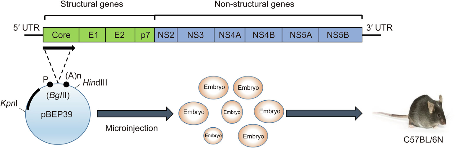
Fig. 2
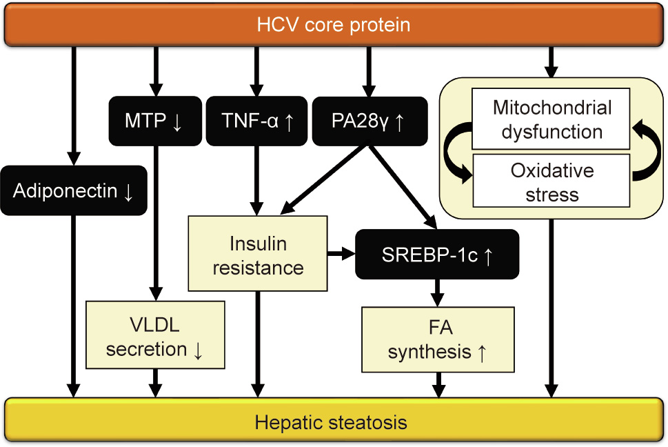
Fig. 3

Fig. 4
References
[ 1 ] Pradat P, Virlogeux V, Trépo E. Epidemiology and elimination of HCV-related liver disease. Viruses 2018;10(10):545. link1
[ 2 ] Kiyosawa K, Sodeyama T, Tanaka E, Gibo Y, Yoshizawa K, Nakano Y, et al. Interrelationship of blood transfusion, non-A, non-B hepatitis and hepatocellular carcinoma: analysis by detection of antibody to hepatitis C virus. Hepatology 1990;12(4):671–5. link1
[ 3 ] Moradpour D, Penin F. Hepatitis C virus proteins: from structure to function. Curr Top Microbiol Immunol 2013;369:113–42. link1
[ 4 ] Gawlik K, Gallay PA. HCV core protein and virus assembly: what we know without structures. Immunol Res 2014;60(1):1–10. link1
[ 5 ] Khaliq S, Jahan S, Pervaiz A. Sequence variability of HCV core region: important predictors of HCV induced pathogenesis and viral production. Infect Genet Evol 2011;11(3):543–56. link1
[ 6 ] Hino K, Nishina S, Sasaki K, Hara Y. Mitochondrial damage and iron metabolic dysregulation in hepatitis C virus infection. Free Radical Biol Med 2019;133:193–9. link1
[ 7 ] Sevastianos VA, Voulgaris TA, Dourakis SP. Hepatitis C, systemic inflammation and oxidative stress: correlations with metabolic diseases. Expert Rev Gastroenterol Hepatol 2020;14(1):27–37. link1
[ 8 ] Wang X, Tanaka N, Hu X, Kimura T, Lu Y, Jia F, et al. A high-cholesterol diet promotes steatohepatitis and liver tumorigenesis in HCV core gene transgenic mice. Arch Toxicol 2019;93(6):1713–25. link1
[ 9 ] Diao P, Wang X, Jia F, Kimura T, Hu X, Shirotori S, et al. A saturated fatty acidrich diet enhances hepatic lipogenesis and tumorigenesis in HCV core gene transgenic mice. J Nutr Biochem 2020;85:108460. link1
[10] Hu X, Wang X, Jia F, Tanaka N, Kimura T, Nakajima T, et al. A trans-fatty acidrich diet promotes liver tumorigenesis in HCV core gene transgenic mice. Carcinogenesis 2020;41(2):159–70.
[11] Jia F, Diao P, Wang X, Hu X, Kimura T, Nakamuta M, et al. Dietary restriction suppresses steatosis-associated hepatic tumorigenesis in hepatitis C virus core gene transgenic mice. Liver Cancer 2020;9(5):529–48. link1
[12] Wang CC, Cheng PN, Kao JH. Systematic review: chronic viral hepatitis and metabolic derangement. Aliment Pharmacol Ther 2020;51(2):216–30. link1
[13] Majumder M, Steele R, Ghosh AK, Zhou XY, Thornburg L, Ray R, et al. Expression of hepatitis C virus non-structural 5A protein in the liver of transgenic mice. FEBS Lett 2003;555(3):528–32.
[14] Koike K, Moriya K, Ishibashi K, Matsuura Y, Suzuki T, Saito I, et al. Expression of hepatitis C virus envelope proteins in transgenic mice. J Gen Virol 1995;76 (12):3031–8. link1
[15] Wang AG, Moon HB, Kim JM, Hwang SB, Yu DY, Lee DS. Expression of hepatitis C virus nonstructural 4B in transgenic mice. Exp Mol Med 2006;38(3):241–6. link1
[16] Moriya K, Fujie H, Shintani Y, Yotsuyanagi H, Tsutsumi T, Ishibashi K, et al. The core protein of hepatitis C virus induces hepatocellular carcinoma in transgenic mice. Nat Med 1998;4(9):1065–7. link1
[17] McLauchlan J. Properties of the hepatitis C virus core protein: a structural protein that modulates cellular processes. J Viral Hepat 2000;7(1):2–14. link1
[18] Boulant S, Montserret R, Hope RG, Ratinier M, Targett-Adams P, Lavergne JP, et al. Structural determinants that target the hepatitis C virus core protein to lipid droplets. J Biol Chem 2006;281(31):22236–47. link1
[19] Liu C, Qu A, Han X, Wang Y. HCV core protein represses the apoptosis and improves the autophagy of human hepatocytes. Int J Clin Exp Med 2015;8 (9):15787–93. link1
[20] Cho JW, Baek WK, Yang SH, Chang J, Sung YC, Suh MH. HCV core protein modulates Rb pathway through pRb down-regulation and E2F–1 upregulation. Biochim Biophys Acta 2001;1538(1):59–66. link1
[21] Lee SK, Park SO, Joe CO, Kim YS. Interaction of HCV core protein with 14-3-3e protein releases Bax to activate apoptosis. Biochem Biophys Res Commun 2007;352(3):756–62. link1
[22] Liu J, Ding X, Tang J, Cao Y, Hu P, Zhou F, et al. Enhancement of canonical Wnt/ b-catenin signaling activity by HCV core protein promotes cell growth of hepatocellular carcinoma cells. PLoS ONE 2011;6(11):e27496. link1
[23] Huang S, Xie Y, Yang P, Chen P, Zhang L, Pfeffer S. HCV core protein-induced down-regulation of microRNA-152 promoted aberrant proliferation by regulating Wnt1 in HepG2 cells. PLoS ONE 2014;9(1):e81730. link1
[24] Shao YY, Hsieh MS, Wang HY, Li YS, Lin H, Hsu HW, et al. Hepatitis C virus core protein potentiates proangiogenic activity of hepatocellular carcinoma cells. Oncotarget 2017;8(49):86681–92. link1
[25] Abe M, Koga H, Yoshida T, Masuda H, Iwamoto H, Sakata M, et al. Hepatitis C virus core protein upregulates the expression of vascular endothelial growth factor via the nuclear factor-jB/hypoxia-inducible factor-1a axis under hypoxic conditions. Hepatol Res 2012;42(6):591–600.
[26] Tan Y, Li Y. HCV core protein promotes hepatocyte proliferation and chemoresistance by inhibiting NR4A1. Biochem Biophys Res Commun 2015;466(3):592–8. link1
[27] Benzoubir N, Lejamtel C, Battaglia S, Testoni B, Benassi B, Gondeau C, et al. HCV core-mediated activation of latent TGF-b via thrombospondin drives the crosstalk between hepatocytes and stromal environment. J Hepatol 2013;59 (6):1160–8. link1
[28] Loizides-Mangold U, Clément S, Alfonso-Garcia A, Branche E, Conzelmann S, Parisot C, et al. HCV 3a core protein increases lipid droplet cholesteryl ester content via a mechanism dependent on sphingolipid biosynthesis. PLoS ONE 2014;9(12):e115309. link1
[29] Alberstein M, Zornitzki T, Zick Y, Knobler H. Hepatitis C core protein impairs insulin downstream signalling and regulatory role of IGFBP-1 expression. J Viral Hepat 2012;19(1):65–71. link1
[30] Lewitt MS, Dent MS, Hall K. The insulin-like growth factor system in obesity, insulin resistance and type 2 diabetes mellitus. J Clin Med 2014;3 (4):1561–74. link1
[31] Anggakusuma, Frentzen A, Gürlevik E, Yuan Q, Steinmann E, Ott M, et al. Control of hepatitis C virus replication in mouse liver-derived cells by MAVSdependent production of type I and type III interferons. J Virol 2015;89 (7):3833–45. link1
[32] Lan HY, Zhao Y, Yang J, Sun MN, Lei YF, Yao M, et al. Establishment of a novel triple-transgenic mouse: conditionally and liver-specifically expressing hepatitis C virus NS3/4A protease. Mol Biol Rep 2014;41(11):7349–59. link1
[33] Arrieta JJ, Rodríguez-Iñigo E, Ortiz-Movilla N, Bartolomé J, Pardo M, Manzarbeitia F, et al. In situ detection of hepatitis C virus RNA in salivary glands. Am J Pathol 2001;158(1):259–64. link1
[34] Bansal R, Frelin L, Brenndörfer ED, Storm G, Prakash J, Sällberg M, et al. Hepatitis C virus nonstructural 3/4A protein dampens inflammation and contributes to slow fibrosis progression during chronic fibrosis in vivo. PLoS ONE 2015;10(6):e0128466. link1
[35] Cristina J, Moreno MDP, Moratorio G. Hepatitis C virus genetic variability in patients undergoing antiviral therapy. Virus Res 2007;127(2):185–94. link1
[36] Strosberg AD, Kota S, Takahashi V, Snyder JK, Mousseau G. Core as a novel viral target for hepatitis C drugs. Viruses 2010;2(8):1734–51. link1
[37] Koike K, Moriya K, Kimura S. Role of hepatitis C virus in the development of hepatocellular carcinoma: transgenic approach to viral hepatocarcinogenesis. J Gastroenterol Hepatol 2002;17(4):394–400. link1
[38] Koike K. Hepatitis C virus contributes to hepatocarcinogenesis by modulating metabolic and intracellular signaling pathways. J Gastroenterol Hepatol 2007;22(Suppl 1):S108–11. link1
[39] Hirano J, Yoshio S, Sakai Y, Songling L, Suzuki T, Itoh Y, et al. Hepatitis C virus modulates signal peptide peptidase to alter host protein processing. Proc Natl Acad Sci USA 2021;118(22):e2026184118.
[40] Moriya K, Yotsuyanagi H, Shintani Y, Fujie H, Ishibashi K, Matsuura Y, et al. Hepatitis C virus core protein induces hepatic steatosis in transgenic mice. J Gen Virol 1997;78(7):1527–31.
[41] Harada S, Watanabe Y, Takeuchi K, Suzuki T, Katayama T, Takebe Y, et al. Expression of processed core protein of hepatitis C virus in mammalian cells. J Virol 1991;65(6):3015–21. link1
[42] Kahn BB. Type 2 diabetes: when insulin secretion fails to compensate for insulin resistance. Cell 1998;92(5):593–6. link1
[43] Cavaghan MK, Ehrmann DA, Polonsky KS. Interactions between insulin resistance and insulin secretion in the development of glucose intolerance. J Clin Invest 2000;106(3):329–33. link1
[44] Shintani Y, Fujie H, Miyoshi H, Tsutsumi T, Tsukamoto K, Kimura S, et al. Hepatitis C virus infection and diabetes: direct involvement of the virus in the development of insulin resistance. Gastroenterology 2004;126(3):840–8. link1
[45] Miyamoto H, Moriishi K, Moriya K, Murata S, Tanaka K, Suzuki T, et al. Involvement of the PA28c-dependent pathway in insulin resistance induced by hepatitis C virus core protein. J Virol 2007;81(4):1727–35. link1
[46] Kadowaki T. Insights into insulin resistance and type 2 diabetes from knockout mouse models. J Clin Invest 2000;106(4):459–65. link1
[47] Tanaka N, Nagaya T, Komatsu M, Horiuchi A, Tsuruta G, Shirakawa H, et al. Insulin resistance and hepatitis C virus: a case-control study of non-obese, non-alcoholic and non-steatotic hepatitis virus carriers with persistently normal serum aminotransferase. Liver Int 2008;28(8):1104–11.
[48] Lefkowitch JH, Schiff ER, Davis GL, Perrillo RP, Lindsay K, Bodenheimer HC, et al. Pathological diagnosis of chronic hepatitis C: a multicenter comparative study with chronic hepatitis B. Gastroenterology 1993;104(2):595–603. link1
[49] Bach N, Thung SN, Schaffner F. The histological features of chronic hepatitis C and autoimmune chronic hepatitis: a comparative analysis. Hepatology 1992;15(4):572–7. link1
[50] Ohata K, Hamasaki K, Toriyama K, Matsumoto K, Saeki A, Yanagi K, et al. Hepatic steatosis is a risk factor for hepatocellular carcinoma in patients with chronic hepatitis C virus infection. Cancer 2003;97(12):3036–43. link1
[51] Chang ML, Yeh HC, Tsou YK, Wang CJ, Cheng HY, Sung CM, et al. HCV coreinduced nonobese hepatic steatosis is associated with hypoadiponectinemia and is ameliorated by adiponectin administration. Obesity 2012;20 (7):1474–80.
[52] Mori Y, Moriishi K, Matsuura Y. Hepatitis C virus core protein: its coordinate roles with PA28c in metabolic abnormality and carcinogenicity in the liver. Int J Biochem Cell Biol 2008;40(8):1437–42. link1
[53] Koike K, Moriya K. Metabolic aspects of hepatitis C viral infection: steatohepatitis resembling but distinct from NASH. J Gastroenterol 2005;40 (4):329–36. link1
[54] Moriishi K, Mochizuki R, Moriya K, Miyamoto H, Mori Y, Abe T, et al. Critical role of PA28c in hepatitis C virus-associated steatogenesis and hepatocarcinogenesis. Proc Natl Acad Sci USA 2007;104(5):1661–6. link1
[55] Tsutsumi T, Suzuki T, Shimoike T, Suzuki R, Moriya K, Shintani Y, et al. Interaction of hepatitis C virus core protein with retinoid X receptor alpha modulates its transcriptional activity. Hepatology 2002;35(4):937–46. link1
[56] Perlemuter G, Sabile A, Letteron P, Vona G, Topilco A, Chrétien Y, et al. Hepatitis C virus core protein inhibits microsomal triglyceride transfer protein activity and very low density lipoprotein secretion: a model of viralrelated steatosis. FASEB J 2002;16(2):185–94. link1
[57] Roingeard P, Hourioux C. Hepatitis C virus core protein, lipid droplets and steatosis. J Viral Hepat 2008;15(3):157–64. link1
[58] Tanaka N, Moriya K, Kiyosawa K, Koike K, Gonzalez FJ, Aoyama T. PPARa activation is essential for HCV core protein-induced hepatic steatosis and hepatocellular carcinoma in mice. J Clin Invest 2008;118(2):683–94. link1
[59] Tanaka N, Aoyama T, Kimura S, Gonzalez FJ. Targeting nuclear receptors for the treatment of fatty liver disease. Pharmacol Ther 2017;179:142–57. link1
[60] Koike K, Tsutsumi T, Yotsuyanagi H, Moriya K. Lipid metabolism and liver disease in hepatitis C viral infection. Oncology 2010;78(Suppl 1):24–30. link1
[61] Shiode Y, Hikita H, Tanaka S, Shirai K, Doi A, Sakane S, et al. Hepatitis C virus enhances Rubicon expression, leading to autophagy inhibition and intracellular innate immune activation. Sci Rep 2020;10(1):15290. link1
[62] Jiang TX, Zou JB, Zhu QQ, Liu CH, Wang GF, Du TT, et al. SIP/CacyBP promotes autophagy by regulating levels of BRUCE/Apollon, which stimulates LC3-I degradation. Proc Natl Acad Sci USA 2019;116(27):13404–13. link1
[63] Singh R, Kaushik S, Wang Y, Xiang Y, Novak I, Komatsu M, et al. Autophagy regulates lipid metabolism. Nature 2009;458(7242):1131–5. link1
[64] Hara Y, Yanatori I, Ikeda M, Kiyokage E, Nishina S, Tomiyama Y, et al. Hepatitis C virus core protein suppresses mitophagy by interacting with parkin in the context of mitochondrial depolarization. Am J Pathol 2014;184(11):3026–39. link1
[65] Moriya K, Nakagawa K, Santa T, Shintani Y, Fujie H, Miyoshi H, et al. Oxidative stress in the absence of inflammation in a mouse model for hepatitis C virusassociated hepatocarcinogenesis. Cancer Res 2001;61(11):4365–70. link1
[66] Moriya K, Todoroki T, Tsutsumi T, Fujie H, Shintani Y, Miyoshi H, et al. Increase in the concentration of carbon 18 monounsaturated fatty acids in the liver with hepatitis C: analysis in transgenic mice and humans. Biochem Biophys Res Commun 2001;281(5):1207–12. link1
[67] Vidali M, Tripodi MF, Ivaldi A, Zampino R, Occhino G, Restivo L, et al. Interplay between oxidative stress and hepatic steatosis in the progression of chronic hepatitis C. J Hepatol 2008;48(3):399–406. link1
[68] Takenaka K, Adachi E, Nishizaki T, Hiroshige K, Ikeda T, Tsuneyoshi M, et al. Possible multicentric occurrence of hepatocellular carcinoma: a clinicopathological study. Hepatology 1994;19(4):889–94. link1
[69] Oikawa T, Ojima H, Yamasaki S, Takayama T, Hirohashi S, Sakamoto M. Multistep and multicentric development of hepatocellular carcinoma: histological analysis of 980 resected nodules. J Hepatol 2005;42(2):225–9. link1
[70] Caporaso N, Romano M, Marmo R, de Sio I, Morisco F, Minerva A, et al. Hepatitis C virus infection is an additive risk factor for development of hepatocellular carcinoma in patients with cirrhosis. J Hepatol 1991;12 (3):367–71. link1
[71] Hoofnagle JH. Hepatitis C: the clinical spectrum of disease. Hepatology 1997;26(S3):15S–20S. link1
[72] Wang Y, Nakajima T, Gonzalez FJ, Tanaka N. PPARs as metabolic regulators in the liver: lessons from liver-specific PPAR-null mice. Int J Mol Sci 2020;21 (6):2061. link1
[73] Brocker CN, Yue J, Kim D, Qu A, Bonzo JA, Gonzalez FJ. Hepatocyte-specific PPARA expression exclusively promotes agonist-induced cell proliferation without influence from nonparenchymal cells. Am J Physiol Gastrointest Liver Physiol 2017;312(3):G283–99. link1
[74] Tanaka N, Moriya K, Kiyosawa K, Koike K, Aoyama T. Hepatitis C virus core protein induces spontaneous and persistent activation of peroxisome proliferator-activated receptor alpha in transgenic mice: implications for HCV-associated hepatocarcinogenesis. Int J Cancer 2008;122(1):124–31. link1
[75] Shrivastava A, Manna SK, Ray R, Aggarwal BB. Ectopic expression of hepatitis C virus core protein differentially regulates nuclear transcription factors. J Virol 1998;72(12):9722–8. link1
[76] Tsutsumi T, Suzuki T, Moriya K, Yotsuyanagi H, Shintani Y, Fujie H, et al. Alteration of intrahepatic cytokine expression and AP-1 activation in transgenic mice expressing hepatitis C virus core protein. Virology 2002;304(2):415–24. link1
[77] Lu W, Lo SY, Chen M, Wu KJ, Fung YK, Ou JH. Activation of p53 tumor suppressor by hepatitis C virus core protein. Virology 1999;264(1):134–41. link1
[78] Pan Z, Bhat MB, Nieminen AL, Ma J. Synergistic movements of Ca2+ and Bax in cells undergoing apoptosis. J Biol Chem 2001;276(34):32257–63. link1
[79] Benali-Furet NL, Chami M, Houel L, De Giorgi F, Vernejoul F, Lagorce D, et al. Hepatitis C virus core triggers apoptosis in liver cells by inducing ER stress and ER calcium depletion. Oncogene 2005;24(31):4921–33. link1
[80] Umemura A, He F, Taniguchi K, Nakagawa H, Yamachika S, Font-Burgada J, et al. P62, upregulated during preneoplasia, induces hepatocellular carcinogenesis by maintaining survival of stressed HCC-initiating cells. Cancer Cell 2016;29(6):935–48. link1
[81] Kato T, Miyamoto M, Date T, Yasui K, Taya C, Yonekawa H, et al. Repeated hepatocyte injury promotes hepatic tumorigenesis in hepatitis C virus transgenic mice. Cancer Sci 2003;94(8):679–85. link1
[82] Ioannou GN, Morrow OB, Connole ML, Lee SP. Association between dietary nutrient composition and the incidence of cirrhosis or liver cancer in the United States population. Hepatology 2009;50(1):175–84. link1
[83] Yu L, Morishima C, Ioannou GN. Dietary cholesterol intake is associated with progression of liver disease in patients with chronic hepatitis C: analysis of the hepatitis C antiviral long-term treatment against cirrhosis trial. Clin Gastroenterol Hepatol 2013;11(12):1661–6.e3. link1
[84] Ostapowicz G, Watson KJ, Locarnini SA, Desmond PV. Role of alcohol in the progression of liver disease caused by hepatitis C virus infection. Hepatology 1998;27(6):1730–5. link1
[85] Kimura T, Kobayashi A, Tanaka N, Sano K, Komatsu M, Fujimori N, et al. Clinicopathological characteristics of non-B non-C hepatocellular carcinoma without past hepatitis B virus infection. Hepatol Res 2017;47(5):405–18. link1
[86] Tanaka N, Kimura T, Fujimori N, Nagaya T, Komatsu M, Tanaka E. Current status, problems, and perspectives of non-alcoholic fatty liver disease research. World J Gastroenterol 2019;25(2):163–77. link1
[87] Duarte-Salles T, Fedirko V, Stepien M, Aleksandrova K, Bamia C, Lagiou P, et al. Dietary fat, fat subtypes and hepatocellular carcinoma in a large European cohort. Int J Cancer 2015;137(11):2715–28. link1
[88] Zˇácˇek P, Bukowski M, Mehus A, Johnson L, Zeng H, Raatz S, et al. Dietary saturated fatty acid type impacts obesity-induced metabolic dysfunction and plasma lipidomic signatures in mice. J Nutr Biochem 2019;64:32–44. link1
[89] Zelber-Sagi S, Ivancovsky-Wajcman D, Fliss Isakov N, Webb M, Orenstein D, Shibolet O, et al. High red and processed meat consumption is associated with non-alcoholic fatty liver disease and insulin resistance. J Hepatol 2018;68 (6):1239–46. link1
[90] Freedman ND, Cross AJ, McGlynn KA, Abnet CC, Park Y, Hollenbeck AR, et al. Association of meat and fat intake with liver disease and hepatocellular carcinoma in the NIH-AARP cohort. J Natl Cancer Inst 2010;102(17): 1354–65. link1
[91] Moriya K, Miyoshi H, Shinzawa S, Tsutsumi T, Fujie H, Goto K, et al. Hepatitis C virus core protein compromises iron-induced activation of antioxidants in mice and HepG2 cells. J Med Virol 2010;82(5):776–92. link1
[92] Tsutsumi T, Suzuki T, Moriya K, Shintani Y, Fujie H, Miyoshi H, et al. Hepatitis C virus core protein activates ERK and p38 MAPK in cooperation with ethanol in transgenic mice. Hepatology 2003;38(4):820–8. link1
[93] Ghebreyesus TA, Frieden TR. REPLACE: a roadmap to make the world trans fat free by 2023. Lancet 2018;391(10134):1978–80. link1
[94] Mozaffarian D, Katan MB, Ascherio A, Stampfer MJ, Willett WC. Trans fatty acids and cardiovascular disease. N Engl J Med 2006;354(15):1601–13. link1
[95] Micha R, Mozaffarian D. Trans fatty acids: effects on metabolic syndrome, heart disease and diabetes. Nat Rev Endocrinol 2009;5(6):335–44. link1
[96] Hu X, Tanaka N, Guo R, Lu Yu, Nakajima T, Gonzalez FJ, et al. PPARa protects against trans-fatty-acid-containing diet-induced steatohepatitis. J Nutr Biochem 2017;39:77–85. link1
[97] Clayton ZS, Fusco E, Kern M. Egg consumption and heart health: a review. Nutrition 2017;37:79–85. link1
[98] Sozen E, Ozer NK. Impact of high cholesterol and endoplasmic reticulum stress on metabolic diseases: an updated mini-review. Redox Biol 2017;12:456–61. link1
[99] Ioannou GN. The role of cholesterol in the pathogenesis of NASH. Trends Endocrinol Metab 2016;27(2):84–95. link1
[100] Huff MW. Dietary cholesterol, cholesterol absorption, postprandial lipemia and atherosclerosis. Can J Clin Pharmacol 2003;10 Suppl A:26A–32A.
[101] Dev S, Babitt JL. Overview of iron metabolism in health and disease. Hemodial Int 2017;21(S1):S6–S20. link1
[102] Tanaka N, Horiuchi A, Yamaura T, Komatsu M, Tanaka E, Kiyosawa K. Efficacy and safety of 6-month iron reduction therapy in patients with hepatitis C virus-related cirrhosis: a pilot study. J Gastroenterol 2007;42(1):49–55. link1
[103] Tanaka N, Kiyosawa K. Phlebotomy: a promising treatment for chronic hepatitis C. J Gastroenterol 2004;39(6):601–3. link1
[104] Tanaka N, Horiuchi A, Yamaura T, Komatsu M, Yokoyama T, Okaniwa S, et al. Efficacy and safety of addition of minor bloodletting (petit phlebotomy) in hepatitis C virus-infected patients receiving regular glycyrrhizin injections. J Gastroenterol 2009;44(6):577–82. link1
[105] Kimura T, Tanaka N, Fujimori N, Sugiura A, Yamazaki T, Joshita S, et al. Mild drinking habit is a risk factor for hepatocarcinogenesis in non-alcoholic fatty liver disease with advanced fibrosis. World J Gastroenterol 2018;24 (13):1440–50. link1
[106] Ceni E, Mello T, Galli A. Pathogenesis of alcoholic liver disease: role of oxidative metabolism. World J Gastroenterol 2014;20(47):17756–72. link1
[107] Vandenbulcke H, Moreno C, Colle I, Knebel JF, Francque S, Sersté T, et al. Alcohol intake increases the risk of HCC in hepatitis C virus-related compensated cirrhosis: a prospective study. J Hepatol 2016;65(3):543–51. link1
[108] Brandhorst S, Longo VD. Fasting and caloric restriction in cancer prevention and treatment. Recent Results Cancer Res 2016;207:241–66. link1
[109] Barnosky AR, Hoddy KK, Unterman TG, Varady KA. Intermittent fasting vs daily calorie restriction for type 2 diabetes prevention: a review of human findings. Transl Res 2014;164(4):302–11. link1
[110] Fujimoto M, Tsuneyama K, Nakanishi Y, Salunga TL, Nomoto K, Sasaki Y, et al. A dietary restriction influences the progression but not the initiation of MSG-induced nonalcoholic steatohepatitis. J Med Food 2014;17(3): 374–83. link1
[111] Holmer M, Lindqvist C, Petersson S, Moshtaghi-Svensson J, Tillander V, Brismar TB, et al. Treatment of NAFLD with intermittent calorie restriction or low-carb high-fat diet—a randomised controlled trial. JHEP Rep 2021;3 (3):100256. link1


 京公网安备 11010502051620号


