
2023, Volume 24, Issue 5
Engineering >> 2023, Volume 24, Issue 5 doi: 10.1016/j.eng.2022.10.009
Single-Molecule Methods for Characterizing Different DNA Higher-Order Structures
a School of Molecular Medicine, Hangzhou Institute for Advanced Study, University of Chinese Academy of Sciences, Hangzhou 310024, China
b The Cancer Hospital of the University of Chinese Academy of Sciences, Institute of Basic Medicine and Cancer (IBMC), Chinese Academy of Sciences, Hangzhou 310022, China
c Academy of Medical Engineering and Translational Medicine (AMT), Tianjin University, Tianjin 300072, China
d Institute of Nano Biomedicine and Engineering, Department of Instrument Science and Engineering, School of Electronic Information and Electrical Engineering, Shanghai Jiao Tong University, Shanghai 200240, China
Next Previous
Abstract
DNA is considered to be not only a carrier of the genetic information of life but also a highly programmable and self-assembled nanomaterial. Different DNA structures are related to their biological and chemical functions. Hence, understanding the physical and chemical properties of various DNA structures is of great importance in biology and nanochemistry. However, the bulk assay ignores the heterogeneity of DNA structures in solution. Single-molecule methods are powerful tools for observing the behavior of individual molecules and probing the high heterogeneity of free energy states. In this review, we introduce single-molecule methods, including single-molecule detection and manipulation methods, and discuss how these methods can be conducive to measuring the molecular properties of single-/double-stranded DNA (ss/dsDNA), DNA higher-order structures, and DNA nanostructures. We conclude by providing a new perspective on the combination of DNA nanotechnology and single-molecule methods to understand the biophysical properties of DNA and other bio-matter and soft matter.
Keywords
Single-molecule methods ; DNA structure ; Mechanical properties ; Conformational transitions
Figures
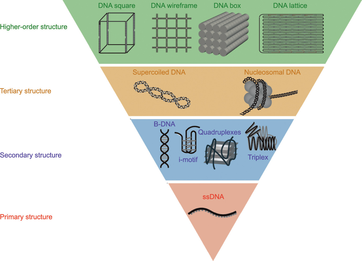
Fig. 1

Fig. 2

Fig. 3

Fig. 4

Fig. 5
Fig. 6
Fig. 7
Fig. 8

Fig. 9
Fig. 10

Fig. 11

Fig. 12

Fig. 13
References
[ 1 ] Aaij C, Borst P. The gel electrophoresis of DNA. Biochim Biophys Acta Nucleic Acids Protein Synth 1972;269(2):192‒200. link1
[ 2 ] Xiao W, Oefner PJ. Denaturing high-performance liquid chromatography: a review. Hum Mutat 2001;17(6):439‒74. link1
[ 3 ] Kay LE. NMR studies of protein structure and dynamics. 2005. J Magn Reson 2011;213(2):477‒91.
[ 4 ] Scott LG, Hennig M. RNA structure determination by NMR. Methods Mol Biol 2008;452:29‒61. link1
[ 5 ] Joo C, Balci H, Ishitsuka Y, Buranachai C, Ha T. Advances in single-molecule fluorescence methods for molecular biology. Annu Rev Biochem 2008;77(1):51‒76. link1
[ 6 ] Deamer DW, Branton D. Characterization of nucleic acids by nanopore analysis. Acc Chem Res 2002;35(10):817‒25. link1
[ 7 ] Cattoni DI, Fiche JB, Nöllmann M. Single-molecule super-resolution imaging in bacteria. Curr Opin Microbiol 2012;15(6):758‒63. link1
[ 8 ] Gosse C, Croquette V. Magnetic tweezers: micromanipulation and force measurement at the molecular level. Biophys J 2002;82(6):3314‒29. link1
[ 9 ] Polimeno P, Magazzù A, Iatì MA, Patti F, Saija R, Esposti Boschi CD, et al. Optical tweezers and their applications. J Quant Spectrosc Radiat Transf 2018;218:131‒50. link1
[10] Binnig G, Quate CF, Gerber C. Atomic force microscope. Phys Rev Lett 1986;56(9):930‒3. link1
[11] Heidarsson PO, Naqvi MM, Otazo MR, Mossa A, Kragelund BB, Cecconi C. Direct single-molecule observation of calcium-dependent misfolding in human neuronal calcium sensor-1. Proc Natl Acad Sci USA 2014;111(36):13069‒74. link1
[12] Friedrichs J, Taubenberger A, Franz CM, Muller DJ. Cellular remodelling of individual collagen fibrils visualized by time-lapse AFM. J Mol Biol 2007;372(3):594‒607. link1
[13] Watson JD, Crick FH. Genetical implications of the structure of deoxyribonucleic acid. Nature 1953;171(4361):964‒7. link1
[14] Lipps HJ, Rhodes D. G-quadruplex structures: in vivo evidence and function. Trends Cell Biol 2009;19(8):414‒22. link1
[15] Guéron M, Leroy JL. The i-motif in nucleic acids. Curr Opin Struct Biol 2000;10(3):326‒31. link1
[16] Li F, Lin Y, Le XC. Binding-induced formation of DNA three-way junctions and its application to protein detection and DNA strand displacement. Anal Chem 2013;85(22):10835‒41. link1
[17] Duckett DR, Murchie AI, Diekmann S, von Kitzing E, Kemper B, Lilley DM. The structure of the Holliday junction, and its resolution. Cell 1988;55(1):79‒89. link1
[18] Frank-Kamenetskii MD, Mirkin SM. Triplex DNA structures. Annu Rev Biochem 1995;64(1):65‒95. link1
[19] Hansma HG, Revenko I, Kim K, Laney DE. Atomic force microscopy of long and short double-stranded, single-stranded and triple-stranded nucleic acids. Nucleic Acids Res 1996;24(4):713‒20. link1
[20] Ullsperger CJ, Vologodskii AV, Cozzarelli NR. Unlinking of DNA by topoisomerases during DNA replication. In: Eckstein F, Lilley DMJ, editors. Nucleic acids and molecular biology. Heidelberg: Springer; 1995. p. 115‒42. link1
[21] Muthurajan UM, Park YJ, Edayathumangalam RS, Suto RK, Chakravarthy S, Dyer PN, et al. Structure and dynamics of nucleosomal DNA. Biopolymers 2003;68(4):547‒56. link1
[22] Gerling T, Wagenbauer KF, Neuner AM, Dietz H. Dynamic DNA devices and assemblies formed by shape-complementary, non-base pairing 3D components. Science 2015;347(6229):1446‒52. link1
[23] Kuzyk A, Schreiber R, Fan Z, Pardatscher G, Roller EM, Högele A, et al. DNA-based self-assembly of chiral plasmonic nanostructures with tailored optical response. Nature 2012;483(7389):311‒4. link1
[24] Rothemund PW. Folding DNA to create nanoscale shapes and patterns. Nature 2006;440(7082):297‒302. link1
[25] Liu Y, Cheng J, Fan S, Ge H, Luo T, Tang L, et al. Modular reconfigurable DNA origami: from two-dimensional to three-dimensional structures. Angew Chem Int Ed Engl 2020;59(51):23277‒82. link1
[26] Baldock BL, Hutchison JE. UV-visible spectroscopy-based quantification of unlabeled DNA bound to gold nanoparticles. Anal Chem 2016;88(24):12072‒80. link1
[27] Jangir DK, Dey SK, Kundu S, Mehrotra R. Assessment of amsacrine binding with DNA using UV-visible, circular dichroism and Raman spectroscopic techniques. J Photochem Photobiol B 2012;114:38‒43. link1
[28] Charak S, Shandilya M, Tyagi G, Mehrotra R. Spectroscopic and molecular docking studies on chlorambucil interaction with DNA. Int J Biol Macromol 2012;51(4):406‒11. link1
[29] Masiero S, Trotta R, Pieraccini S, De Tito S, Perone R, Randazzo A, et al. A non-empirical chromophoric interpretation of CD spectra of DNA G-quadruplex structures. Org Biomol Chem 2010;8(12):2683‒92. link1
[30] Rahman KM, James CH, Thurston DE. Observation of the reversibility of a covalent pyrrolobenzodiazepine (PBD) DNA adduct by HPLC/MS and CD spectroscopy. Org Biomol Chem 2011;9(5):1632‒41. link1
[31] Tucker BA, Gabriel S, Sheardy RD. A CD spectroscopic investigation of intermolecular and intramolecular DNA quadruplexes. In: Frontiers in nucleic acids. American Chemical Society; 2011. p. 51‒67. link1
[32] Strey HH, Parsegian VA, Podgornik R. Equation of state for DNA liquid crystals: fluctuation enhanced electrostatic double layer repulsion. Phys Rev Lett 1997;78(5):895‒8. link1
[33] Aihara H, Ito Y, Kurumizaka H, Yokoyama S, Shibata T. The N-terminal domain of the human Rad51 protein binds DNA: structure and a DNA binding surface as revealed by NMR. J Mol Biol 1999;290(2):495‒504. link1
[34] Takizawa Y, Tanaka H, Machida S, Koyama M, Maehara K, Ohkawa Y, et al. Cryo-EM structure of the nucleosome containing the ALB1 enhancer DNA sequence. Open Biol 2018;8(3):170255. link1
[35] Evans E, Ritchie K, Merkel R. Sensitive force technique to probe molecular adhesion and structural linkages at biological interfaces. Biophys J 1995;68(6):2580‒7. link1
[36] Kim S, Blainey PC, Schroeder CM, Xie XS. Multiplexed single-molecule assay for enzymatic activity on flow-stretched DNA. Nat Methods 2007;4(5):397‒9. link1
[37] Smith SB, Finzi L, Bustamante C. Direct mechanical measurements of the elasticity of single DNA molecules by using magnetic beads. Science 1992;258(5085):1122‒6. link1
[38] Cluzel P, Lebrun A, Heller C, Lavery R, Viovy JL, Chatenay D, et al. DNA: an extensible molecule. Science 1996;271(5250):792‒4. link1
[39] Hodeib S, Raj S, Manosas M, Zhang W, Bagchi D, Ducos B, et al. Single molecule studies of helicases with magnetic tweezers. Methods 2016;105:3‒15. link1
[40] Ashkin A. Atomic-beam deflection by resonance-radiation pressure. Phys Rev Lett 1970;25(19):1321‒4. link1
[41] Ashkin A, Dziedzic JM, Bjorkholm JE, Chu S. Observation of a single-beam gradient force optical trap for dielectric particles. Opt Lett 1986;11(5):288. link1
[42] Neuman KC, Nagy A. Single-molecule force spectroscopy: optical tweezers, magnetic tweezers and atomic force microscopy. Nat Methods 2008;5(6):491‒505. link1
[43] Neuman KC, Chadd EH, Liou GF, Bergman K, Block SM. Characterization of photodamage to Escherichia coli in optical traps. Biophys J 1999;77(5):2856‒63. link1
[44] Sacconi L, Tolić-Nørrelykke IM, Stringari C, Antolini R, Pavone FS. Optical micromanipulations inside yeast cells. Appl Opt 2005;44(11):2001‒7. link1
[45] Cherney DP, Bridges TE, Harris JM. Optical trapping of unilamellar phospholipid vesicles: investigation of the effect of optical forces on the lipid membrane shape by confocal-Raman microscopy. Anal Chem 2004;76(17):4920‒8. link1
[46] Ishii Y, Yanagida T. Single molecule detection in life sciences. Single Mol 2000;1(1):5‒16. link1
[47] Stryer L, Haugland RP. Energy transfer: a spectroscopic ruler. Proc Natl Acad Sci USA 1967;58(2):719‒26. link1
[48] Ritort F. Single-molecule experiments in biological physics: methods and applications. J Phys Condens Matter 2006;18(32):R531‒83. link1
[49] Tsukanov R, Tomov TE, Masoud R, Drory H, Plavner N, Liber M, et al. Detailed study of DNA hairpin dynamics using single-molecule fluorescence assisted by DNA origami. J Phys Chem B 2013;117(40):11932‒42. link1
[50] Akeson M, Branton D, Kasianowicz JJ, Brandin E, Deamer DW. Microsecond time-scale discrimination among polycytidylic acid, polyadenylic acid, and polyuridylic acid as homopolymers or as segments within single RNA molecules. Biophys J 1999;77(6):3227‒33. link1
[51] Meller A, Nivon L, Brandin E, Golovchenko J, Branton D. Rapid nanopore discrimination between single polynucleotide molecules. Proc Natl Acad Sci USA 2000;97(3):1079‒84. link1
[52] Kawano R, Osaki T, Sasaki H, Takinoue M, Yoshizawa S, Takeuchi S. Rapid detection of a cocaine-binding aptamer using biological nanopores on a chip. J Am Chem Soc 2011;133(22):8474‒7. link1
[53] Butler TZ, Pavlenok M, Derrington IM, Niederweis M, Gundlach JH. Single-molecule DNA detection with an engineered MspA protein nanopore. Proc Natl Acad Sci USA 2008;105(52):20647‒52. link1
[54] Storm AJ, Storm C, Chen J, Zandbergen H, Joanny JF, Dekker C. Fast DNA translocation through a solid-state nanopore. Nano Lett 2005;5(7):1193‒7. link1
[55] Boškovic´ F, Zhu J, Chen K, Keyser UF. Monitoring G-quadruplex formation with DNA carriers and solid-state nanopores. Nano Lett 2019;19(11):7996‒8001. link1
[56] Mazidi H, Lu J, Nehorai A, Lew MD. Minimizing structural bias in single-molecule super-resolution microscopy. Sci Rep 2018;8(1):13133. link1
[57] Izeddin I, El Beheiry M, Andilla J, Ciepielewski D, Darzacq X, Dahan M. PSF shaping using adaptive optics for three-dimensional single-molecule super-resolution imaging and tracking. Opt Express 2012;20(5):4957‒67. link1
[58] Jungmann R, Steinhauer C, Scheible M, Kuzyk A, Tinnefeld P, Simmel FC. Single-molecule kinetics and super-resolution microscopy by fluorescence imaging of transient binding on DNA origami. Nano Lett 2010;10(11):4756‒61. link1
[59] Steinhauer C, Jungmann R, Sobey TL, Simmel FC, Tinnefeld P. DNA origami as a nanoscopic ruler for super-resolution microscopy. Angew Chem Int Ed Engl 2009;48(47):8870‒3. link1
[60] Blom H, Widengren J. Stimulated emission depletion microscopy. Chem Rev 2017;117(11):7377‒427. link1
[61] Funatsu T, Harada Y, Higuchi H, Tokunaga M, Saito K, Ishii Y, et al. Imaging and nano-manipulation of single biomolecules. Biophys Chem 1997;68(1‒3):63‒72.
[62] Long X, Parks JW, Bagshaw CR, Stone MD. Mechanical unfolding of human telomere G-quadruplex DNA probed by integrated fluorescence and magnetic tweezers spectroscopy. Nucleic Acids Res 2013;41(4):2746‒55. link1
[63] Lee M, Kim SH, Hong SC. Minute negative superhelicity is sufficient to induce the B-Z transition in the presence of low tension. Proc Natl Acad Sci USA 2010;107(11):4985‒90. link1
[64] Ngo TTM, Zhang Q, Zhou R, Yodh JG, Ha T. Asymmetric unwrapping of nucleosomes under tension directed by DNA local flexibility. Cell 2015;160(6):1135‒44. link1
[65] Mitra J, Makurath MA, Ngo TTM, Troitskaia A, Chemla YR, Ha T. Extreme mechanical diversity of human telomeric DNA revealed by fluorescence-force spectroscopy. Biophys Comput Biol 2019;116(17):8350‒9. link1
[66] Wasserman MR, Schauer GD, O’Donnell ME, Liu S. Replication fork activation is enabled by a single-stranded DNA gate in CMG helicase. Cell 2019;178(3):600‒11. link1
[67] Ye S, Chen Z, Zhang X, Li F, Guo L, Hou XM, et al. Proximal single-stranded RNA destabilizes human telomerase RNA G-quadruplex and induces its distinct conformers. J Phys Chem Lett 2021;12(13):3361‒6. link1
[68] Lee S, Hohng S. An optical trap combined with three-color FRET. J Am Chem Soc 2013;135(49):18260‒3. link1
[69] Kang J, Jung J, Kim SK. Flexibility of single-stranded DNA measured by single-molecule FRET. Biophys Chem 2014;195:49‒52. link1
[70] Mortusewicz O, Evers B, Helleday T. PC4 promotes genome stability and DNA repair through binding of ssDNA at DNA damage sites. Oncogene 2016;35(6):761‒70. link1
[71] Yusufzai T, Kong X, Yokomori K, Kadonaga JT. The annealing helicase HARP is recruited to DNA repair sites via an interaction with RPA. Genes Dev 2009;23(20):2400‒4. link1
[72] Ke C, Humeniuk M, S-Gracz H, Marszalek PE. Direct measurements of base stacking interactions in DNA by single-molecule atomic-force spectroscopy. Phys Rev Lett 2007;99(1):018302. link1
[73] Tinland B, Pluen A, Sturm J, Weill G. Persistence length of single-stranded DNA. Macromolecules 1997;30(19):5763‒5. link1
[74] Chen H, Meisburger SP, Pabit SA, Sutton JL, Webb WW, Pollack L. Ionic strength-dependent persistence lengths of single-stranded RNA and DNA. Biophys Comput Biol 2012;109(3):799‒804. link1
[75] Viader-Godoy X, Manosas M, Ritort F. Sugar-pucker force-induced transition in single-stranded DNA. Int J Mol Sci 2021;22(9):4745. link1
[76] De Lorenzo S, Ribezzi-Crivellari M, Arias-Gonzalez JR, Smith SB, Ritort F. A temperature-jump optical trap for single-molecule manipulation. Biophys J 2015;108(12):2854‒64. link1
[77] Danilowicz C, Lee CH, Coljee VW, Prentiss M. Effects of temperature on the mechanical properties of single stranded DNA. Phys Rev E Stat Nonlin Soft Matter Phys 2007;75(3 Pt 1):030902. link1
[78] Watson JD, Crick FH. Molecular structure of nucleic acids: a structure for deoxyribose nucleic acid. Nature 1953;171(4356):737‒8. link1
[79] Klug A. Rosalind Franklin and the discovery of the structure of DNA. Nature 1968;219(5156):808‒10. link1
[80] Maier B, Bensimon D, Croquette V. Replication by a single DNA polymerase of a stretched single-stranded DNA. Proc Natl Acad Sci USA 2000;97(22):12002‒7. link1
[81] Dessinges MN, Maier B, Zhang Y, Peliti M, Bensimon D, Croquette V. Stretching single stranded DNA, a model polyelectrolyte. Phys Rev Lett 2002;89(24):248102. link1
[82] Hugel T, Rief M, Seitz M, Gaub HE, Netz RR. Highly stretched single polymers: atomic-force-microscope experiments versus ab-initio theory. Phys Rev Lett 2005;94(4):048301. link1
[83] Zhou Z. Stretching instability of a two-dimensional freely rotating chain. Chin J Phys 2018;56(6):2967‒76. link1
[84] Livadaru L, Netz RR, Kreuzer HJ. Stretching response of discrete semiflexible polymers. Macromolecules 2003;36(10):3732‒44. link1
[85] Dobrynin AV, Carrillo JM, Rubinstein M. Chains are more flexible under tension. Macromolecules 2010;43(21):9181‒90. link1
[86] Radiom M, Borkovec M. Influence of ligand-receptor interactions on force-extension behavior within the freely jointed chain model. Phys Rev E 2017;96(6):062501. link1
[87] Smith SB, Finzi L, Bustamante C. Direct mechanical measurements of the elasticity of single DNA molecules by using magnetic beads. Science 1992;258(5085):1122‒6. link1
[88] Kratky O, Porod G. X-ray investigiation of chain molecules in solution. Recl Trav Chim Pays Bas 1949;68:1106‒22. link1
[89] Marko JF, Siggia ED. Stretching DNA. Macromolecules 1995;28(26):8759‒70. link1
[90] Wang MD, Yin H, Landick R, Gelles J, Block SM. Stretching DNA with optical tweezers. Biophys J 1997;72(3):1335‒46. link1
[91] Smith SB, Cui Y, Bustamante C. Overstretching B-DNA: the elastic response of individual double-stranded and single-stranded DNA molecules. Science 1996;271(5250):795‒9. link1
[92] Rief M, Clausen-Schaumann H, Gaub HE. Sequence-dependent mechanics of single DNA molecules. Nat Struct Biol 1999;6(4):346‒9.
[93] Essevaz-Roulet B, Bockelmann U, Heslot F. Mechanical separation of the complementary strands of DNA. Proc Natl Acad Sci USA 1997;94(22):11935‒40. link1
[94] Bockelmann U, Thomen P, Essevaz-Roulet B, Viasnoff V, Heslot F. Unzipping DNA with optical tweezers: high sequence sensitivity and force flips. Biophys J 2002;82(3):1537‒53. link1
[95] Woodside MT, Behnke-Parks WM, Larizadeh K, Travers K, Herschlag D, Block SM. Nanomechanical measurements of the sequence-dependent folding landscapes of single nucleic acid hairpins. Biophys Comput Biol 2006;103(16):6190‒5. link1
[96] Johnson CA, Bloomingdale RJ, Ponnusamy VE, Tillinghast CA, Znosko BM, Lewis M. Computational model for predicting experimental RNA and DNA nearest-neighbor free energy rankings. J Phys Chem B 2011;115(29):9244‒51. link1
[97] Mak CH. Unraveling base stacking driving forces in DNA. J Phys Chem B 2016;120(26):6010‒20. link1
[98] Sattin BD, Pelling AE, Goh MC. DNA base pair resolution by single molecule force spectroscopy. Nucleic Acids Res 2004;32(16):4876‒83. link1
[99] Kilchherr F, Wachauf C, Pelz B, Rief M, Zacharias M, Dietz H. Single-molecule dissection of stacking forces in DNA. Science 2016;353(6304):aaf5508. link1
[100] Luo Z, Xiao H, Peng X, Li Y, Zhu Z, Tian Y, et al. Long-range ordered water correlations between A-T/C‒G nucleotides. Matter 2020;3(3):794‒804. link1
[101] Clegg RM, Murchie AI, Lilley DM. The solution structure of the four-way DNA junction at low-salt conditions: a fluorescence resonance energy transfer analysis. Biophys J 1994;66(1):99‒109. link1
[102] Duckett DR, Murchie AI, Lilley DM. The role of metal ions in the conformation of the four-way DNA junction. EMBO J 1990;9(2):583‒90. link1
[103] Hyeon C, Lee J, Yoon J, Hohng S, Thirumalai D. Hidden complexity in the isomerization dynamics of Holliday junctions. Nat Chem 2012;4(11):907‒14. link1
[104] McKinney SA, Déclais AC, Lilley DM, Ha T. Structural dynamics of individual Holliday junctions. Nat Struct Biol 2003;10(2):93‒7. link1
[105] Lushnikov AY, Bogdanov A, Lyubchenko YL. DNA recombination: Holliday junctions dynamics and branch migration. J Biol Chem 2003;278(44):43130‒4. link1
[106] Karymov M, Lyubchenko YL, Daniel D, Sankey OF. Holliday junction dynamics and branch migration: single-molecule analysis. Proc Natl Acad Sci USA 2005;102(23):8186‒91. link1
[107] Hohng S, Zhou R, Nahas MK, Yu J, Schulten K, Lilley DMJ, et al. Fluorescence-force spectroscopy maps two-dimensional reaction landscape of the Holliday junction. Science 2007;318(5848):279‒83. link1
[108] Nickels PC, Wünsch B, Holzmeister P, Bae W, Kneer LM, Grohmann D, et al. Molecular force spectroscopy with a DNA origami-based nanoscopic force clamp. Science 2016;354(6310):305‒7. link1
[109] You H, Zhou Y, Yan J. Using magnetic tweezers to unravel the mechanism of the G-quadruplex binding and unwinding activities of DHX36 helicase. Methods Mol Biol 2021;2209:175‒91. link1
[110] Gellert M, Lipsett MN, Davies DR. Helix formation by guanylic acid. Proc Natl Acad Sci USA 1962;48(12):2013‒8. link1
[111] Parkinson GN, Lee MP, Neidle S. Crystal structure of parallel quadruplexes from human telomeric DNA. Nature 2002;417(6891):876‒80. link1
[112] Phan AT. Human telomeric G-quadruplex: structures of DNA and RNA sequences. FEBS J 2010;277(5):1107‒17. link1
[113] Cheng Y, Zhang Y, Gong Z, Zhang X, Li Y, Shi X, et al. High mechanical stability and slow unfolding rates are prevalent in parallel-stranded DNA G-quadruplexes. J Phys Chem Lett 2020;11(19):7966‒71. link1
[114] Dai J, Carver M, Yang D. Polymorphism of human telomeric quadruplex structures. Biochimie 2008;90(8):1172‒83. link1
[115] Ying L, Ying L, Green JJ, Li H, Klenerman D, Balasubramanian S, et al. Studies on the structure and dynamics of the human telomeric G quadruplex by single-molecule fluorescence resonance energy transfer. Chemistry 2003;100(25):14629‒34. link1
[116] Lee JY, Okumus B, Kim DS, Ha T. Extreme conformational diversity in human telomeric DNA. Proc Natl Acad Sci USA 2005;102(52):18938‒43. link1
[117] Shirude PS, Balasubramanian S. Single molecule conformational analysis of DNA G-quadruplexes. Biochimie 2008;90(8):1197‒206. link1
[118] Long X, Stone MD. Kinetic partitioning modulates human telomere DNA G-quadruplex structural polymorphism. PLoS One 2013;8(12):e83420. link1
[119] Noer SL, Preus S, Gudnason D, Aznauryan M, Mergny JL, Birkedal V. Folding dynamics and conformational heterogeneity of human telomeric G-quadruplex structures in Na+ solutions by single molecule FRET microscopy. Nucleic Acids Res 2016;44(1):464‒71. link1
[120] Wang S, Liang L, Tang J, Cai Y, Zhao C, Fang S, et al. Label-free single-molecule identification of telomere G-quadruplexes with a solid-state nanopore sensor. RSC Adv 2020;10(45):27215‒24. link1
[121] Shim J, Gu LQ. Single-molecule investigation of G-quadruplex using a nanopore sensor. Methods 2012;57(1):40‒6. link1
[122] You H, Zeng X, Xu Y, Lim CJ, Efremov AK, Phan AT, et al. Dynamics and stability of polymorphic human telomeric G-quadruplex under tension. Nucleic Acids Res 2014;42(13):8789‒95. link1
[123] You H, Wu J, Shao F, Yan J. Stability and kinetics of c-MYC promoter G-quadruplexes studied by single-molecule manipulation. J Am Chem Soc 2015;137(7):2424‒7. link1
[124] Yu Z, Koirala D, Cui Y, Easterling LF, Zhao Y, Mao H. Click chemistry assisted single-molecule fingerprinting reveals a 3D biomolecular folding funnel. J Am Chem Soc 2012;134(30):12338‒41. link1
[125] Cheng Y, Tang Q, Li Y, Zhang Y, Zhao C, Yan J, et al. Folding/unfolding kinetics of G-quadruplexes upstream of the P1 promoter of the human BCL-2 oncogene. J Biol Chem 2019;294(15):5890‒5. link1
[126] Zhang Y, Cheng Y, Chen J, Zheng K, You H. Mechanical diversity and folding intermediates of parallel-stranded G-quadruplexes with a bulge. Nucleic Acids Res 2021;49(12):7179‒88. link1
[127] Williams DE, Eisenman J, Baird A, Rauch C, Van Ness K, March CJ, et al. Identification of a ligand for the c-kit proto-oncogene. Cell 1990;63(1):167‒74. link1
[128] Ceschi S, Sissi C. KIT promoter: structure, function and targeting. In: Neidle S, editor. Annual reports in medicinal chemistry. New York City: Academic Press; 2020. p. 409‒39. link1
[129] Da Ros S, Nicoletto G, Rigo R, Ceschi S, Zorzan E, Dacasto M, et al. G-quadruplex modulation of SP1 functional binding sites at the KIT proximal promoter. Int J Mol Sci 2020;22(1):E329. link1
[130] Buglione E, Salerno D, Marrano CA, Cassina V, Vesco G, Nardo L, et al. Nanomechanics of G-quadruplexes within the promoter of the KIT oncogene. Nucleic Acids Res 2021;49(8):4564‒73. link1
[131] King JJ, Irving KL, Evans CW, Chikhale RV, Becker R, Morris CJ, et al. DNA Gquadruplex and i-motif structure formation is interdependent in human cells. J Am Chem Soc 2020;142(49):20600‒4. link1
[132] Mir B, Serrano I, Buitrago D, Orozco M, Escaja N, González C. Prevalent sequences in the human genome can form mini i-motif structures at physiological pH. J Am Chem Soc 2017;139(40):13985‒8. link1
[133] Wright EP, Huppert JL, Waller ZAE. Identification of multiple genomic DNA sequences which form i-motif structures at neutral pH. Nucleic Acids Res 2017;45(6):2951‒9. link1
[134] Wang F, Liu X, Willner I. DNA switches: from principles to applications. Angew Chem Int Ed Engl 2015;54(4):1098‒129. link1
[135] Zeraati M, Langley DB, Schofield P, Moye AL, Rouet R, Hughes WE, et al. I-motif DNA structures are formed in the nuclei of human cells. Nat Chem 2018;10(6):631‒7. link1
[136] Niu K, Zhang X, Deng H, Wu F, Ren Y, Xiang H, et al. BmILF and i-motif structure are involved in transcriptional regulation of BmPOUM2 in Bombyx mori. Nucleic Acids Res 2018;46(4):1710‒23. link1
[137] Abou Assi H, Garavís M, González C, Damha MJ. i-motif DNA: structural features and significance to cell biology. Nucleic Acids Res 2018;46(16):8038‒56. link1
[138] Dhakal S, Schonhoft JD, Koirala D, Yu Z, Basu S, Mao H. Coexistence of an ILPR i-motif and a partially folded structure with comparable mechanical stability revealed at the single-molecule level. J Am Chem Soc 2010;132(26):8991‒7. link1
[139] Megalathan A, Cox BD, Wilkerson PD, Kaur A, Sapkota K, Reiner JE, et al. Single-molecule analysis of i-motif within self-assembled DNA duplexes and nanocircles. Nucleic Acids Res 2019;47(14):7199‒212. link1
[140] Megalathan A, Wijesinghe KM, Ranson L, Dhakal S. Single-molecule analysis of nanocircle-embedded i-motifs under crowding. J Phys Chem B 2021;125(9):2193‒201. link1
[141] Paul S, Hossain SS, Samanta A. Insights into the folding pathway of a c-MYC-promoter-based i-motif DNA in crowded environments at the singlemolecule level. J Phys Chem B 2020;124(5):763‒70. link1
[142] Ding Y, Fleming AM, He L, Burrows CJ. Unfolding kinetics of the human telomere i-motif under a 10 pN force imposed by the α-hemolysin nanopore identify transient folded-state lifetimes at physiological pH. J Am Chem Soc 2015;137(28):9053‒60. link1
[143] Xi D, Cui M, Zhou X, Zhuge X, Ge Y, Wang Y, et al. Nanopore-based single-molecule investigation of DNA sequences with potential to form i-motif structures. ACS Sens 2021;6(7):2691‒9. link1
[144] Jonchhe S, Shrestha P, Ascencio K, Mao H. A new concentration jump strategy reveals the lifetime of i-motif at physiological pH without force. Anal Chem 2018;90(5):3205‒10. link1
[145] Doronina SO, Behr JP. Towards a general triple helix mediated DNA recognition scheme. Chem Soc Rev 1997;26(1):63‒71. link1
[146] Ling L, Butt HJ, Berger R. Rupture force between the third strand and the double strand within a triplex DNA. J Am Chem Soc 2004;126(43):13992‒7. link1
[147] Lee IB, Lee JY, Lee NK, Hong SC. Direct observation of the formation of DNA triplexes by single-molecule FRET measurements. Curr Appl Phys 2012;12(4):1027‒32. link1
[148] Li N, Wang J, Ma K, Liang L, Mi L, Huang W, et al. The dynamics of forming a triplex in an artificial telomere inferred by DNA mechanics. Nucleic Acids Res 2019;47(15):e86. link1
[149] Fuller FB. Decomposition of the linking number of a closed ribbon: a problem from molecular biology. Proc Natl Acad Sci USA 1978;75(8):3557‒61. link1
[150] Forth S, Deufel C, Sheinin MY, Daniels B, Sethna JP, Wang MD. Abrupt buckling transition observed during the plectoneme formation of individual DNA molecules. Phys Rev Lett 2008;100(14):148301. link1
[151] Postow L, Crisona NJ, Peter BJ, Hardy CD, Cozzarelli NR. Topological challenges to DNA replication: conformations at the fork. Proc Natl Acad Sci USA 2001;98(15):8219‒26. link1
[152] Liu LF, Wang JC. Supercoiling of the DNA template during transcription. Proc Natl Acad Sci USA 1987;84(20):7024‒7. link1
[153] Wu HY, Shyy SH, Wang JC, Liu LF. Transcription generates positively and negatively supercoiled domains in the template. Cell 1988;53(3):433‒40. link1
[154] Strick TR, Allemand JF, Bensimon D, Croquette V. Behavior of supercoiled DNA. Biophys J 1998;74(4):2016‒28. link1
[155] Strick TR, Allemand JF, Bensimon D, Bensimon A, Croquette V. The elasticity of a single supercoiled DNA molecule. Science 1996;271(5257):1835‒7. link1
[156] Vlijm R, Torre VD, Dekker JC. Counterintuitive DNA sequence dependence in supercoiling-induced DNA melting. PLoS One 2015;10(10):e0141576. link1
[157] Kim SH, Jung HJ, Lee IB, Lee NK, Hong SC. Sequence-dependent cost for Z-form shapes the torsion-driven B-Z transition via close interplay of Z-DNA and DNA bubble. Nucleic Acids Res 2021;49(7):3651‒60. link1
[158] Sheinin MY, Forth S, Marko JF, Wang MD. Underwound DNA under tension: structure, elasticity, and sequence-dependent behaviors. Phys Rev Lett 2011;107(10):108102. link1
[159] Deufel C, Forth S, Simmons CR, Dejgosha S, Wang MD. Nanofabricated quartz cylinders for angular trapping: DNA supercoiling torque detection. Nat Methods 2007;4(3):223‒5. link1
[160] Le TT, Gao X, Park SH, Lee J, Inman JT, Lee JH, et al. Synergistic coordination of chromatin torsional mechanics and topoisomerase activity. Cell 2019;179(3):619‒31. link1
[161] Kornberg RD. Structure of chromatin. Annu Rev Biochem 1977;46(1):931‒54. link1
[162] Luger K, Mäder AW, Richmond RK, Sargent DF, Richmond TJ. Crystal structure of the nucleosome core particle at 2.8 A resolution. Nature 1997;389(6648):251‒60. link1
[163] Fu H, Freedman BS, Lim CT, Heald R, Yan J. Atomic force microscope imaging of chromatin assembled in Xenopus laevis egg extract. Chromosoma 2011;120(3):245‒54. link1
[164] Kotova S, Li M, Dimitriadis EK, Craigie R. Nucleoprotein intermediates in HIV-1 DNA integration visualized by atomic force microscopy. J Mol Biol 2010;399(3):491‒500. link1
[165] Montel F, Castelnovo M, Menoni H, Angelov D, Dimitrov S, Faivre-Moskalenko C. RSC remodeling of oligo-nucleosomes: an atomic force microscopy study. Nucleic Acids Res 2011;39(7):2571‒9. link1
[166] Kilic S, Felekyan S, Doroshenko O, Boichenko I, Dimura M, Vardanyan H, et al. Single-molecule FRET reveals multiscale chromatin dynamics modulated by HP1a. Nat Commun 2018;9(1):235. link1
[167] Claudet C, Angelov D, Bouvet P, Dimitrov S, Bednar J. Histone octamer instability under single molecule experiment conditions. J Biol Chem 2005;280(20):19958‒65. link1
[168] Kruithof M, Chien FT, Routh A, Logie C, Rhodes D, van Noort J. Single-molecule force spectroscopy reveals a highly compliant helical folding for the 30-nm chromatin fiber. Nat Struct Mol Biol 2009;16(5):534‒40. link1
[169] Brower-Toland BD, Smith CL, Yeh RC, Lis JT, Peterson CL, Wang MD. Mechanical disruption of individual nucleosomes reveals a reversible multistage release of DNA. Biophys Comput Biol 2002;99(4):1960‒5. link1
[170] Leuba SH, Karymov MA, Tomschik M, Ramjit R, Smith P, Zlatanova J. Assembly of single chromatin fibers depends on the tension in the DNA molecule: magnetic tweezers study. Biophys Comput Biol 2003;100(2):495‒500. link1
[171] Gupta P, Zlatanova J, Tomschik M. Nucleosome assembly depends on the torsion in the DNA molecule: a magnetic tweezers study. Biophys J 2009;97(12):3150‒7. link1
[172] Xiao X, Liu C, Pei Y, Wang YZ, Kong J, Lu K, et al. Histone H2A ubiquitination reinforces mechanical stability and asymmetry at the single-nucleosome level. J Am Chem Soc 2020;142(7):3340‒5. link1
[173] Belotserkovskaya R, Oh S, Bondarenko VA, Orphanides G, Studitsky VM, Reinberg D. FACT facilitates transcription-dependent nucleosome alteration. Science 2003;301(5636):1090‒3. link1
[174] Mason PB, Struhl K. FACT complex travels with elongating RNA polymerase II and is important for the fidelity of transcriptional initiation in vivo. Science 2003;23(22):8323‒33. link1
[175] Chen P, Dong L, Hu M, Wang YZ, Xiao X, Zhao Z, et al. Functions of FACT in breaking the nucleosome and maintaining its integrity at the single-nucleosome level. Mol Cell 2018;71(2):284‒93. link1
[176] Wang YZ, Liu C, Zhao J, Yu J, Luo A, Xiao X, et al. H2A mono-ubiquitination differentiates FACT’s functions in nucleosome assembly and disassembly. Nucleic Acids Res 2022;50(2):833‒46. link1
[177] Yu L, Cheng J, Wang D, Pan V, Chang S, Song J, et al. Stress in DNA gridiron facilitates the formation of two-dimensional crystalline structures. J Am Chem Soc 2022;144(22):9747‒52. link1
[178] Fan S, Ji B, Liu Y, Zou K, Tian Z, Dai B, et al. Spatiotemporal control of molecular cascade reactions by a reconfigurable DNA origami domino array. Angew Chem Int Ed Engl 2022;61(9):e202116324. link1
[179] Fan S, Wang D, Cheng J, Liu Y, Luo T, Cui D, et al. Information coding in a reconfigurable DNA origami domino array. Angew Chem Int Ed Engl 2020;59(31):12991‒7. link1
[180] Fan S, Cheng J, Liu Y, Wang D, Luo T, Dai B, et al. Proximity-induced pattern operations in reconfigurable DNA origami domino array. J Am Chem Soc 2020;142(34):14566‒73. link1
[181] Platnich CM, Rizzuto FJ, Cosa G, Sleiman HF. Single-molecule methods in structural DNA nanotechnology. Chem Soc Rev 2020;49(13):4220‒33. link1
[182] Ji J, Karna D, Mao H. DNA origami nano-mechanics. Chem Soc Rev 2021;50(21):11966‒78. link1
[183] Seeman NC, Sleiman HF. DNA nanotechnology. Nat Rev Mater 2017;3(1):17068. link1
[184] Chen JH, Seeman NC. Synthesis from DNA of a molecule with the connectivity of a cube. Nature 1991;350(6319):631‒3. link1
[185] Oliveira CL, Juul S, Jørgensen HL, Knudsen B, Tordrup D, Oteri F, et al. Structure of nanoscale truncated octahedral DNA cages: variation of single-stranded linker regions and influence on assembly yields. ACS Nano 2010;4(3):1367‒76. link1
[186] He Y, Su M, Fang PA, Zhang C, Ribbe AE, Jiang W, et al. On the chirality of self-assembled DNA octahedra. Angew Chem Int Ed Engl 2010;49(4):748‒51. link1
[187] Andersen FF, Knudsen B, Oliveira CL, Frøhlich RF, Krüger D, Bungert J, et al. Assembly and structural analysis of a covalently closed nano-scale DNA cage. Nucleic Acids Res 2008;36(4):1113‒9. link1
[188] Shrestha P, Jonchhe S, Emura T, Hidaka K, Endo M, Sugiyama H, et al. Confined space facilitates G-quadruplex formation. Nat Nanotechnol 2017;12(6):582‒8. link1
[189] Jonchhe S, Pandey S, Karna D, Pokhrel P, Cui Y, Mishra S, et al. Duplex DNA is weakened in nanoconfinement. J Am Chem Soc 2020;142(22):10042‒9. link1
[190] Jonchhe S, Pandey S, Emura T, Hidaka K, Hossain MA, Shrestha P, et al. Decreased water activity in nanoconfinement contributes to the folding of G-quadruplex and i-motif structures. Proc Natl Acad Sci USA 2018;115(38):9539‒44. link1
[191] Rajendran A, Endo M, Hidaka K, Tran PL, Mergny JL, Sugiyama H. Controlling the stoichiometry and strand polarity of a tetramolecular G-quadruplex structure by using a DNA origami frame. Nucleic Acids Res 2013;41(18):8738‒47. link1
[192] Sannohe Y, Endo M, Katsuda Y, Hidaka K, Sugiyama H. Visualization of dynamic conformational switching of the G-quadruplex in a DNA nanostructure. J Am Chem Soc 2010;132(46):16311‒3. link1
[193] Endo M. AFM-based single-molecule observation of the conformational changes of DNA structures. Methods 2019;169:3‒10. link1
[194] Rajendran A, Endo M, Hidaka K, Sugiyama H. Direct and single-molecule visualization of the solution-state structures of G-hairpin and G-triplex intermediates. Angew Chem Int Ed Engl 2014;53(16):4107‒12. link1
[195] Feng Y, Hashiya F, Hidaka K, Sugiyama H, Endo M. Direct observation of dynamic interactions between orientation-controlled nucleosomes in a DNA origami frame. Chemistry 2020;26(66):15282‒9. link1
[196] Rajendran A, Endo M, Hidaka K, Sugiyama H. Direct and real-time observation of rotary movement of a DNA nanomechanical device. J Am Chem Soc 2013;135(3):1117‒23. link1
[197] Endo M, Yang Y, Suzuki Y, Hidaka K, Sugiyama H. Single-molecule visualization of the hybridization and dissociation of photoresponsive oligonucleotides and their reversible switching behavior in a DNA nanostructure. Angew Chem Int Ed Engl 2012;51(42):10518‒22. link1
[198] Shrestha P, Emura T, Koirala D, Cui Y, Hidaka K, Maximuck WJ, et al. Mechanical properties of DNA origami nanoassemblies are determined by Holliday junction mechanophores. Nucleic Acids Res 2016;44(14):6574‒82. link1
[199] Bae W, Kim K, Min D, Ryu JK, Hyeon C, Yoon TY. Programmed folding of DNA origami structures through single-molecule force control. Nat Commun 2014;5(1):5654. link1
[200] Saccà B, Ishitsuka Y, Meyer R, Sprengel A, Schöneweiß EC, Nienhaus GU, et al. Reversible reconfiguration of DNA origami nanochambers monitored by single-molecule FRET. Angew Chem Int Ed Engl 2015;54(12):3592‒7. link1
[201] Goodman RP, Heilemann M, Doose S, Erben CM, Kapanidis AN, Turberfield AJ. Reconfigurable, braced, three-dimensional DNA nanostructures. Nat Nanotechnol 2008;3(2):93‒6. link1
[202] Andersen ES, Dong M, Nielsen MM, Jahn K, Subramani R, Mamdouh W, et al. Self-assembly of a nanoscale DNA box with a controllable lid. Nature 2009;459(7243):73‒6. link1
[203] Jepsen MDE, Sørensen RS, Maffeo C, Aksimentiev A, Kjems J, Birkedal V. Single molecule analysis of structural fluctuations in DNA nanostructures. Nanoscale 2019;11(39):18475‒82. link1
[204] Smith MJ, Marshall CB, Theillet FX, Binolfi A, Selenko P, Ikura M. Real-time NMR monitoring of biological activities in complex physiological environments. Curr Opin Struct Biol 2015;32:39‒47. link1
[205] Luo T, Fan S, Liu Y, Song J. Information processing based on DNA toehold-mediated strand displacement (TMSD) reaction. Nanoscale 2021;13(4):2100‒12. link1
[206] Zhang Y, Hu J, Yang XY, Zhang CY. Combination of bidirectional strand displacement amplification with single-molecule detection for multiplexed DNA glycosylases assay. Talanta 2021;235:122805. link1
[207] Scheible MB, Pardatscher G, Kuzyk A, Simmel FC. Single molecule characterization of DNA binding and strand displacement reactions on lithographic DNA origami microarrays. Nano Lett 2014;14(3):1627‒33. link1
[208] Chao J, Wang J, Wang F, Ouyang X, Kopperger E, Liu H, et al. Solving mazes with single-molecule DNA navigators. Nat Mater 2019;18(3):273‒9. link1
[209] Pei Y, Bian T, Liu Y, Liu Y, Xie Y, Song J. Single-molecule resettable DNA computing via magnetic tweezers. Nano Lett 2022;22(7):3003‒10. link1
[210] Koirala D, Shrestha P, Emura T, Hidaka K, Mandal S, Endo M, et al. Single-molecule mechanochemical sensing using DNA origami nanostructures. Angew Chem Int Ed Engl 2014;53(31):8137‒41. link1
[211] Mandal S, Zhang X, Pandey S, Mao H. Single-molecule topochemical analyses for large-scale multiplexing tasks. Anal Chem 2019;91(21):13485‒93. link1
[212] Baylin SB. DNA methylation and gene silencing in cancer. Nat Clin Pract Oncol 2005;2(1 Suppl 1):S4‒11. link1
[213] Marnett LJ, Plastaras JP. Endogenous DNA damage and mutation. Trends Genet 2001;17(4):214‒21. link1
[214] Yang YJ, Dong HL, Qiang XW, Fu H, Zhou EC, Zhang C, et al. Cytosine methylation enhances DNA condensation revealed by equilibrium measurements using magnetic tweezers. J Am Chem Soc 2020;142(20):9203‒9. link1
[215] McCauley MJ, Furman L, Dietrich CA, Rouzina I, Núñez ME, Williams MC. Quantifying the stability of oxidatively damaged DNA by single-molecule DNA stretching. Nucleic Acids Res 2018;46(8):4033‒43. link1
[216] Ferreira-Bravo IA, DeStefano JJ. Xeno-nucleic acid (XNA) 2′-fluoro-arabino nucleic acid (FANA) aptamers to the receptor binding domain of SARS-CoV-2 S protein block ACE2 binding. Viruses 2021;13(10):1983. link1
[217] Li X, Li Z, Yu H. Selection of threose nucleic acid aptamers to block PD-1/PDL1 interaction for cancer immunotherapy. Chem Commun 2020;56(93):14653‒6. link1
[218] Elayadi AN, Corey DR. Application of PNA and LNA oligomers to chemotherapy. Curr Opin Investig Drugs 2001;2(4):558‒61.
[219] Morihiro K, Kasahara Y, Obika S. Biological applications of xeno nucleic acids. Mol Biosyst 2017;13(2):235‒45. link1
[220] Assi HA, Harkness 5th RW, Martin-Pintado N, Wilds CJ, Campos-Olivas R, Mittermaier AK, et al. Stabilization of i-motif structures by 2′-β-fluorination of DNA. Nucleic Acids Res 2016;44(11):4998‒5009. link1
[221] Martín-Pintado N, Yahyaee-Anzahaee M, Deleavey GF, Portella G, Orozco M, Damha MJ, et al. Dramatic effect of furanose C20 substitution on structure and stability: directing the folding of the human telomeric quadruplex with a single fluorine atom. J Am Chem Soc 2013;135(14):5344‒7. link1
[222] Chaput JC. Redesigning the genetic polymers of life. Acc Chem Res 2021;54(4):1056‒65. link1
[223] Nikoomanzar A, Dunn MR, Chaput JC. Evaluating the rate and substrate specificity of laboratory evolved XNA polymerases. Anal Chem 2017;89(23):12622‒5. link1
[224] Yang Y, Xiang J, Sun H, Chen H, Zhang H, Tang Y. New insights into the effect of molecular crowding environment induced by dimethyl sulfoxide on the conformation and stability of G-quadruplex. Chin Chem Lett 2023;34(3):107695. link1
[225] Syrchina MS, Shakhov AM, Aybush AV, Nadtochenko VA. Optical trapping of nucleolus reveals viscoelastic properties of nucleoplasm inside mouse germinal vesicle oocytes. 2020. bioRxiv: 2020.03.19.999342. link1
[226] Keizer VIP, Grosse-Holz S, Woringer M, Zambon L, Aizel K, Bongaerts M, et al. Live-cell micromanipulation of a genomic locus reveals interphase chromatin mechanics. Biophysics 2022;377(6605):489‒95. link1


 京公网安备 11010502051620号


