
2021年 第7卷 第10期
《工程(英文)》 >> 2021年 第7卷 第10期 doi: 10.1016/j.eng.2020.09.010
三维仿生支架平台的刚度和层尺寸对癌细胞分离的影响
Department of Electrical Engineering, Center for Biosystems, Neuroscience, and Nanotechnology, City University of Hong Kong, Hong Kong 999077, China
下一篇 上一篇
摘要
癌细胞分离是癌症诊断和治疗的理想手段。除了生化方法,工程化平台是根据癌细胞响应周围微环境物理变化的独特特性将癌细胞与正常细胞分离的有效选择。本研究根据精确控制的设计参数(包括刚度、层数和结构布局),开发了三维(3D)仿生支架平台,用于分离鼻咽癌(NPC43)细胞与永生化鼻咽上皮(NP460)细胞。支架平台上NPC43细胞和NP460细胞的迁移特征表明,NPC43细胞可以挤进10 µm宽、15 µm深的沟槽,而NP460细胞不能。迁移行为的不同主要是由细胞与周围微环境的交互作用不同所导致。NPC43细胞具有丝状伪足样突触,而NP460细胞呈片状形态。使用这些3D仿生平台进行研究发现,在较硬的双层支架平台[顶层为40/10 μm沟脊/沟槽(R/T)栅格,底层为20/10 μm R/T网格]上,NPC43细胞与NP460细胞的分离效率可达89%。此外,通过添加活性条件培养基(ACM)可使细胞具有更高的运动性和变形性,从而将分离效率进一步提高到93%。这些结果表明,研究人员可以利用设计适当的仿生工程化平台分离癌细胞和正常细胞,从而辅助实现癌症诊断和治疗。
图片

图1

图2
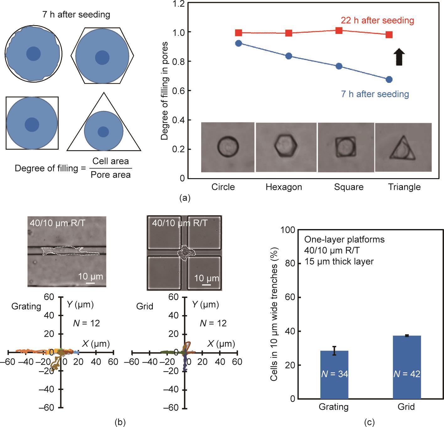
图3
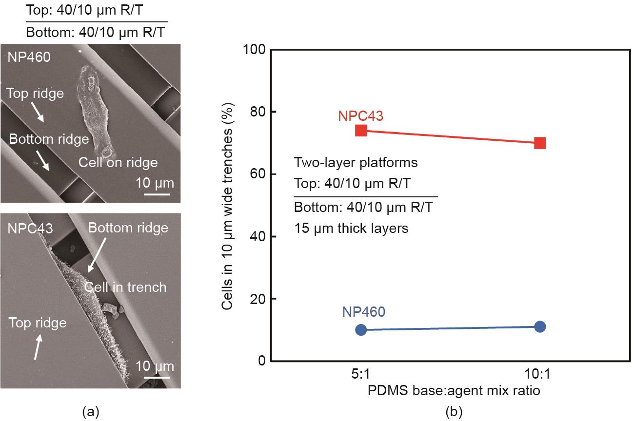
图4

图5
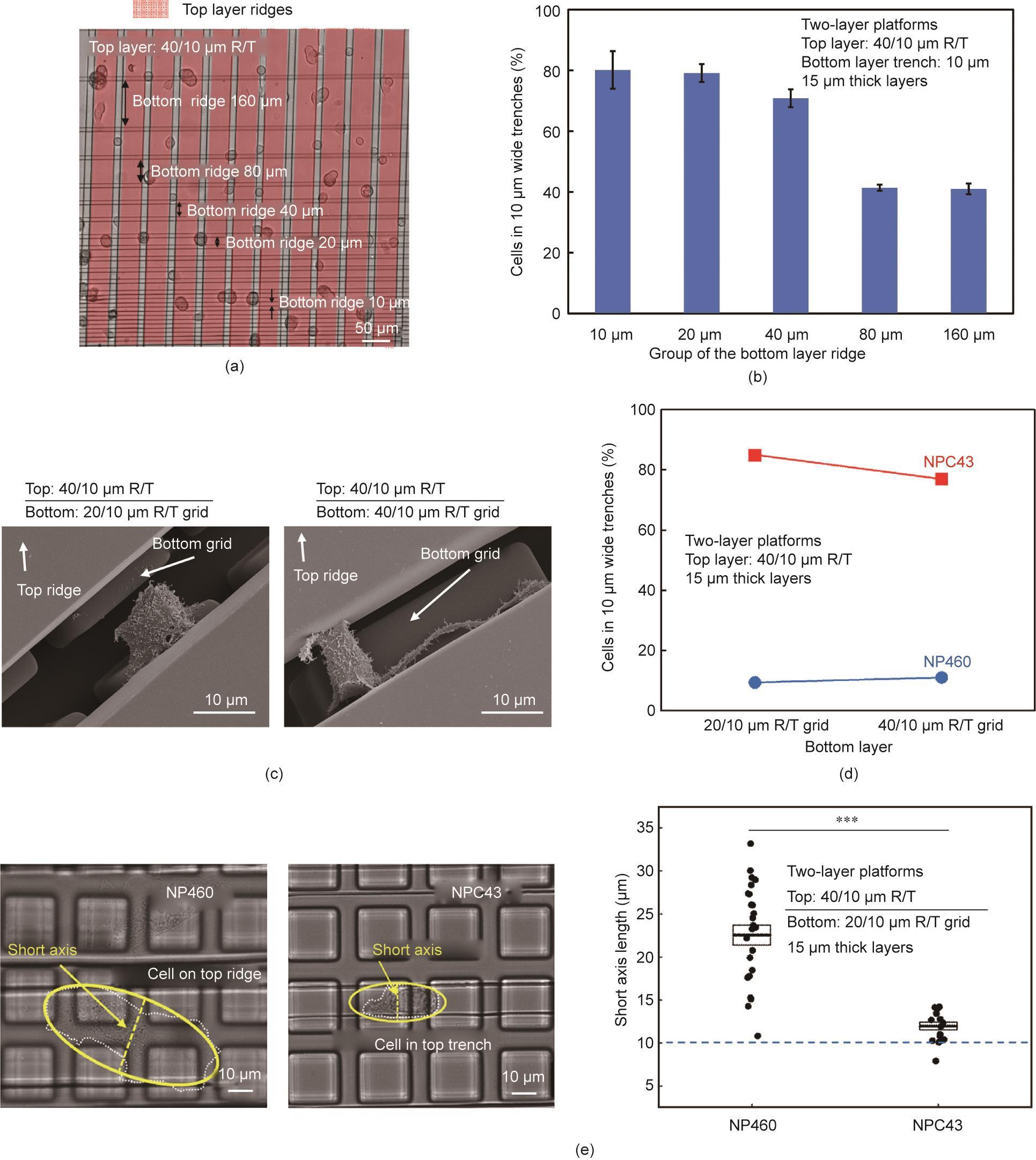
图6

图7
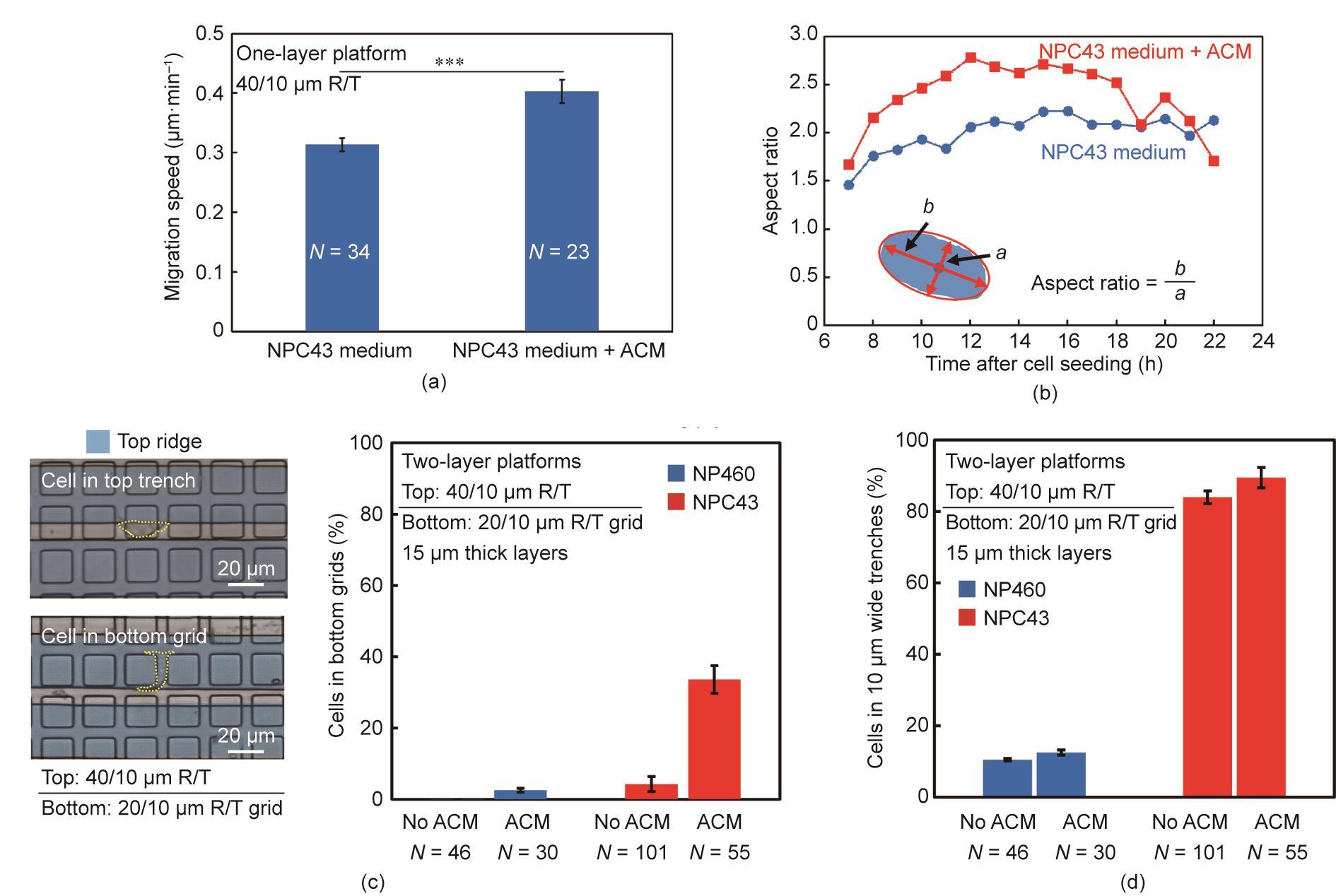
图8
参考文献
[ 1 ] Gossett DR, Weaver WM, Mach AJ, Hur SC, Tse HTK, Lee W, et al. Label-free cell separation and sorting in microfluidic systems. Anal Bioanal Chem 2010;397 (8):3249–67. 链接1
[ 2 ] Fiddler M. Fetal cell based prenatal diagnosis: perspectives on the present and future. J Clin Med 2014;3(3):972–85. 链接1
[ 3 ] Blainey PC, Quake SR. Dissecting genomic diversity, one cell at a time. Nat Methods 2014;11(1):19–21. 链接1
[ 4 ] Schor SL, Schor AM. Phenotypic and genetic alterations in mammary stroma: implications for tumour progression. Breast Cancer Res 2001;3(6):373–9. 链接1
[ 5 ] Guo KT, SchÄfer R, Paul A, Gerber A, Ziemer G, Wendel HP. A new technique for the isolation and surface immobilization of mesenchymal stem cells from whole bone marrow using high-specific DNA aptamers. Stem Cells 2006;24 (10):2220–31. 链接1
[ 6 ] Cho SH, Chen CH, Tsai FS, Godin JM, Lo YH. Human mammalian cell sorting using a highly integrated micro-fabricated fluorescence-activated cell sorter (lFACS). Lab Chip 2010;10(12):1567–73. 链接1
[ 7 ] Pasut A, Oleynik P, Rudnicki MA. Isolation of muscle stem cells by fluorescence activated cell sorting cytometry. Methods Mol Biol 2012;798:53–64. 链接1
[ 8 ] Schulz KR, Danna EA, Krutzik PO, Nolan GP. Single-cell phospho-protein analysis by flow cytometry. Curr Protoc Immunol 2012;96(1):8.17.1-20. 链接1
[ 9 ] Wu M, Singh AK. Single-cell protein analysis. Curr Opin Biotechnol 2012;23 (1):83–8. 链接1
[10] Miltenyi S, Müller W, Weichel W, Radbruch A. High gradient magnetic cell separation with MACS. Cytometry 1990;11(2):231–8. 链接1
[11] Allan AL, Vantyghem SA, Tuck AB, Chambers AF, Chin-Yee IH, Keeney M. Detection and quantification of circulating tumor cells in mouse models of human breast cancer using immunomagnetic enrichment and multiparameter flow cytometry. Cytometry A 2005;65(1):4–14. 链接1
[12] Hejazian M, Li W, Nguyen NT. Lab on a chip for continuous-flow magnetic cell separation. Lab Chip 2015;15(4):959–70. 链接1
[13] Holt LM, Olsen ML. Novel applications of magnetic cell sorting to analyze celltype specific gene and protein expression in the central nervous system. PLoS ONE 2016;11(2):e0150290. 链接1
[14] Citri A, Pang ZP, Südhof TC, Wernig M, Malenka RC. Comprehensive qPCR profiling of gene expression in single neuronal cells. Nat Protoc 2011;7 (1):118–27. 链接1
[15] Paiè P, Zandrini T, Vázquez RM, Osellame R, Bragheri F. Particle manipulation by optical forces in microfluidic devices. Micromachines 2018;9(5):200. 链接1
[16] Eberwine J, Yeh H, Miyashiro K, Cao Y, Nair S, Finnell R, et al. Analysis of gene expression in single live neurons. Proc Natl Acad Sci USA 1992;89 (7):3010–4. 链接1
[17] Yousuff CM, Ho ETW, Hussain KI, Hamid NHB. Microfluidic platform for cell isolation and manipulation based on cell properties. Micromachines 2017;8 (1):15. 链接1
[18] Tai CH, Hsiung SK, Chen CY, Tsai ML, Lee GB. Automatic microfluidic platform for cell separation and nucleus collection. Biomed Microdevices 2007;9 (4):533–43. 链接1
[19] Yun H, Kim K, Lee WG. Cell manipulation in microfluidics. Biofabrication 2013;5(2):022001. 链接1
[20] Shields IV CW, Reyes CD, López GP. Microfluidic cell sorting: a review of the advances in the separation of cells from debulking to rare cell isolation. Lab Chip 2015;15(5):1230–49. 链接1
[21] Ji HM, Samper V, Chen Y, Heng CK, Lim TM, Yobas L. Silicon-based microfilters for whole blood cell separation. Biomed Microdevices 2008;10(2):251–7. 链接1
[22] Huang LR, Cox EC, Austin RH, Sturm JC. Continuous particle separation through deterministic lateral displacement. Science 2004;304(5673):987–90. 链接1
[23] Kuntaegowdanahalli SS, Bhagat AAS, Kumar G, Papautsky I. Inertial microfluidics for continuous particle separation in spiral microchannels. Lab Chip 2009;9(20):2973–80. 链接1
[24] Zheng S, Lin H, Liu JQ, Balic M, Datar R, Cote RJ, et al. Membrane microfilter device for selective capture, electrolysis and genomic analysis of human circulating tumor cells. J Chromatogr A 2007;1162(2):154–61. 链接1
[25] Takagi J, Yamada M, Yasuda M, Seki M. Continuous particle separation in a microchannel having asymmetrically arranged multiple branches. Lab Chip 2005;5(7):778–84. 链接1
[26] Yamada M, Kano K, Tsuda Y, Kobayashi J, Yamato M, Seki M, et al. Microfluidic devices for size-dependent separation of liver cells. Biomed Microdevices 2007;9(5):637–45. 链接1
[27] Kuo JS, Zhao Y, Schiro PG, Ng L, Lim DSW, Shelby JP, et al. Deformability considerations in filtration of biological cells. Lab Chip 2010;10(7):837–42. 链接1
[28] Mohamed H, Turner JN, Caggana M. Biochip for separating fetal cells from maternal circulation. J Chromatogr A 2007;1162(2):187–92. 链接1
[29] Hsu CH, Di Carlo D, Chen C, Irimia D, Toner M. Microvortex for focusing, guiding and sorting of particles. Lab Chip 2008;8(12):2128–34. 链接1
[30] Chen DF, Du H, Li WH. A 3D paired microelectrode array for accumulation and separation of microparticles. J Micromech Microeng 2006;16(7):1162. 链接1
[31] Cui HH, Voldman J, He XF, Lim KM. Separation of particles by pulsed dielectrophoresis. Lab Chip 2009;9(16):2306–12. 链接1
[32] MacDonald MP, Spalding GC, Dholakia K. Microfluidic sorting in an optical lattice. Nature 2003;426(6965):421–4. 链接1
[33] Milne G, Rhodes D, MacDonald M, Dholakia K. Fractionation of polydisperse colloid with acousto-optically generated potential energy landscapes. Opt Lett 2007;32(9):1144–6. 链接1
[34] McFaul SM, Lin BK, Ma H. Cell separation based on size and deformability using microfluidic funnel ratchets. Lab Chip 2012;12(13):2369–76. 链接1
[35] Preira P, Grandné V, Forel JM, Gabriele S, Camara M, Theodoly O. Passive circulating cell sorting by deformability using a microfluidic gradual filter. Lab Chip 2013;13(1):161–70. 链接1
[36] Lu X, Martin A, Soto F, Angsantikul P, Li J, Chen C, et al. Parallel label-free isolation of cancer cells using arrays of acoustic microstreaming traps. Adv Mater Technol 2019;4(2):1800374. 链接1
[37] Tang QY, Tong WY, Shi J, Shi P, Lam YW, Pang SW. Influence of engineered surface on cell directionality and motility. Biofabrication 2014;6(1):015011. 链接1
[38] Zhou SF, Gopalakrishnan S, Xu YH, Yang J, Lam YW, Pang SW. A unidirectional cell switching gate by engineering grating length and bending angle. PLoS ONE 2016;11(1):e0147801. 链接1
[39] Wei WI, Sham JST. Nasopharyngeal carcinoma. Lancet 2005;365 (9476):2041–54. 链接1
[40] Chan KCA, Hung ECW, Woo JKS, Chan PKS, Leung SF, Lai FPT, et al. Early detection of nasopharyngeal carcinoma by plasma Epstein–Barr virus DNA analysis in a surveillance program. Cancer 2013;119(10):1838–44. 链接1
[41] Siva Sankar P, Mat MFC, Muniandy K, Xiang BLS, Ling PS, Hoe SLL, et al. Modeling nasopharyngeal carcinoma in three dimensions. Oncol Lett 2017;13 (4):2034–44. 链接1
[42] Yip YL, Lin W, Deng W, Jia L, Lo KW, Busson P, et al. Establishment of a nasopharyngeal carcinoma cell line capable of undergoing lytic Epstein–Barr virus reactivation. Lab Invest 2018;98(8):1093–104. 链接1
[43] Dittmer DP, Hilscher CJ, Gulley ML, Yang EV, Chen M, Glaser R. Multiple pathways for Epstein–Barr virus episome loss from nasopharyngeal carcinoma. Int J Cancer 2008;123(9):2105–12. 链接1
[44] Lin W, Yip YL, Jia L, Deng W, Zheng H, Dai W, et al. Establishment and characterization of new tumor xenografts and cancer cell lines from EBVpositive nasopharyngeal carcinoma. Nat Commun 2018;9(1):4663. 链接1
[45] Wang Y, Wang G, Luo X, Qiu J, Tang C. Substrate stiffness regulates the proliferation, migration, and differentiation of epidermal cells. Burns 2012;38 (3):414–20. 链接1
[46] Bangasser BL, Shamsan GA, Chan CE, Opoku KN, Tüzel E, Schlichtmann BW, et al. Shifting the optimal stiffness for cell migration. Nat Commun 2017;8 (1):15313. 链接1
[47] Pathak A, Kumar S. Independent regulation of tumor cell migration by matrix stiffness and confinement. Proc Natl Acad Sci USA 2012;109(26):10334–9. 链接1
[48] Zhong Y, Ji B. Impact of cell shape on cell migration behavior on elastic substrate. Biofabrication 2013;5(1):015011. 链接1
[49] Huang L, Chua MLK. Surgery as an alternative to radiotherapy in early-stage nasopharyngeal carcinoma: innovation at the expense of uncertainty. Cancer Commun 2020;40(2–3):119–21. 链接1
[50] Zhao L, Lu YT, Li F, Wu K, Hou S, Yu J, et al. High-purity prostate circulating tumor cell isolation by a polymer nanofiber-embedded microchip for whole exome sequencing. Adv Mater 2013;25(21):2897–902. 链接1
[51] van Zijl F, Krupitza G, Mikulits W. Initial steps of metastasis: cell invasion and endothelial transmigration. Mutat Res 2011;728(1–2):23–34. 链接1
[52] Bange J, Prechtl D, Cheburkin Y, Specht K, Harbeck N, Schmitt M, et al. Cancer progression and tumor cell motility are associated with the FGFR4 Arg388 allele. Cancer Res 2002;62(3):840–7. 链接1
[53] Olson MF, Sahai E. The actin cytoskeleton in cancer cell motility. Clin Exp Metastasis 2009;26(4):273–87. 链接1
[54] Luga V, Zhang L, Viloria-Petit AM, Ogunjimi AA, Inanlou MR, Chiu E, et al. Exosomes mediate stromal mobilization of autocrine Wnt–PCP signaling in breast cancer cell migration. Cell 2012;151(7):1542–56. 链接1
[55] Zhang L, Luga V, Armitage SK, Musiol M, Won A, Yip CM, et al. A lateral signalling pathway coordinates shape volatility during cell migration. Nat Commun 2016;7(1):11714. 链接1
[56] Zhou SF, Gopalakrishnan S, Xu YH, To SKY, Wong AST, Pang SW, et al. Substrates with patterned topography reveal metastasis of human cancer cells. Biomed Mater 2017;12(5):055001. 链接1


 京公网安备 11010502051620号


