
2022年 第16卷 第9期
《工程(英文)》 >> 2022年 第16卷 第9期 doi: 10.1016/j.eng.2021.05.007
基于一种全新的优化多变量非等距模型对中国汉族成人左心室多普勒超声心动图测值生理性变异的校正方法
a Key Laboratory of Cardiovascular Remodeling and Function Research, Chinese Ministry of Education & Key Laboratory of Cardiovascular Remodeling and Function Research, Chinese National Health Commission and Chinese Academy of Medical Sciences & the State and Shandong Province Joint Key Laboratory of Translational Cardiovascular Medicine & Department of Cardiology, Qilu Hospital, Shandong University, Jinan 250012, China
b Department of Cardiology, Qilu Hospital (Qingdao), Shandong University, Qingdao 266053, China
c Department of Biostatistics, School of Public Health, Shandong University, Jinan 250012, China
d Ultrasonography Department, Shenzhen People’s Hospital, Shenzhen 518020, China
e School of Microelectronics, Shandong University, Jinan 250101, China
f School of Mathematical Sciences, Ocean University of China, Qingdao 266100, China
下一篇 上一篇
摘要
多数左心室(LV)多普勒超声心动图参数测值随年龄和性别显著变化,因此有必要对其生理性差异进行校正。本研究旨在验证:不同的多普勒参数测值与生物学特征变量间呈异速非线性相关,且其校正常数与校正指数各不同。共测量了1224名健康成人的23个LV多普勒参数。随机选择70%数据(A组)建立优化多变量非线性模型(OMAM),在30%数据(B组)和183名超重人群数据(C组)中验证OMAM的可靠性,并与基于体表面积(BSA)的单变量等距模型(SVIM)进行比较。结果显示,校正前,23个LV多普勒参数均与一个或多个生物学特征变量显著相关,B组中47.8% (11/23)的参数存在性别间差异,经OMAM校正后,81.8% (9/11)的参数消除了性别间差异。OMAM对B组和C组数据的校正成功率分别为100% (23/23)和82.6% (19/23),建立了独立于生物学特征变量的多普勒参数的OMAM参考值,而基于BSA的SVIM校正成功率为零。不同的LV多普勒参数与一个或多个生物学特征变量呈异速非线性相关;本研究建立的OMAM成功校正了因生物学特征变量差异对健康和超重人群多普勒测值的生理性影响,其校正效果显著优于传统的SVIM。然而,OMAM针对其他种族、肥胖和疾病状态人群的适用性仍需进一步探究
补充材料
图片
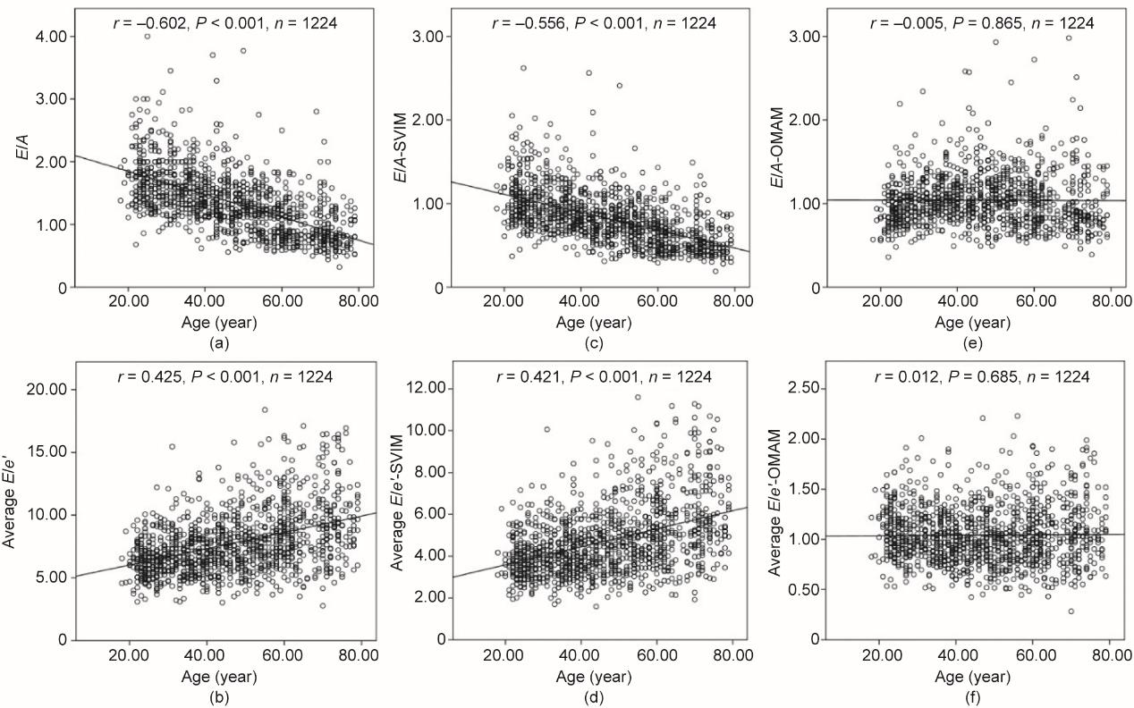
图1
参考文献
[ 1 ] Nagueh SF, Smiseth OA, Appleton CP, Byrd BF3rd, Dokainish H, Edvardsen T, et al.; Houston, Texas; Oslo, Norway; Phoenix, Arizona; Nashville, Tennessee; Hamilton, Ontario, Canada; Uppsala, Sweden; Ghent and Liège, Belgium; Cleveland, Ohio; Novara, Italy; Rochester, Minnesota; Bucharest, Romania; and LouisSt., Missouri. Recommendations for the evaluation of left ventricular diastolic function by echocardiography: an update from the American Society of Echocardiography and the European Association of Cardiovascular Imaging. Eur Heart J Cardiovasc Imaging 2016;17(12):1321‒60. 链接1
[ 2 ] Lang RM, Badano LP, Mor-Avi V, Afilalo J, Armstrong A, Ernande L, et al. Recommendations for cardiac chamber quantification by echocardiography in adults: an update from the American Society of Echocardiography and the European Association of Cardiovascular Imaging. Eur Heart J Cardiovasc Imaging 2015;16(3):233‒70. 链接1
[ 3 ] Caballero L, Kou S, Dulgheru R, Gonjilashvili N, Athanassopoulos GD, Barone D, et al. Echocardiographic reference ranges for normal cardiac Doppler data: results from the NORRE Study. Eur Heart J Cardiovasc Imaging 2015;16(9):1031‒41.
[ 4 ] Yao GH, Zhang M, Yin LX, Zhang C, Xu MJ, Deng Y, et al.; the Echocardiographic Measurements in Normal Chinese Adults (EMINCA) Study Investigators. Doppler echocardiographic measurements in normal Chinese adults (EMINCA): a prospective, nationwide, and multicentre study. Eur Heart J Cardiovasc Imaging 2016;17(5):512‒22. 链接1
[ 5 ] Daimon M, Watanabe H, Abe Y, Hirata K, Hozumi T, Ishii K, et al.; the JAMP Study Investigators. Normal values of echocardiographic parameters in relation to age in a healthy Japanese population: the JAMP study. Circ J 2008;72(11):1859‒66. 链接1
[ 6 ] Roberson DA, Cui W, Chen Z, Madronero LF, Cuneo BF. Annular and septal Doppler tissue imaging in children: normal z-score tables and effects of age, heart rate, and body surface area. J Am Soc Echocardiogr 2007;20(11):1276‒84. 链接1
[ 7 ] Cantinotti M, Lopez L. Nomograms for blood flow and tissue Doppler velocities to evaluate diastolic function in children: a critical review. J Am Soc Echocardiogr 2013;26(2):126‒41. 链接1
[ 8 ] Yao GH, Chen XY, Zhang Q, Zeng XY, Li XL, Zhang S, et al. A novel mathematical model for correcting the physiologic variances of two-dimensional echocardiographic measurements in healthy Chinese adults. J Am Soc Echocardiogr 2019;32(7):876‒83.e11. 链接1
[ 9 ] Yao GH, Deng Y, Liu Y, Xu MJ, Zhang C, Deng YB, et al.; the Echocardiographic Measurements in Normal Chinese Adults (EMINCA) Study Investigators. Echocardiographic measurements in normal Chinese adults focusing on cardiac chambers and great arteries: a prospective, nationwide, and multicenter study. J Am Soc Echocardiogr 2015;28(5):570‒9. 链接1
[10] Nagueh SF, Appleton CP, Gillebert TC, Marino PN, Oh JK, Smiseth OA, et al. Recommendations for the evaluation of left ventricular diastolic function by echocardiography. J Am Soc Echocardiogr 2009;22(2):107‒33. 链接1
[11] Neilan TG, Pradhan AD, Weyman AE. Derivation of a size-independent variable for scaling of cardiac dimensions in a normal adult population. J Am Soc Echocardiogr 2008;21(7):779‒85. 链接1
[12] Du Bois D, Du Bois EF. A formula to estimate the approximate surface area if height and weight be known. Arch Intern Med 1916;17:863‒71.
[13] Satumora S. Ultrasonic Doppler method for the inspection of cardiac function. J Acoust Soc Am 1957;29(11):1181‒5. 链接1
[14] Popovic´ ZB, Sun JP, Yamada H, Drinko J, Mauer K, Greenberg NL, et al. Differences in left ventricular long-axis function from mice to humans follow allometric scaling to ventricular size. J Physiol 2005;568(Pt 1):255‒65. 链接1
[15] Henry WL, Ware J, Gardin JM, Hepner SI, McKay J, Weiner M. Echocardiographic measurements in normal subjects. Growth-related changes that occur between infancy and early adulthood. Circulation 1978;57(2):278‒85. 链接1
[16] Dalen H, Thorstensen A, Vatten LJ, Aase SA, Stoylen A. Reference values and distribution of conventional echocardiographic Doppler measures and longitudinal tissue Doppler velocities in a population free from cardiovascular disease. Circ Cardiovasc Imaging 2010;3(5):614‒22. 链接1
[17] Patey O, Carvalho JS, Thilaganathan B. Intervendor discordance of fetal and neonatal myocardial tissue Doppler and speckle-tracking measurements. J Am Soc Echocardiogr 2019;32(10):1339‒49.e23. 链接1
[18] Kuznetsova T, Herbots L, López B, Jin Y, Richart T, Thijs L, et al. Prevalence of left ventricular diastolic dysfunction in a general population. Circ Heart Fail 2009;2(2):105‒12. 链接1
[19] Asch FM, Miyoshi T, Addetia K, Citro R, Daimon M, Desale S, et al.; the WASE Investigators. Similarities and differences in left ventricular size and function among races and nationalities. Results of the World Alliance of Societies of Echocardiography (WASE) normal values study. J Am Soc Echocardiogr 2019;32(11):1396‒406.e2.


 京公网安备 11010502051620号


