2017, Volume 3, Issue 3
Engineering >> 2017, Volume 3, Issue 3 doi: 10.1016/J.ENG.2017.03.014
Facile and Scalable Preparation of Fluorescent Carbon Dots for Multifunctional Applications
a Beijing Advanced Innovation Center for Soft Matter Science and Engineering & State Key Laboratory of Organic-Inorganic Composites, Beijing University of Chemical Technology, Beijing 100029, China
b Center of Advanced Science and Engineering for Carbon (Case4Carbon), Department of Macromolecular Science and Engineering, Case School of Engineering, Case Western Reserve University, Cleveland, OH 44106, USA
c SCNU-ZJU Joint Research Center of Photonics, South China Academy of Advanced Optoelectronics, South China Normal University, Guangzhou 510006, China
d Department of Chemical Engineering, Curtin University, Perth, WA 6845, Australia
Next Previous
Abstract
The synthesis of fluorescent nanomaterials has received considerable attention due to the great potential of these materials for a wide range of applications, from chemical sensing through bioimaging to optoelectronics. Herein, we report a facile and scalable approach to prepare fluorescent carbon dots (FCDs) via a one-pot reaction of citric acid with ethylenediamine at 150 °C under ambient air pressure. The resultant FCDs possess an optical bandgap of 3.4 eV and exhibit strong excitation-wavelength-independent blue emission (λEm = 450 nm) under either one- or two-photon excitation. Owing to their low cytotoxicity and long fluorescence lifetime, these FCDs were successfully used as internalized fluorescent probes in human cancer cell lines (HeLa cells) for two-photon excited imaging of cells by fluorescence lifetime imaging microscopy with high-contrast resolution. They were also homogenously mixed with commercial inks and used to draw fluorescent patterns on normal papers and on many other substrates (e.g., certain flexible plastic films, textiles, and clothes). Thus, these nanomaterials are promising for use in solid-state fluorescent sensing, security labeling, and wearable optoelectronics.
Keywords
Scalable ; Carbon dots ; Two-photon ; Fluorescence lifetime imaging ; Patterning
SupplementaryMaterials
Figures
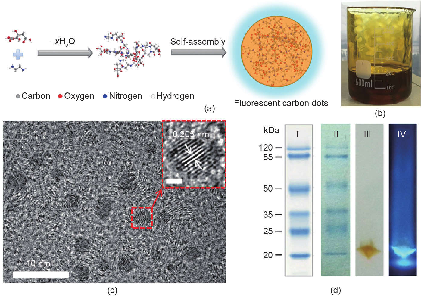
Fig. 1
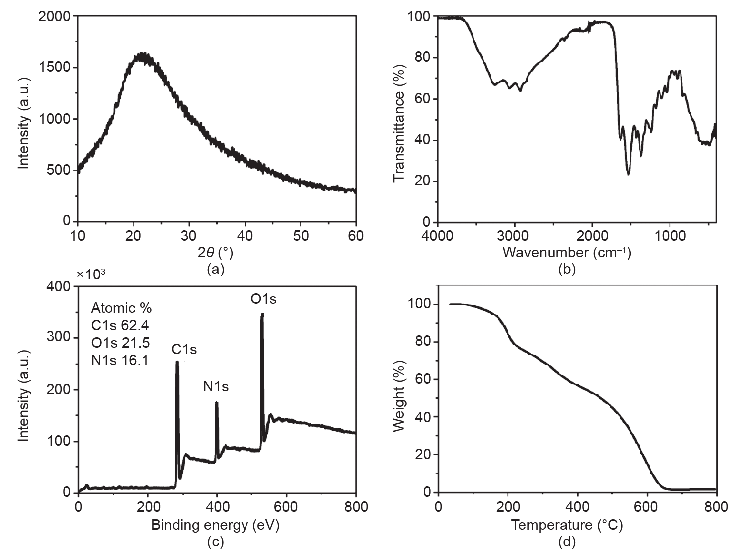
Fig. 2
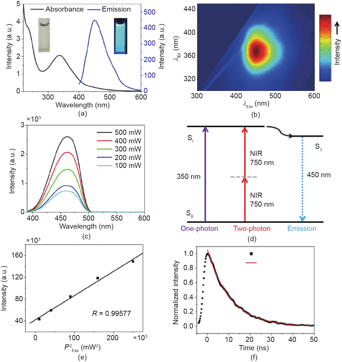
Fig. 3
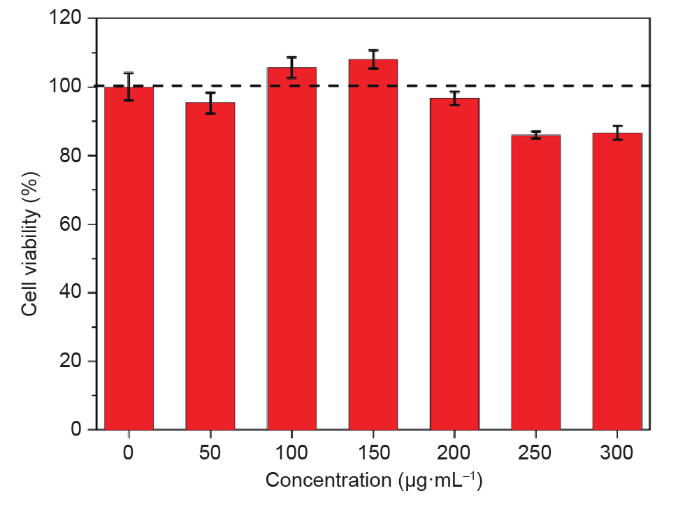
Fig. 4

Fig. 5

Fig. 6
References
[ 1 ] Yao J, Yang M, Duan Y. Chemistry, biology, and medicine of fluorescent nanomaterials and related systems: New insights into biosensing, bioimaging, genomics, diagnostics, and therapy. Chem Rev 2014;114(12):6130–78 link1
[ 2 ] Bruchez Jr M, Moronne M, Gin P, Weiss S, Alivisatos AP. Semiconductor nanocrystals as fluorescent biological labels. Science 1998;281(5385):2013–6 link1
[ 3 ] Wang D, Qian J, Cai F, He S, Han S, Mu Y. ‘Green’-synthesized near-infrared PbS quantum dots with silica-PEG dual-layer coating: Ultrastable and biocompatible optical probes for in vivo animal imaging. Nanotechnology 2012;23(24):245701 link1
[ 4 ] Chen JF, Ding HM, Wang JX, Shao L. Preparation and characterization of porous hollow silica nanoparticles for drug delivery application. Biomaterials 2004;25(4):723–7 link1
[ 5 ] Wang D, Qian J, He S, Park JS, Lee KS, Han S, et al.. Aggregation-enhanced fluorescence in PEGylated phospholipid nanomicelles for in vivo imaging. Biomaterials 2011;32(25):5880–8 link1
[ 6 ] Wang D, Qian J, Qin W, Qin A, Tang BZ, He S. Biocompatible and photostable AIE dots with red emission for in vivo two-photon bioimaging. Sci Rep 2014;4(3):4279
[ 7 ] Bharali DJ, Klejbor I, Stachowiak EK, Dutta P, Roy I, Kaur N, et al.. Organically modified silica nanoparticles: A nonviral vector for in vivo gene delivery and expression in the brain. Proc Natl Acad Sci 2005;102(32):11539–44 link1
[ 8 ] Chen G, Qiu H, Prasad PN, Chen X. Upconversion nanoparticles: Design, nanochemistry, and applications in theranostics. Chem Rev 2014;114(10):5161–214 link1
[ 9 ] Wang D, Zhu L, Chen JF, Dai L. Liquid marbles based on magnetic upconversion nanoparticles as magnetically and optically responsive miniature reactors for photocatalysis and photodynamic therapy. Angew Chem Int Ed 2016;55(36):10795–9 link1
[10] Xing Y, Dai L. Nanodiamonds for nanomedicine. Nanomedicine 2009;4(2):207–18 link1
[11] Wang D, Chen JF, Dai L. Recent advances in graphene quantum dots for fluorescence bioimaging from cells through tissues to animals. Part Part Syst Charact 2015;32(5):515–23 link1
[12] Baker SN, Baker GA. Luminescent carbon nanodots: Emergent nanolights. Angew Chem Int Ed 2010;49(38):6726–44 link1
[13] Zhu S, Song Y, Zhao X, Shao J, Zhang J, Yang B. The photoluminescence mechanism in carbon dots (graphene quantum dots, carbon nanodots, and polymer dots): Current state and future perspective. Nano Res 2015;8(2):355–81 link1
[14] Zhang H, Huang Y, Hu S, Huang Q, Wei C, Zhang W, et al.. Fluorescent probes for “off-on” sensitive and selective detection of mercury ions and L-cysteine based on graphitic carbon nitride nanosheets. J Mater Chem C 2015;3(9):2093–100 link1
[15] Yang ST, Cao L, Luo PG, Lu F, Wang X, Wang H, et al.. Carbon dots for optical imaging in vivo. J Am Chem Soc 2009;131(32):11308–9 link1
[16] Liu J, Zhu W, Yu S, Yan X. Three dimensional carbogenic dots/TiO2 nanoheterojunctions with enhanced visible light-driven photocatalytic activity. Carbon 2014;79(1):369–79 link1
[17] Zhang YQ, Ma DK, Zhang YG, Chen W, Huang SM. N-doped carbon quantum dots for TiO2-based photocatalysts and dye-sensitized solar cells. Nano Energy 2013;2(5):545–52 link1
[18] Jiang K, Sun S, Zhang L, Lu Y, Wu A, Cai C, et al.. Red, green, and blue luminescence by carbon dots: Full-color emission tuning and multicolor cellular imaging. Angew Chem Int Ed 2015;54(18):5360–3 link1
[19] ]Liu J, Liu Y, Liu N, Han Y, Zhang X, Huang H, et al.. Metal-free efficient photocatalyst for stable visible water splitting via a two-electron pathway. Science 2015;347(6225):970–4 link1
[20] Mirtchev P, Henderson EJ, Soheilnia N, Yip CM, Ozin GA. Solution phase synthesis of carbon quantum dots as sensitizers for nanocrystalline TiO2 solar cells. J Mater Chem 2012;22(4):1265–9 link1
[21] Wu ZL, Zhang P, Gao MX, Liu CF, Wang W, Leng F, et al.. One-pot hydrothermal synthesis of highly luminescent nitrogen-doped amphoteric carbon dots for bioimaging from Bombyx mori silk-natural proteins. J Mater Chem B 2013;1(22):2868–73 link1
[22] Qu D, Zheng M, Zhang L, Zhao H, Xie Z, Jing X, et al.. Formation mechanism and optimization of highly luminescent N-doped graphene quantum dots. Sci Rep 2014;4(9):5294
[23] Devaraju MK, Honma I. Hydrothermal and solvothermal process towards development of LiMPO4 (M= Fe, Mn) nanomaterials for lithium-ion batteries. Adv Energy Mater 2012;2(3):284–97 link1
[24] Anastas PT, Warner JC. Principles of green chemistry. In:Green chemistry: Theory and practice. New York: Oxiford University Press; 1998. p. 30.
[25] Wang D, Zhu L, Mccleese C, Bruda C, Chen JF, Dai L. Fluorescent carbon dots from milk by microwave cooking. RSC Advances 2016;6(47):41516–21 link1
[26] Zhou J, Booker C, Li R, Zhou X, Sham TK, Sun X, et al.. An electrochemical avenue to blue luminescent nanocrystals from multiwalled carbon nanotubes (MWCNTs). J Am Chem Soc 2007;129(4):744–5 link1
[27] Li Y, Zhao Y, Cheng H, Hu Y, Shi G, Dai L, et al.. Nitrogen-doped graphene quantum dots with oxygen-rich functional groups. J Am Chem Soc 2012;134(1):15–8 link1
[28] Dong Y, Shao J, Chen C, Li H, Wang R, Chi Y, et al.. Blue luminescent graphene quantum dots and graphene oxide prepared by tuning the carbonization degree of citric acid. Carbon 2012;50(12):4738–43 link1
[29] Resch-Genger U, Grabolle M, Cavaliere-Jaricot S, Nitschke R, Nann T. Quantum dots versus organic dyes as fluorescent labels. Nat Methods 2008;5(9):763–75 link1
[30] Wang D, Zhu L, Chen JF, Dai L. Can graphene quantum dots cause DNA damage in cells? Nanoscale 2015;7(21):9894–901 link1
[31] Qian J, Wang D, Cai FH, Xi W, Peng L, Zhu ZF, et al.. Observation of multiphoton-induced fluorescence from graphene oxide nanoparticles and applications in in vivo functional bioimaging. Angew Chem Int Ed 2012;51(42):10570–5 link1
[32] Shi L, Li Y, Li X, Wen X, Zhang G, Yang J, et al.. Facile and eco-friendly synthesis of green fluorescent carbon nanodots for applications in bioimaging, patterning and staining. Nanoscale 2015;7(16):7394–401 link1
[33] Sun H, Zhang J, Zhang KY, Liu S, Liang H, Lv W, et al.. Development of two-channel phosphorescent core-shell nanoprobe for ratiometric and time-resolved luminescence imaging of intracellular oxygen levels. Part Part Syst Charact 2015;32(1):48–53 link1
[34] Orte A, Alvarez-Pez JM, Ruedas-Rama MJ. Fluorescence lifetime imaging microscopy for the detection of intracellular pH with quantum dot nanosensors. ACS Nano 2013;7(7):6387–95 link1


 京公网安备 11010502051620号


