
2022, Volume 10, Issue 3
Engineering >> 2022, Volume 10, Issue 3 doi: 10.1016/j.eng.2020.07.031
Rotation of Biological Cells: Fundamentals and Applications
a Division of Materials Science, Nara Institute of Science and Technology, Nara 630-0192, Japan
b Institute of Innovation for Future Society, Nagoya University, Aichi 464-8603, Japan
c Department of Precision Mechanics, Faculty of Science and Engineering, Chuo University, Tokyo 112-8551, Japan
d Graduate School of Frontier Biosciences, Osaka University, Osaka 565-0871, Japan
e RIKEN Center for Biosystems Dynamics Research, Osaka 565-0871, Japan
f School of Mechanical, Materials, Mechatronic, and Biomedical Engineering, University of Wollongong, Wollongong 2522, Australia
g School of Engineering, Macquarie University, Sydney 2109, Australia
Next Previous
Abstract
Cell rotation is one of the most important techniques for cell manipulation in modern bioscience, as it not only permits cell observation from any arbitrary angle, but also simplifies the procedures for analyzing the mechanical properties of cells, characterizing cell physiology, and performing microsurgery. Numerous approaches have been reported for rotating cells in a wide range of academic and industrial applications. Among them, the most popular are micro-robot-based direct contact manipulation and field-based non-contact methods (e.g., optical, magnetic, electric, acoustic, and hydrodynamic methods). This review first summarizes the fundamental mechanisms, merits, and demerits of these six main groups of approaches, and then discusses their differences and limitations in detail. We aim to bridge the gap between each method and illustrate the development progress, current advances, and prospects in the field of cell rotation.
Keywords
Cell rotation ; Cell reorientation ; Micromanipulation ; Microfluidics
Figures
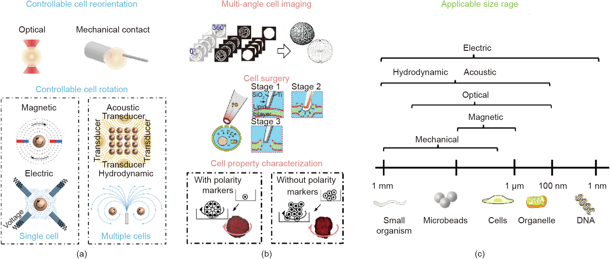
Fig. 1

Fig. 2

Fig. 3

Fig. 4

Fig. 5

Fig. 6

Fig. 7
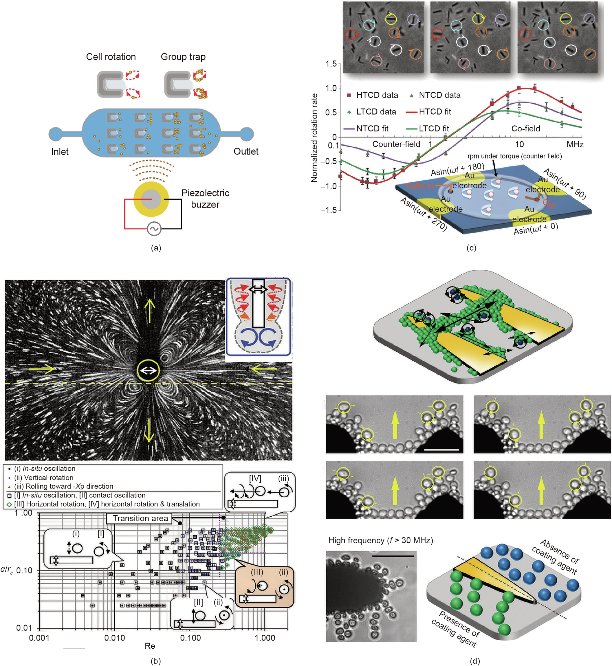
Fig. 8
References
[ 1 ] Habaza M, Kirschbaum M, Guernth-Marschner C, Dardikman G, Barnea I, Korenstein R, et al. Rapid 3D refractive-index imaging of live cells in suspension without labeling using dielectrophoretic cell rotation. Adv Sci 2017;4(2):1600205. link1
[ 2 ] Wollrab V, Thiagarajan R, Wald A, Kruse K, Riveline D. Still and rotating myosin clusters determine cytokinetic ring constriction. Nat Commun 2016;7 (1):11860. link1
[ 3 ] Leung C, Lu Z, Zhang XP, Sun Y. Three-dimensional rotation of mouse embryos. IEEE Trans Biomed Eng 2012;59(4):1049–56. link1
[ 4 ] Zhao Q, Sun M, Cui M, Yu J, Qin Y, Zhao X. Robotic cell rotation based on the minimum rotation force. IEEE Trans Autom Sci Eng 2015;12(4):1504–15. link1
[ 5 ] Xie M, Shakoor A, Shen Y, Mills JK, Sun D. Out-of-plane rotation control of biological cells with a robot-tweezers manipulation system for orientationbased cell surgery. IEEE Trans Biomed Eng 2019;66(1):199–207. link1
[ 6 ] Abu Ajamieh I, Benhabib B, Mills JK. Automatic system for the blastocyst embryo manipulation and rotation. Ann Biomed Eng 2020;48(1):426–36. link1
[ 7 ] Huang L, Zhao P, Wang W. 3D cell electrorotation and imaging for measuring multiple cellular biophysical properties. Lab Chip 2018;18(16):2359–68. link1
[ 8 ] Benhal P, Chase JG. System identification and stochastic estimation of dielectric properties of a spherical particle using AC-induced electrorotation. In: Proceedings of the 2015 20th International Conference on Process Control; 2015 Jul 9–12; Strbske Pleso, Slovakia.
[ 9 ] Carmon G, Feingold M. Rotation of single bacterial cells relative to the optical axis using optical tweezers. Opt Lett 2011;36(1):40–2. link1
[10] Merola F, Miccio L, Memmolo P, Di Caprio G, Galli A, Puglisi R, et al. Digital holography as a method for 3D imaging and estimating the biovolume of motile cells. Lab Chip 2013;13(23):4512–6. link1
[11] Hosseini SM, Moulavi F, Asgari V, Shirazi A, Abazari-Kia AH, Ghanaei HR, et al. Simple, fast, and efficient method of manual oocyte enucleation using a pulled Pasteur pipette. Vitr Cell Dev Biol Anim 2013;49:569–75. link1
[12] Elbez R, McNaughton BH, Patel L, Pienta KJ, Kopelman R, Chan C. Nanoparticle induced cell magneto-rotation: monitoring morphology, stress and drug sensitivity of a suspended single cancer cell. PLoS ONE 2011;6(12):e28475. link1
[13] Lee K, Yi Yi, Yu Y. Remote control of T cell activation using magnetic janus particles. Angew Chem Int Ed Engl 2016;55(26):7384–7. link1
[14] Rohani A, Varhue W, Su YH, Swami NS. Electrical tweezer for highly parallelized electrorotation measurements over a wide frequency bandwidth. Electrophoresis 2014;35(12–3):1795–802. link1
[15] Han SI, Joo YD, Han KH. An electrorotation technique for measuring the dielectric properties of cells with simultaneous use of negative quadrupolar dielectrophoresis and electrorotation. Analyst (Lond) 2013;138(5):1529–37. link1
[16] Ahmed D, Ozcelik A, Bojanala N, Nama N, Upadhyay A, Chen Y, et al. Rotational manipulation of single cells and organisms using acoustic waves. Nat Commun 2016;7(1):11085. link1
[17] Ozcelik A, Nama N, Huang PH, Kaynak M, McReynolds MR, Hanna-Rose W, et al. Acoustofluidic rotational manipulation of cells and organisms using oscillating solid structures. Small 2016;12(37):5120–5. link1
[18] Yalikun Y, Kanda Y, Morishima K. Hydrodynamic vertical rotation method for a single cell in an open space. Microfluid Nanofluidics 2016;20(5):74. link1
[19] Shelby JP, Chiu DT. Controlled rotation of biological micro- and nano-particles in microvortices. Lab Chip 2004;4(3):168–70. link1
[20] Rodrigo JA, Soto JM, Alieva T. Fast label-free microscopy technique for 3D dynamic quantitative imaging of living cells. Biomed Opt Express 2017;8 (12):5507–17. link1
[21] Umezawa K, Yoshida M, Kamiya M, Yamasoba T, Urano Y. Rational design of reversible fluorescent probes for live-cell imaging and quantification of fast glutathione dynamics. Nat Chem 2017;9(3):279–86. link1
[22] Wang T, Chen J, Zhou T, Song L. Fabricating microstructures on glass for microfluidic chips by glass molding process. Micromachines (Basel) 2018;9 (6):1–15. link1
[23] Zheng C, Zhao G, Liu W, Chen Y, Zhang Z, Jin L, et al. Three-dimensional superresolved live cell imaging through polarized multi-angle TIRF. Opt Lett 2018;43(7):1423–6. link1
[24] Fiolka R, Shao L, Rego EH, Davidson MW, Gustafsson MGL. Time-lapse twocolor 3D imaging of live cells with doubled resolution using structured illumination. Proc Natl Acad Sci USA 2012;109(14):5311–5. link1
[25] Läubli NF, Shamsudhin N, Vogler H, Munglani G, Grossniklaus U, Ahmed D, et al. 3D Manipulation and imaging of plant cells using acoustically activated microbubbles. Small Methods 2019;3(3):1800527. link1
[26] Habaza M, Gilboa B, Roichman Y, Shaked NT. Tomographic phase microscopy with 180 rotation of live cells in suspension by holographic optical tweezers. Opt Lett 2015;40(8):1881–4. link1
[27] Kim K, Yoon J, Park YK. Optical diffraction tomography for simultaneous 3D visualization and tracking of optically trapped particles. In: Proceedings of the Asia Communications and Photonics Conference 2015; 2015 Nov 19–23; Hong Kong, China.
[28] Cao B, Kelbauskas L, Chan S, Shetty RM, Smith D, Meldrum DR. Rotation of single live mammalian cells using dynamic holographic optical tweezers. Opt Lasers Eng 2017;92:70–5. link1
[29] Zhang S, Gibson LJ, Stilgoe AB, Nieminen TA, Rubinsztein-Dunlop H. Measuring local properties inside a cell-mimicking structure using rotating optical tweezers. J Biophotonics 2019;12(7):e201900022. link1
[30] Dai C, Zhang Z, Lu Y, Shan G, Wang X, Zhao Q, et al. Robotic manipulation of deformable cells for orientation control. IEEE Trans Robot 2020;36 (1):271–83. link1
[31] Zhao Y, Sun H, Sha X, Gu L, Zhan Z, Li WJ. A review of automated microinjection of zebrafish embryos. Micromachines (Basel) 2018;10(1):E7. link1
[32] Laffafian I, Hallett MB. Lipid-assisted microinjection: introducing material into the cytosol and membranes of small cells. Biophys J 1998;75(5):2558–63. link1
[33] Tanner K, Mori H, Mroue R, Bruni-Cardoso A, Bissell MJ. Coherent angular motion in the establishment of multicellular architecture of glandular tissues. Proc Natl Acad Sci USA 2012;109(6):1973–8. link1
[34] Tang Q, Liang F, Huang L, Zhao P, Wang W. On-chip simultaneous rotation of large-scale cells by acoustically oscillating bubble array. Biomed Microdevices 2020;22(1):13. link1
[35] Wu T-H, Teslaa T, Kalim S, French CT, Moghadam S, Wall R, et al. Photothermal nanoblade for large cargo delivery into mammalian cells. Anal Chem 2011;83(4):1321–7. link1
[36] Huang L, Zhao P, Liang F, Wang W. Single-cell 3D electro-rotation. Methods Cell Biol 2018;148:97–116. link1
[37] Lebel P, Basu A, Oberstrass FC, Tretter EM, Bryant Z. Gold rotor bead tracking for high-speed measurements of DNA twist, torque and extension. Nat Methods 2014;11(4):456–62. link1
[38] Xin Q, Li P, He Y, Shi C, Qiao Y, Bian X, et al. Magnetic tweezers for the mechanical research of DNA at the single molecule level. Anal Methods 2017;9(39):5720–30. link1
[39] Ding X, Lin SCS, Kiraly B, Yue H, Li S, Chiang IK, et al. On-chip manipulation of single microparticles, cells, and organisms using surface acoustic waves. PNAS 2012;109(28):11105–9. link1
[40] Xie Y, Bachman H, Huang TJ. Acoustofluidic methods in cell analysis. Trends Analyt Chem 2019;117:280–90. link1
[41] Tian Z, Yang S, Huang P-H, Wang Z, Zhang P, Gu Y, et al. Wave number-spiral acoustic tweezers for dynamic and reconfigurable manipulation of particles and cells. Sci Adv 2019;5(5):u6062. link1
[42] Kuncova J, Kallio P. Challenges in capillary pressure microinjection. Conf Proc IEEE Eng Med Biol Soc 2004;2004:4998–5001. link1
[43] Iritani A. Micromanipulation of gametes for in vitro assisted fertilization. Mol Reprod Dev 1991;28(2):199–207. link1
[44] Ren D, Wang J, Wang B, You Z. Probes for biomolecules detection based on RET-enhanced fluorescence polarization. Biosens Bioelectron 2016;79:802–9. link1
[45] Ren D, Xia Y, Wang B, You Z. Multiplexed analysis for anti-epidermal growth factor receptor tumor cell growth inhibition based on quantum dot probes. Anal Chem 2016;88(8):4318–27. link1
[46] Wang Z, Feng C, Muruganandam R, Ang WT, Tan SYM, Latt WT. Threedimensional cell rotation with fluidic flow-controlled cell manipulating device. IEEE/ASME Trans Mechatron 2016;21(4):1995–2003. link1
[47] Wang Z, Feng C, Ang WT, Tan SYM, Latt WT. Autofocusing and polar body detection in automated cell manipulation. IEEE Trans Biomed Eng 2017;64 (5):1099–105. link1
[48] Ajamieh IA, Benhabib B, Mills JK. Automated system for cell manipulation and rotation. In: Proceedings of the 2018 IEEE International Conference on Mechatronics and Automation; 2018 Aug 5–8; Changchun, China.
[49] Zhao C, Liu Y, Sun M, Zhao X. Robotic cell rotation based on optimal poking direction. Micromachines (Basel) 2018;9(4):E141. link1
[50] Wang Z, Latt WT, Tan SYM, Ang WT. Visual servoed three-dimensional cell rotation system. IEEE Trans Biomed Eng 2015;62(10):2498–507. link1
[51] Zhuang S, Lin W, Zhong J, Zhang G, Li Li, Qiu J, et al. Visual servoed threedimensional rotation control in zebrafish larva heart microinjection system. IEEE Trans Biomed Eng 2018;65(1):64–73. link1
[52] Aishan Y, Yalikun Y, Funano S-I, Shen Y, Tanaka Yo. Accurate rotation of ultrathin glass chamber for single-cell multidirectional observation. Appl Phys Express 2020;13:2. link1
[53] Aishan Y, Yalikun Y, Amaya S, Shen Y, Tanaka Y. Thin glass micro-dome structure based microlens fabricated by accurate thermal expansion of microcavities. Appl Phys Lett 2019;115(26):263501. link1
[54] Tanaka Y. Electric actuating valves incorporated into an all glass-based microchip exploiting the flexibility of ultra thin glass. RSC Adv 2013;3 (26):10213–20. link1
[55] Bochu W, Hucheng Z, Yiyao L, Yi J, Sakanishi A. The effects of alternative stress on the cell membrane deformability of chrysanthemum callus cells. Colloids Surf B Biointerfaces 2001;20(4):321–5. link1
[56] Ashkin A, Dziedzic J. Optical trapping and manipulation of viruses and bacteria. Science 1987;235(80):1517–20. link1
[57] Ashkin A, Dziedzic JM, Bjorkholm JE, Chu S. Observation of a single-beam gradient force optical trap for dielectric particles. Opt Lett 1986;11(5):288. link1
[58] Omori R, Kobayashi T, Suzuki A. Observation of a single-beam gradient-force optical trap for dielectric particles in air. Opt Lett 1997;22(11):816–8. link1
[59] Guck J, Ananthakrishnan R, Cunningham CC, Käs J. Stretching biological cells with light. J Phys Condens Matter 2002;14(19):311. link1
[60] Neuman KC, Block SM. Optical trapping. Rev Sci Instrum 2004;75 (9):2787–809. link1
[61] Zhang H, Liu KK. Optical tweezers for single cells. J R Soc Interface 2008;5 (24):671–90. link1
[62] Kim SB, Yoon SY, Sung HJ, Kim SS. Cross-type optical particle separation in a microchannel. Anal Chem 2008;80(7):2628–30. link1
[63] Koch M, Rohrbach A. How to calibrate an object-adapted optical trap for force sensing and interferometric shape tracking of asymmetric structures. Opt Express 2014;22(21):25242–57. link1
[64] Koch M, Rohrbach A. Object-adapted optical trapping and shape-tracking of energy-switching helical bacteria. Nat Photonics 2012;6(10):680–6. link1
[65] Chen X, Xiao G, Han X, Xiong W, Luo H, Yao B. Observation of spin and orbital rotation of red blood cell in dual-beam fibre-optic trap with transverse offset. J Opt 2017;19(5):055612. link1
[66] Chen X, Xiao G, Yang K, Xiong W, Luo H. Characteristics of the orbital rotation in dual-beam fiber-optic trap with transverse offset. Opt Express 2016;24 (15):16952–60. link1
[67] Kolb T, Albert S, Haug M, Whyte G. Optofluidic rotation of living cells for single-cell tomography. J Biophotonics 2015;8(3):239–46. link1
[68] Tatarkova SA, Carruthers AE, Dholakia K. One-dimensional optically bound arrays of microscopic particles. Phys Rev Lett 2002;89(28 Pt 1):283901. link1
[69] Kreysing MK, Kießling T, Fritsch A, Dietrich C, Guck JR, Käs JA. The optical cell rotator. Opt Express 2008;16(21):16984–92. link1
[70] Dasgupta R, Ahlawat S, Verma RS, Gupta PK. Optical orientation and rotation of trapped red blood cells with Laguerre–Gaussian mode. Opt Express 2011;19(8):7680–9688. link1
[71] Hosokawa Y, Hagiyama M, Iino T, Murakami Y, Ito A. Noncontact estimation of intercellular breaking force using a femtosecond laser impulse quantified by atomic force microscopy. Proc Natl Acad Sci USA 2011;108(5):1777–82. link1
[72] Hosokawa Y. Applications of the femtosecond laser-induced impulse to cell research. Jpn J Appl Phys 2019;58(11):110102. link1
[73] Koch MD, Shaevitz JW. Introduction to optical tweezers. Methods Mol Biol 2017;1486:3–24. link1
[74] Xie M, Shakoor A, Wu C. Manipulation of biological cells using a robot-aided optical tweezers system. Micromachines (Basel) 2018;9(5):245. link1
[75] Xie M, Chen S, Mills JK, Wang Y, Liu Y, Sun D. Cell out-of-plane rotation control using a cell surgery robotic system equipped with optical tweezers manipulators. In: Proceedings of the 2016 IEEE International Conference on Information and Automation; 2016 Aug 1–3; Ningbo, China.
[76] Wu MC. Optoelectronic tweezers. Nat Photonics 2011;5(6):322–4. link1
[77] Chowdhury S, Thakur A, Wang C, Svec P, Losert W, Gupta SK. Automated indirect manipulation of irregular shaped cells with Optical Tweezers for studying collective cell migration. In: Proceedings of the 2013 IEEE International Conference on Robotics and Automation; 2013 May 6–10; Karlsruhe, Germany.
[78] Chowdhury S, Thakur A, Svec P, Wang C, Losert W, Gupta SK. Automated manipulation of biological cells using gripper formations controlled by optical tweezers. IEEE Trans Autom Sci Eng 2014;11(2):338–47. link1
[79] Thakur A, Chowdhury S, Švec P, Wang C, Losert W, Gupta SK. Indirect pushing based automated micromanipulation of biological cells using optical tweezers. Int J Rob Res 2014;33(8):1098–111. link1
[80] Cheah CC, Ta QM, Haghighi R. Grasping and manipulation of a micro-particle using multiple optical traps. Automatica 2016;68:216–27. link1
[81] Lapotko DO, Zharov VP. Spectral evaluation of laser-induced cell damage with photothermal microscopy. Lasers Surg Med 2005;36(1):22–30. link1
[82] Galli R, Uckermann O, Andresen EF, Geiger KD, Koch E, Schackert G, et al. Intrinsic indicator of photodamage during label-free multiphoton microscopy of cells and tissues. PLoS ONE 2014;9(10):e110295. link1
[83] Liu Y, Sonek GJ, Berns MW, Tromberg BJ. Physiological monitoring of optically trapped cells: assessing the effects of confinement by 1064-nm laser tweezers using microfluorometry. Biophys J 1996;71(4):2158–67. link1
[84] Heymann PGB, Henkenius KSE, Ziebart T, Braun A, Hirthammer K, Halling F, et al. Modulation of tumor cell metabolism by laser photochemotherapy with cisplatin or zoledronic acid in vitro. Anticancer Res 2018;38(3):1291–301. link1
[85] Karu T. Photobiology of low-power laser effects. Health Phys 1989;56 (5):691–704. link1
[86] Doukas AG, Flotte TJ. Physical characteristics and biological effects of laserinduced stress waves. Ultrasound Med Biol 1996;22(2):151–64. link1
[87] Thomsen S. Pathologic analysis of photothermal and photomechanical effects of laser-tissue interactions. Photochem Photobiol 1991;53(6):825–35. link1
[88] Ye Z, Sitti M. Dynamic trapping and two-dimensional transport of swimming microorganisms using a rotating magnetic microrobot. Lab Chip 2014;14 (13):2177–82. link1
[89] Neuman KC, Nagy A. Single-molecule force spectroscopy: optical tweezers, magnetic tweezers and atomic force microscopy. Nat Methods 2008;5 (6):491–505. link1
[90] Li X, Liu C, Chen S, Wang Y, Cheng SH, Sun D. In vivo manipulation of single biological cells with an optical tweezers-based manipulator and a disturbance compensation controller. IEEE Trans Rob 2017;33(5):1200–12. link1
[91] Banerjee AG, Pomerance A, Losert W, Gupta SK. Developing a stochastic dynamic programming framework for optical tweezer-based automated particle transport operations. IEEE Trans Autom Sci Eng 2010;7 (2):218–27. link1
[92] Wu Y, Sun D, Huang W, Xi N. Dynamics analysis and motion planning for automated cell transportation with optical tweezers. IEEE/ASME Trans Mechatron 2013;18(2):706–13. link1
[93] Hayakawa T, Sakuma S, Arai F. On-chip 3D rotation of oocyte based on a vibration-induced local whirling flow. Microsyst Nanoeng 2015;1(1): 15001. link1
[94] Yu Y, Shang L, Guo J, Wang J, Zhao Y. Design of capillary microfluidics for spinning cell-laden microfibers. Nat Protoc 2018;13(11):2557–79. link1
[95] Fang Y, Eglen RM. Three-dimensional cell cultures in drug discovery and development. SLAS Discov 2017;22(5):456–72. link1
[96] Conway JRW, Carragher NO, Timpson P. Developments in preclinical cancer imaging: innovating the discovery of therapeutics. Nat Rev Cancer 2014;14 (5):314–28. link1
[97] Moreira L, Bakir B, Chatterji P, Dantes Z, Reichert M, Rustgi AK. Pancreas 3D organoids: current and future aspects as a research platform for personalized medicine in pancreatic cancer. Cell Mol Gastroenterol Hepatol 2018;5 (3):289–98. link1
[98] Idbaih A. Structural and functional intratumor heterogeneities in glioblastoma: a spacetime odyssey at single-cell level. Ann Oncol 2017;28 (7):1415–7. link1
[99] Cohen AE, Moerner WE. Method for trapping and manipulating nanoscale objects in solution. Appl Phys Lett 2005;86(9):093109. https://doi.org/ 10.1063/1.1872220. link1
[100] Probst R, Shapiro B. Three-dimensional electrokinetic tweezing: device design, modeling, and control algorithms. J Micromech Microeng 2011;21 (2):027004. link1
[101] Soffe R, Tang S-Y, Baratchi S, Nahavandi S, Nasabi M, Cooper JM, et al. Controlled rotation and vibration of patterned cell clusters using dielectrophoresis. Anal Chem 2015;87(4):2389–95. link1
[102] Chen YL, Jiang HR. Electrorotation of a metallic coated Janus particle under AC electric fields. Appl Phys Lett 2016;109(19):19–23. link1
[103] Lannin T, Su WW, Gruber C, Cardle I, Huang C, Thege F, et al. Automated electrorotation shows electrokinetic separation of pancreatic cancer cells is robust to acquired chemotherapy resistance, serum starvation, and EMT. Biomicrofluidics 2016;10(6):064109. link1
[104] Chow YN, Wan Rosli WI. Effects of young corn ear addition on nutritional composition and acceptability of conventional cake. Malays J Nutr 2014;20:93–9. link1
[105] Chow YT, Man T, Acosta-Velez GF, Zhu X, Wen X, Chung PS, et al. Rapid fabrication of multifunctional microcapillary for four-dimensional single cell manipulation. In: Proceedings of the 2018 IEEE Micro Electro Mechanical Systems; 2018 Jan 21–25; Belfast, UK.
[106] Chow YT, Man T, Acosta-Vélez GF, Zhu X, Wen X, Chung PS, et al. Liquid metal-based multifunctional micropipette for 4D single cell manipulation. Adv Sci 2018;5(7):1700711. link1
[107] Benhal P, Chase JG, Gaynor P, Oback B, Wang W. AC electric field induced dipole-based on-chip 3D cell rotation. Lab Chip 2014;14(15):2717–27. link1
[108] Georgieva R, Neu B, Shilov VM, Knippel E, Budde A, Latza R, et al. Low frequency electrorotation of fixed red blood cells. Biophys J 1998;74 (4):2114–20. link1
[109] Hölzel R. Non-invasive determination of bacterial single cell properties by electrorotation. BBA 1999;1450(1):53–60. link1
[110] Walid Rezanoor M, Dutta P. Combined AC electroosmosis and dielectrophoresis for controlled rotation of microparticles. Biomicrofluidics 2016;10(2):024101. link1
[111] Zhao Y, Brcka J, Faguet J, Zhang G. Elucidating the mechanism governing cell rotation under DEP using the volumetric polarization and integration method. Biomed Microdevices 2018;20(3):81. link1
[112] Benhal P, Chase G, Gaynor P, Oback B, Wang W. Multiple-cylindrical electrode system for rotational electric field generation in particle rotation applications. Int J Adv Robot Syst 2015;12(7):84. link1
[113] Huang L, He W, Wang W. A cell electro-rotation micro-device using polarized cells as electrodes. Electrophoresis 2019;40(5):784–91. link1
[114] Huang L, Tu L, Zeng X, Mi L, Li X, Wang W. Study of a microfluidic chip integrating single cell trap and 3D stable rotation manipulation. Micromachines (Basel) 2016;7(8):E141. link1
[115] Dalton C, Goater AD, Burt JPH, Smith HV. Analysis of parasites by electrorotation. J Appl Microbiol 2004;96(1):24–32. link1
[116] Huang L, Liang F, Feng Y. A microfluidic chip for single-cell 3D rotation enabling self-adaptive spatial localization. J Appl Phys 2019;126(23): 234702. link1
[117] Feng Y, Huang L, Zhao P, Liang F, Wang W. A microfluidic device integrating impedance flow cytometry and electric impedance spectroscopy for highefficiency single-cell electrical property measurement. Anal Chem 2019;91 (23):15204–12. link1
[118] Ebrahimian H, Giesguth M, Dietz K-J, Reiss G, Herth S. Magnetic tweezers for manipulation of magnetic particles in single cells. Appl Phys Lett 2014;104 (6):063701. link1
[119] Chen La, Offenhäusser A, Krause H-J. Magnetic tweezers with high permeability electromagnets for fast actuation of magnetic beads. Rev Sci Instrum 2015;86(4):044701. link1
[120] Bausch AR, Möller W, Sackmann E. Measurement of local viscoelasticity and forces in living cells by magnetic tweezers. Biophys J 1999;76(1):573–9. link1
[121] Khan MR, Trlica C, Dickey MD. Recapillarity: electrochemically controlled capillary withdrawal of a liquid metal alloy from microchannels. Adv Funct Mater 2015;25(5):671–8. link1
[122] Romodina MN, Lyubin EV, Fedyanin AA. Detection of brownian torque in a magnetically-driven rotating microsystem. Sci Rep 2016;6(1):21212. link1
[123] Pease C, Wijesinghe HS, Etheridge J, Pierce CJ, Sooryakumar R. Magnetic and hydrodynamic torques: dynamics of superparamagnetic bead doublets. J Magn Magn Mater 2018;466:323–32. link1
[124] Fomin VM, Smith EJ, Karnaushenko DD, Makarov D, Schmidt OG. Asymmetric drag in oscillatory motion: ratchet effect without an asymmetric potential. Phys Rev E Stat Nonlinear Soft Matter Phys 2013;87(5):052122. link1
[125] Tian Z, Ren D, You Z. Self-oscillation-based frequency tracking for the drive and detection of resonance magnetometers. Sensors (Basel) 2016;16(5):744. link1
[126] Lee JH, Kim JW, Levy M, Kao A, Noh SH, Bozovic D, et al. Magnetic nanoparticles for ultrafast mechanical control of inner ear hair cells. ACS Nano 2014;8(7):6590–8. link1
[127] Dobson J. Remote control of cellular behaviour with magnetic nanoparticles. Nat Nanotechnol 2008;3(3):139–43. link1
[128] Petit T, Zhang L, Peyer KE, Kratochvil BE, Nelson BJ. Selective trapping and manipulation of microscale objects using mobile microvortices. Nano Lett 2012;12(1):156–60. link1
[129] Zhang L, Petit T, Peyer KE, Nelson BJ. Targeted cargo delivery using a rotating nickel nanowire. Nanomedicine 2012;8(7):1074–80. link1
[130] Zhou Q, Petit T, Choi H, Nelson BJ, Zhang L. Dumbbell fluidic tweezers for dynamical trapping and selective transport of microobjects. Adv Funct Mater 2017;27(1):1604571. link1
[131] Janssen XJA, Lipfert J, Jager T, Daudey R, Beekman J, Dekker NH. Electromagnetic torque tweezers: a versatile approach for measurement of single-molecule twist and torque. Nano Lett 2012;12(7):3634–9. link1
[132] van Oene MM, Ha S, Jager T, Lee M, Pedaci F, Lipfert J, et al. Quantifying the precision of single-molecule torque and twist measurements using allan variance. Biophys J 2018;114(8):1970–9. link1
[133] Sarkar R, Rybenkov VV. A guide to magnetic tweezers and their applications. Front Phys 2016;4:48. link1
[134] Berndt F, Shah G, Power RM, Brugués J, Huisken J. Dynamic and non-contact 3D sample rotation for microscopy. Nat Commun 2018;9(1):5025. link1
[135] Wu JR. Acoustical tweezers. J Acoust Soc Am 1991;89(5):2140–3. link1
[136] Ozcelik A, Rufo J, Guo F, Gu Y, Li P, Lata J, et al. Acoustic tweezers for the life sciences. Nat Methods 2018;15(12):1021–8. link1
[137] Bernard I, Doinikov AA, Marmottant P, Rabaud D, Poulain C, Thibault P. Controlled rotation and translation of spherical particles or living cells by surface acoustic waves. Lab Chip 2017;17(14):2470–80. link1
[138] Yu K, Wei X, Jiang Z, Lei Y, Zhang F. Surface acoustic wave (SAW)-induced particle rotation and aggregation in microdroplet. In: Proceedings of the 2016 IEEE International Conference on Nano/Molecular Medicine and Engineering; 2016 Oct 30–Nov 2; Macau, China.
[139] Guo F, Li P, French JB, Mao Z, Zhao H, Li S, et al. Controlling cell-cell interactions using surface acoustic waves. Proc Natl Acad Sci USA 2015;112 (1):43–8. link1
[140] Lenshof A, Evander M, Laurell T, Nilsson J. Acoustofluidics 5: building microfluidic acoustic resonators. Lab Chip 2012;12(4):684–95. link1
[141] Marzo A, Seah SA, Drinkwater BW, Sahoo DR, Long B, Subramanian S. Holographic acoustic elements for manipulation of levitated objects. Nat Commun 2015;6(1):8661. link1
[142] Franklin A, Marzo A, Malkin R, Drinkwater BW. Three-dimensional ultrasonic trapping of micro-particles in water with a simple and compact two-element transducer. Appl Phys Lett 2017;111(9):094101. link1
[143] Andrade MAB, Pérez N, Adamowski JC. Review of progress in acoustic levitation. Braz J Phys 2018;48(2):190–213. link1
[144] Melde K, Mark AG, Qiu T, Fischer P. Holograms for acoustics. Nature 2016;537 (7621):518–22. link1
[145] Muelas-Hurtado RD, Ealo JL, Pazos-Ospina JF, Volke-Sepúlveda K. Generation of multiple vortex beam by means of active diffraction gratings. Appl Phys Lett 2018;112(8):084101. link1
[146] Li W, Ke M, Peng S, Liu F, Qiu C, Liu Z. Rotational manipulation by acoustic radiation torque of high-order vortex beams generated by an artificial structured plate. Appl Phys Lett 2018;113(5):051902. link1
[147] Zhang SP, Lata J, Chen C, Mai J, Guo F, Tian Z, et al. Digital acoustofluidics enables contactless and programmable liquid handling. Nat Commun 2018;9 (1):2928. link1
[148] Huang P-H, Nama N, Mao Z, Li P, Rufo J, Chen Y, et al. A reliable and programmable acoustofluidic pump powered by oscillating sharp-edge structures. Lab Chip 2014;14(22):4319–23. link1
[149] Feng L, Song B, Chen Y, Liang S, Dai Y, Zhou Q, et al. On-chip rotational manipulation of microbeads and oocytes using acoustic microstreaming generated by oscillating asymmetrical microstructures. Biomicrofluidics 2019;13(6):064103. link1
[150] Hashmi A, Yu G, Reilly-Collette M, Heiman G, Xu J. Oscillating bubbles: a versatile tool for lab on a chip applications. Lab Chip 2012;12(21):4216–27. link1
[151] Torino S, Iodice M, Rendina I, Coppola G, Schonbrun E. A microfluidic approach for inducing cell rotation by means of hydrodynamic forces. Sensors (Basel) 2016;16(8):E1326. link1
[152] Yalikun Y, Aishan Y, Mosha A, Sumiyama K, Tanaka Yo. Oocyte all-surfaces’ imaging method using micro-scale rotational flow. Micro Nano Lett 2018;13 (3):306–11. link1
[153] Zhang Q, Fan J, Aoyama H. Manipulation of particles based on swirl. Jpn J Appl Phys 2018;57(1):017202. link1
[154] Fuchiwaki O, Tanaka Y, Notsu H, Hyakutake T. Multi-axial non-contact in situ micromanipulation by steady streaming around two oscillating cylinders on holonomic miniature robots. Microfluid Nanofluid 2018;22(8):80. link1
[155] Liu X, Shi Q, Lin Y, Kojima M, Mae Y, Fukuda T, et al. Multifunctional noncontact micromanipulation using whirling flow generated by vibrating a single piezo actuator. Small 2019;15(5):1804421. link1
[156] Zhao L, Zhang L, Ding Y. Analysis of micro-fluidic tweezers in the Stokes regime. Phys Fluids 2018;30(3):032006. link1
[157] Tanyeri M, Johnson-Chavarria EM, Schroeder CM. Hydrodynamic trap for single particles and cells. Appl Phys Lett 2010;96(22):224101. link1
[158] Tanyeri M, Ranka M, Sittipolkul N, Schroeder CM. A microfluidic-based hydrodynamic trap: design and implementation. Lab Chip 2011;11(10):1786–94. link1
[159] Yalikun Y, Akiyama Y, Hoshino T, Morishima K. A bio-manipulation method based on the hydrodynamic force of multiple microfluidic streams. J Rob Mechatronics 2013;25(4):611–8. link1
[160] Yalikun Y, Kanda Y, Morishima K. A method of three-dimensional microrotational flow generation for biological applications. Micromachines (Basel) 2016;7(8):E140. link1
[161] Liu X, Shi Q, Lin Y, Kojima M, Mae Y, Huang Q, et al. Hydrodynamic tweezers: trapping and transportation in microscale using vortex induced by oscillation of a single piezoelectric actuator. Sensors (Basel) 2018;18(7):E2002. link1
[162] Jakobsson O, Antfolk M, Laurell T. Continuous flow two-dimensional acoustic orientation of nonspherical cells. Anal Chem 2014;86(12):6111–4. link1
[163] Rohani A, Moore JH, Su Y-H, Stagnaro V, Warren C, Swami NS. Single-cell electro-phenotyping for rapid assessment of Clostridium difficile heterogeneity under vancomycin treatment at sub-MIC (minimum inhibitory concentration) levels. Sens Actuators B Chem 2018;276:472–80. link1
[164] Curtis JE, Koss BA, Grier DG. Dynamic holographic optical tweezers. Opt Commun 2002;207(1-6):169–75. link1
[165] Lutz BR, Chen J, Schwartz DT. Hydrodynamic tweezers: 1. noncontact trapping of single cells using steady streaming microeddies. Anal Chem 2006;78(15): 5429–35. link1
[166] Shetty RM, Myers JR, Sreenivasulu M, Teller W, Vela J, Houkal J, et al. Characterization and comparison of three microfabrication methods to generate out-of-plane microvortices for single cell rotation and 3D imaging. J Micromech Microeng 2017;27(1):015004. link1
[167] Kim K, Xu X, Guo J, Fan DL. Ultrahigh-speed rotating nanoelectromechanical system devices assembled from nanoscale building blocks. Nat Commun 2014;5(1):3632. link1


 京公网安备 11010502051620号


