2015, Volume 1, Issue 3
Engineering >> 2015, Volume 1, Issue 3 doi: 10.15302/J-ENG-2015079
An Ultrasonic Backscatter Instrument for Cancellous Bone Evaluation in Neonates
1 Department of Electronic Engineering, Fudan University, Shanghai 200433, China
2 Department of Neonatology, Children's Hospital of Fudan University, Shanghai 201102, China
3 Key Laboratory of Medical Imaging Computing and Computer Assisted Intervention (MICCAI) of Shanghai, Shanghai 200032, China
Next Previous
Abstract
Ultrasonic backscatter technique has shown promise as a noninvasive cancellous bone assessment tool. A novel ultrasonic backscatter bone diagnostic (UBBD) instrument and an in vivo application for neonatal bone evaluation are introduced in this study. The UBBD provides several advantages, including noninvasiveness, non-ionizing radiation, portability, and simplicity. In this study, the backscatter signal could be measured within 5 s using the UBBD. Ultrasonic backscatter measurements were performed on 467 neonates (268 males and 199 females) at the left calcaneus. The backscatter signal was measured at a central frequency of 3.5 MHz. The delay (T1) and duration (T2) of the backscatter signal of interest (SOI) were varied, and the apparent integrated backscatter (AIB), frequency slope of apparent backscatter (FSAB), zero frequency intercept of apparent backscatter (FIAB), and spectral centroid shift (SCS) were calculated. The results showed that the SOI selection had a direct influence on cancellous bone evaluation. The AIB and FIAB were positively correlated with the gestational age (|R| up to 0.45, P < 0.001) when T1 was short (< 8 µs), while negative correlations (|R| up to 0.56, P < 0.001) were commonly observed for T1 > 10 µs. Moderate positive correlations (|R| up to 0.45, P < 0.001) were observed for FSAB and SCS with gestational age when T1 was long (> 10 µs). The T2 mainly introduced fluctuations in the observed correlation coefficients. The moderate correlations observed with UBBD demonstrate the feasibility of using the backscatter signal to evaluate neonatal bone status. This study also proposes an explicit standard for in vivo SOI selection and neonatal cancellous bone assessment.
Keywords
ultrasonic backscatter ; cancellous bone evaluation ; signal of interest (SOI) ; backscatter instrument ; neonatal bone status
Figures

Fig. 1
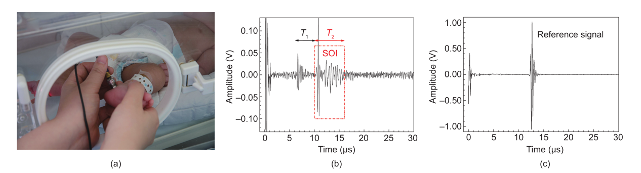
Fig. 2

Fig. 3

Fig. 4

Fig. 5
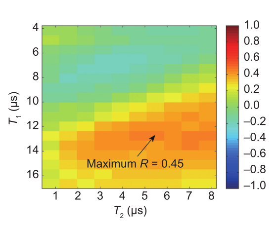
Fig. 6

Fig. 7
References
[ 1 ] K. Engelke, Clinical use of quantitative computed tomography and peripheral quantitative computed tomography in the management of osteoporosis in adults: The 2007 ISCD Official Positions. J. Clin. Densitom., 2008, 11(1): 123–162 link1
[ 2 ] R. Lorente-Ramos, J. Azpeitia-Armán, A. Muñoz-Hernández, J. M. García-Gómez, P. Díez-Martínez, M. Grande-Bárez. Dual-energy X-ray absorptiometry in the diagnosis of osteoporosis: A practical guide. AJR Am. J. Roentgenol., 2011, 196(4): 897–904 link1
[ 3 ] P. Andreopoulou, R. S. Bockman. Management of postmenopausal osteoporosis. Annu. Rev. Med., 2015, 66: 329–342 link1
[ 4 ] C. B. Becker. Sclerostin inhibition for osteoporosis—A new approach. N. Engl. J. Med., 2014, 370(5): 476–477
[ 5 ] T. D. Rachner, S. Khosla, L. C. Hofbauer. Osteoporosis: Now and the future. Lancet, 2011, 377(9773): 1276–1287 link1
[ 6 ] P. Laugier. Quantitative ultrasound of bone: Looking ahead. Joint Bone Spine, 2006, 73(2): 125–128
[ 7 ] D. Mulleman, I. Legroux-Gerot, B. Duquesnoy, X. Marchandise, B. Delcambre, B. Cortet. Quantitative ultrasound of bone in male osteoporosis. Osteoporos. Int., 2002, 13(5): 388–393 link1
[ 8 ] P. H. Nicholson, R. Alkalay. Quantitative ultrasound predicts bone mineral density and failure load in human lumbar vertebrae. Clin. Biomech. (Bristol, Avon), 2007, 22(6): 623–629 link1
[ 9 ] F. Padilla, F. Jenson, V. Bousson, F. Peyrin, P. Laugier. Relationships of trabecular bone structure with quantitative ultrasound parameters: In vitro study on human proximal femur using transmission and backscatter measurements. Bone, 2008, 42(6): 1193–1202 link1
[10] K. A. Wear, S. Nagaraja, M. L. Dreher, S. L. Gibson. Relationships of quantitative ultrasound parameters with cancellous bone microstructure in human calcaneus in vitro. J. Acoust. Soc. Am., 2012, 131(2): 1605–1612
[11] D. Ta, W. Wang, K. Huang, Y. Wang, L. H. Le. Analysis of frequency dependence of ultrasonic backscatter coefficient in cancellous bone. J. Acoust. Soc. Am., 2008, 124(6): 4083–4090
[12] C. Chappard, P. Laugier, B. Fournier, C. Roux, G. Berger. Assessment of the relationship between broadband ultrasound attenuation and bone mineral density at the calcaneus using BUA imaging and DXA. Osteoporos. Int., 1997, 7(4): 316–322 link1
[13] G. Haïat, In vitro speed of sound measurement at intact human femur specimens. Ultrasound Med. Biol., 2005, 31(7): 987–996 link1
[14] D. Hans, Ultrasound velocity of trabecular cubes reflects mainly bone density and elasticity. Calcif. Tissue Int., 1999, 64(1): 18–23 link1
[15] S. Mészáros, E. Tóth, V. Ferencz, E. Csupor, E. Hosszú, C. Horváth. Calcaneous quantitative ultrasound measurements predicts vertebral fractures in idiopathic male osteoporosis. Joint Bone Spine, 2007, 74(1): 79–84
[16] W. Pluskiewicz, B. Drozdzowska. Ultrasonic measurement of the calcaneus in Polish normal and osteoporotic women and men. Bone, 1999, 24(6): 611–617 link1
[17] P. Laugier. An overview of bone sonometry. Int. Congr. Ser., 2004, 1274: 23–32 link1
[18] K. A. Wear. Ultrasonic scattering from cancellous bone: A review. IEEE Trans. Ultrason. Ferroelectr. Freq. Control, 2008, 55(7): 1432–1441 link1
[19] B. K. Hoffmeister, A. P. Holt, S. C. Kaste. Effect of the cortex on ultrasonic backscatter measurements of cancellous bone. Phys. Med. Biol., 2011, 56(19): 6243–6255 link1
[20] B. K. Hoffmeister, Ultrasonic characterization of human cancellous bone in vitro using three different apparent backscatter parameters in the frequency range 0.6−15.0 MHz. IEEE Trans. Ultrason. Ferroelectr. Freq. Control, 2008, 55(7): 1442–1452 link1
[21] K. Il Lee, M. Joo Choi. Frequency-dependent attenuation and backscatter coefficients in bovine trabecular bone from 0.2 to 1.2 MHz. J. Acoust. Soc. Am., 2012, 131(1): EL67–EL73
[22] C. C. Liu, H. J. Han, D. A. Ta, W. Q. Wang. Effect of selected signals of interest on ultrasonic backscattering measurement in cancellous bones. Sci. China Phys. Mech., 2013, 56(7): 1310–1316
[23] C. C. Liu, D. Ta, B. Hu, L. H. Le, W. Wang. The analysis and compensation of cortical thickness effect on ultrasonic backscatter signals in cancellous bone. J. Appl. Phys., 2014, 116(12): 124903
[24] C. C. Liu, The relationship between ultrasonic backscatter and trabecular anisotropic microstructure in cancellous bone. J. Appl. Phys., 2014, 115(6): 064906
[25] F. Padilla, F. Peyrin, P. Laugier. Prediction of backscatter coefficient in trabecular bones using a numerical model of three-dimensional microstructure. J. Acoust. Soc. Am., 2003, 113(2): 1122–1129
[26] K. A. Wear, A. Laib. The dependence of ultrasonic backscatter on trabecular thickness in human calcaneus: Theoretical and experimental results. IEEE Trans. Ultrason. Ferroelectr. Freq. Control, 2003, 50(8): 979–986 link1
[27] K. A. Wear, A. P. Stuber, J. C. Reynolds. Relationships of ultrasonic backscatter with ultrasonic attenuation, sound speed and bone mineral density in human calcaneus. Ultrasound Med. Biol., 2000, 26(8): 1311–1316 link1
[28] B. S. Garra, M. Locher, S. Felker, K. A. Wear. Measurements of ultrasonic backscattered spectral centroid shift from spine in vivo: Methodology and preliminary results. Ultrasound Med. Biol., 2009, 35(1): 165–168 link1
[29] K. Huang, D. Ta, W. Wang, L. H. Le. Simplified inverse filter tracking algorithm for estimating the mean trabecular bone spacing. IEEE Trans. Ultrason. Ferroelectr. Freq. Control, 2008, 55(7): 1453–1464 link1
[30] W. C. Pereira, S. L. Bridal, A. Coron, P. Laugier. Singular spectrum analysis applied to backscattered ultrasound signals from in vitro human cancellous bone specimens. IEEE Trans. Ultrason. Ferroelectr. Freq. Control, 2004, 51(3): 302–312 link1
[31] Y. Q. Jiang, Analysis of apparent integrated backscatter coefficient and backscattered spectral centroid shift in Calcaneus in vivo for the ultrasonic evaluation of osteoporosis. Ultrasound Med. Biol., 2014, 40(6): 1307–1317 link1
[32] R. Zhang, D. Ta, C. Liu, C. Chen. Feasibility of bone assessment with ultrasonic backscatter signals in neonates. Ultrasound Med. Biol., 2013, 39(10): 1751–1759 link1
[33] J. Litniewski, L. Cieslik, M. Lewandowski, R. Tymkiewicz, B. Zienkiewicz, A. Nowicki. Ultrasonic scanner for in vivo measurement of cancellous bone properties from backscattered data. IEEE Trans. Ultrason. Ferroelectr. Freq. Control, 2012, 59(7): 1470–1477 link1
[34] J. P. Karjalainen, Multi-site bone ultrasound measurements in elderly women with and without previous hip fractures. Osteoporos. Int., 2012, 23(4): 1287–1295 link1
[35] C. Liu, Signal of interest selection standard for ultrasonic backscatter in cancellous bone evaluation. Ultrasound Med. Biol., 2015, 41(10): 2714–2721 link1
[36] M. S. Fewtrell, T. J. Cole, N. J. Bishop, A. Lucas. Neonatal factors predicting childhood height in preterm infants: Evidence for a persisting effect of early metabolic bone disease? J. Pediatr., 2000, 137(5): 668–673
[37] M. C. Backström, A. L. Kuusela, R. Mäki. Metabolic bone disease of prematurity. Ann. Med., 1996, 28(4): 275–282 link1
[38] A. Lucas, O. G. Brooke, B. A. Baker, N. Bishop, R. Morley. High alkaline phosphatase activity and growth in preterm neonates. Arch. Dis. Child., 1989, 64(7 Spec No): 902–909 link1
[39] J. E. Teitelbaum, Quantitative ultrasound in the evaluation of bone status in premature and full-term infants. J. Clin. Densitom., 2006, 9(3): 358–362 link1
[40] M. Catache, C. R. Leone. Role of plasma and urinary calcium and phosphorus measurements in early detection of phosphorus deficiency in very low birthweight infants. Acta Paediatr., 2003, 92(1): 76–80
[41] J. Faerk, B. Peitersen, S. Petersen, K. F. Michaelsen. Bone mineralisation in premature infants cannot be predicted from serum alkaline phosphatase or serum phosphate. Arch. Dis. Child. Fetal Neonatal Ed., 2002, 87(2): F133–F136 link1
[42] W. W. K. Koo, J. Walters, A. J. Bush, R. W. Chesney, S. E. Carlson. Dual-energy X-ray absorptiometry studies of bone mineral status in newborn infants. J. Bone Miner. Res., 1996, 11(7): 997–1002
[43] H. McDevitt, S. F. Ahmed. Quantitative ultrasound assessment of bone health in the neonate. Neonatology, 2007, 91(1): 2–11 link1
[44] L. Pereda, T. Ashmeade, J. Zaritt, J. D. Carver. The use of quantitative ultrasound in assessing bone status in newborn preterm infants. J. Perinatol., 2003, 23(8): 655–659 link1
[45] A. Omar, S. Turan, A. Bereket. Reference data for bone speed of sound measurement by quantitative ultrasound in healthy children. Arch. Osteoporos., 2006, 1(1−2): 37–41
[46] P. A. Narayana, J. Ophir. A closed form method for the measurement of attenuation in nonlinearly dispersive media. Ultrason. Imaging, 1983, 5(1): 17–21 link1
[47] B. Rack, Ultrasound for the assessment of bone quality in preterm and term infants. J. Perinatol., 2012, 32(3): 218–226 link1


 京公网安备 11010502051620号


