
2023, Volume 26, Issue 7
Engineering >> 2023, Volume 26, Issue 7 doi: https://doi.org/10.1016/j.eng.2023.02.016
Removable Dyes—The Missing Link for In-Depth N-Glycan Analysis via Multi-Method Approaches
a Max Planck Institute for Dynamics of Complex Technical Systems, Magdeburg 39106, Germany
b glyXera GmbH, Magdeburg 39120, Germany
c Otto-von-Guericke University, Chair of Bioprocess Engineering, Magdeburg 39106, Germany
Next Previous
Abstract
As the roles of glycans in health and disease continue to be unraveled, it is becoming apparent that glycans' immense complexity cannot be ignored. To fully delineate glycan structures, we developed an integrative approach combining a set of cost-effective, widespread, and easy-to-handle analytical methods. The key feature of our workflow is the exploitation of a removable fluorescent label—exemplified by 9-fluorenylmethyl chloroformate (Fmoc)—to bridge the gap between diverse glycoanalytical methods, especially multiplexed capillary gel electrophoresis with laser-induced fluorescence detection (xCGE-LIF) and matrix-assisted laser desorption/ionization time-of-flight mass spectrometry (MALDI-TOF-MS). Through the detailed structural analysis of selected, dauntingly complex N-glycans from chicken ovalbumin, horse serum, and bovine transferrin, we illustrate the capabilities of the presented strategy. Moreover, this approach "visualizes" N-glycans that have been difficult to identify thus far—such as the sulfated glycans on human immunoglobulin A—including minute changes in glycan structures, potentially providing useful new targets for biomarker discovery.
Keywords
Glycoproteins ; N-glycans ; Reversible labeling ; Hydrophilic interaction liquid chromatography ; Capillary gel electrophoresis ; Mass spectrometry
SupplementaryMaterials
Figures
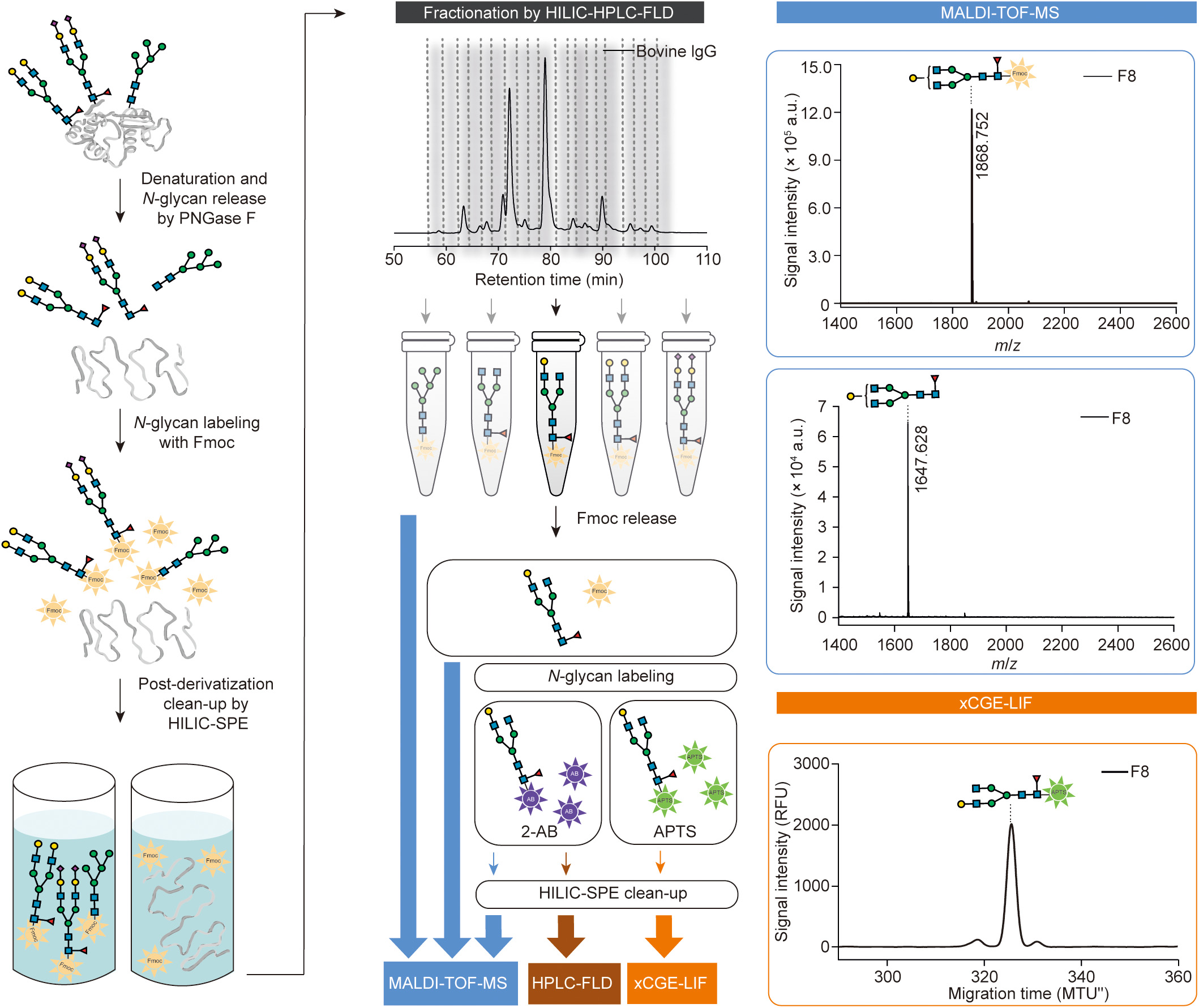
Fig. 1

Fig. 2

Fig. 3
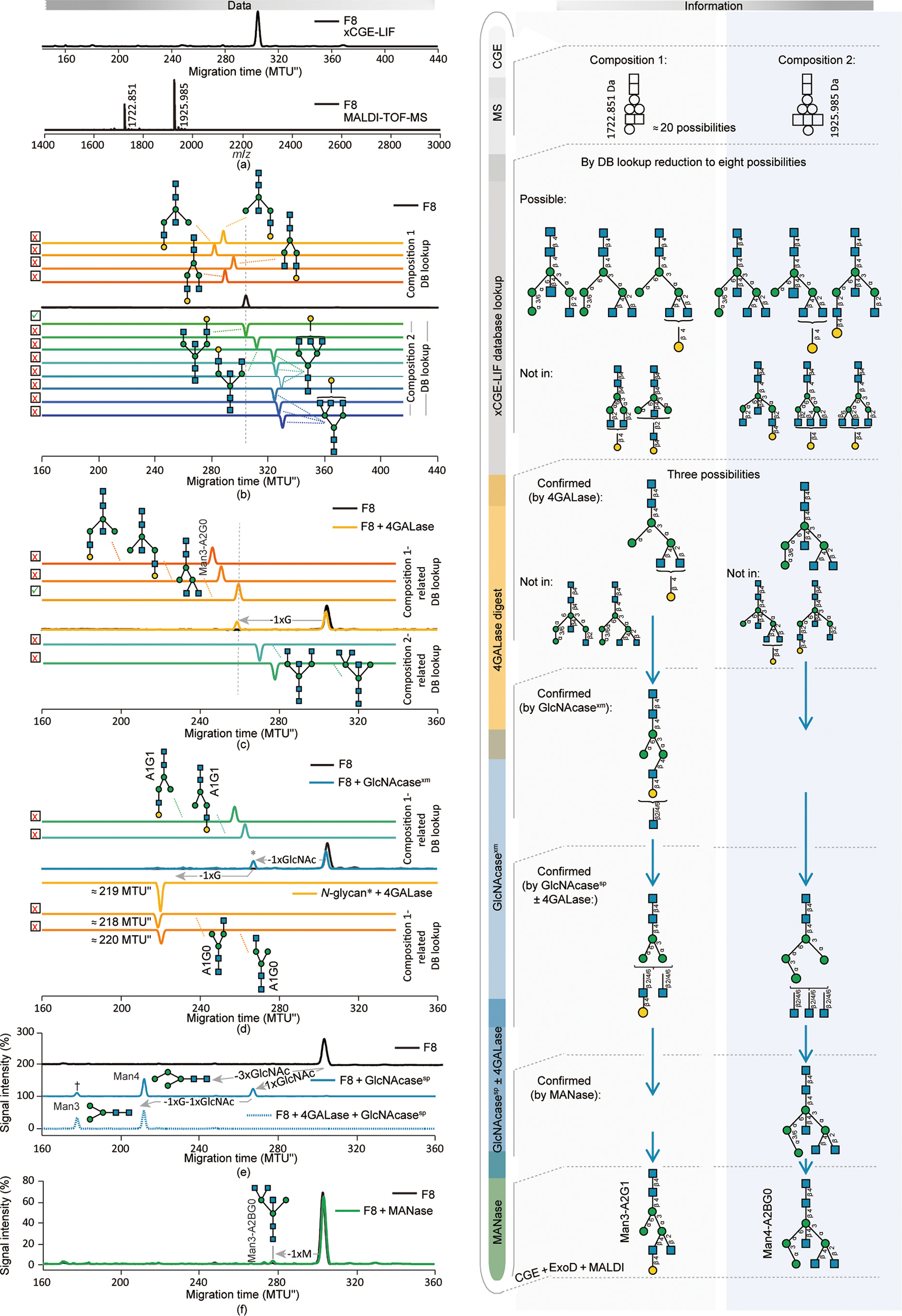
Fig. 4

Fig. 5
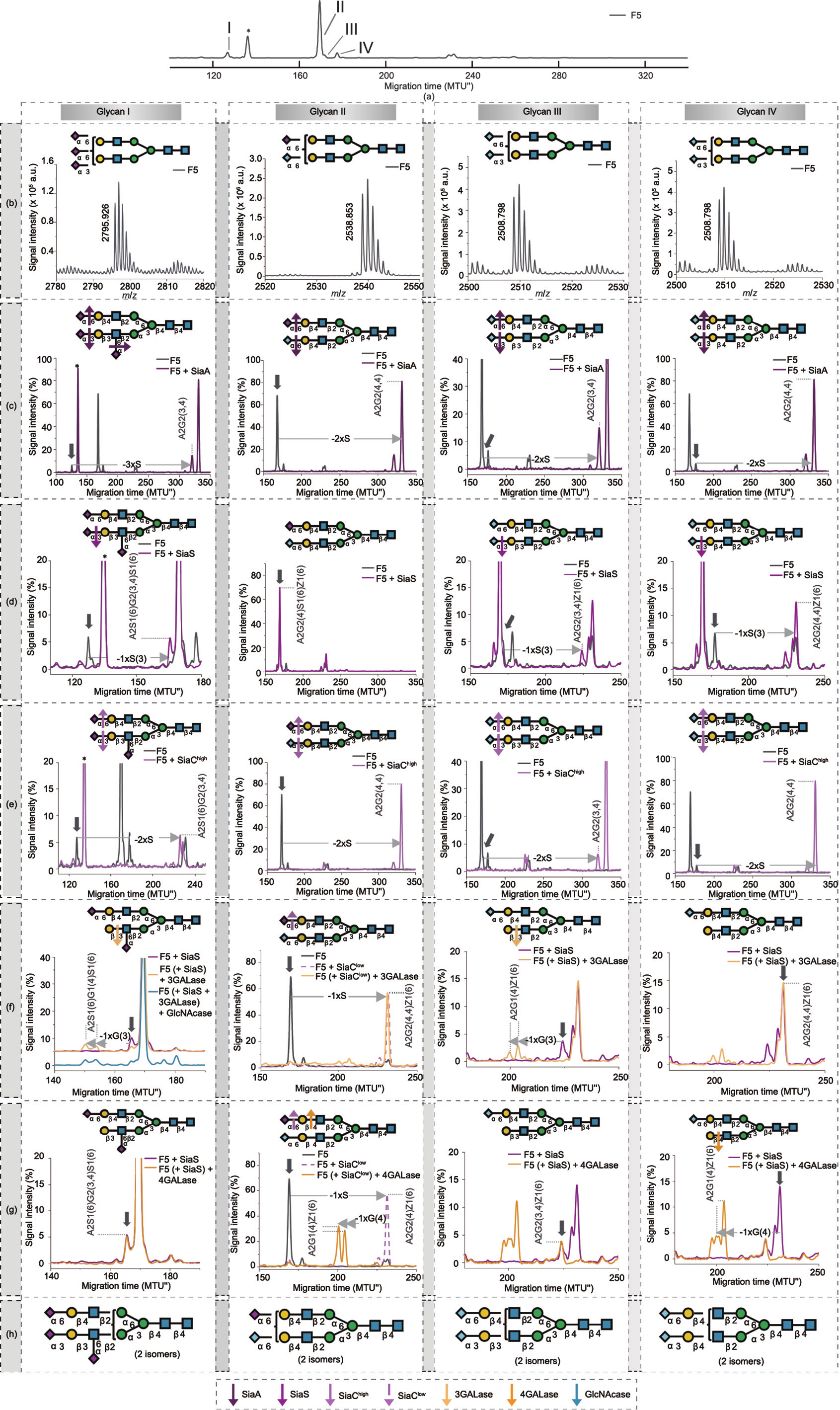
Fig. 6
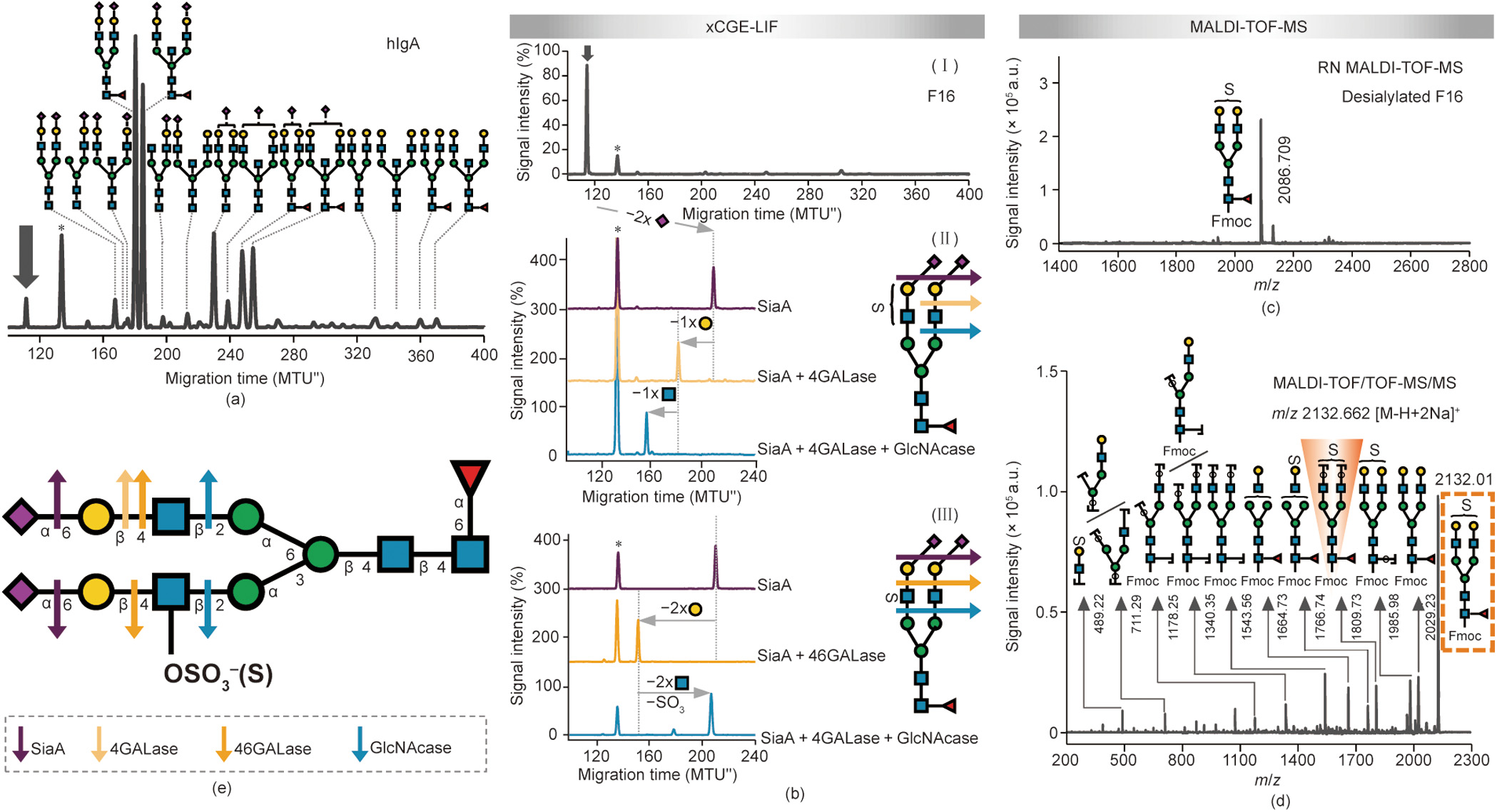
Fig. 7
References
[ 1 ] Watkins WM. The ABO blood group system: historical background. Transfus Med 2001;11(4):243–65. link1
[ 2 ] Olsson ML, Clausen H. Modifying the red cell surface: towards an ABOuniversal blood supply. Br J Haematol 2008;140(1):3–12. link1
[ 3 ] Walt D, Aoki-Kinoshita KF, Bertozzi CR, Boons GJ, Darvill A, et al. Transforming glycoscience: a roadmap for the future. Washington DC: National Academies Press; 2012. link1
[ 4 ] Dennis JW, Laferté S, Waghorne C, Breitman ML, Kerbel RS. Beta 1–6 branching of Asn-linked oligosaccharides is directly associated with metastasis. Science 1987;236(4801):582–5. link1
[ 5 ] Przybyło M, Pochec´ E, Link-Lenczowski P, Lityn´ ska A. Beta1–6 branching of cell surface glycoproteins may contribute to uveal melanoma progression by upregulating cell motility. Mol Vis 2008;14:625–66. link1
[ 6 ] Taniguchi N, Kizuka Y. Glycans and cancer: role of N-glycans in cancer biomarker, progression and metastasis, and therapeutics. Adv Cancer Res 2015;126:11–51. link1
[ 7 ] Mechref Y, Muzikar J, Novotny MV. Comprehensive assessment of N-glycans derived from a murine monoclonal antibody: a case for multimethodological approach. Electrophoresis 2005;26(10):2034–46. link1
[ 8 ] Mitra I, Zhuang Z, Zhang Y, Yu CY, Hammoud ZT, Tang H, et al. N-glycan profiling by microchip electrophoresis to differentiate disease states related to esophageal adenocarcinoma. Anal Chem 2012;84(8):3621–7. link1
[ 9 ] Gennaro LA, Salas-Solano O. On-line CE-LIF-MS technology for the direct characterization of N-linked glycans from therapeutic antibodies. Anal Chem 2008;80(10):3838–45. link1
[10] Cajic S, Hennig R, Burock R, Rapp E. Capillary (gel) electrophoresis-based methods for immunoglobulin (G) glycosylation analysis. In: Pezer M, editor. Antibody glycosylation. Experientia supplementum, vol. 112. Cham: Springer; 2021. p. 137–72. link1
[11] Pabst M, Kolarich D, Pöltl G, Dalik T, Lubec G, Hofinger A, et al. Comparison of fluorescent labels for oligosaccharides and introduction of a new postlabeling purification method. Anal Biochem 2009;384(2):263–73. link1
[12] Ruhaak LR, Zauner G, Huhn C, Bruggink C, Deelder AM, Wuhrer M. Glycan labeling strategies and their use in identification and quantification. Anal Bioanal Chem 2010;397(8):3457–81. link1
[13] Carpino LA. The 9-fluorenylmethyloxycarbonyl family of base-sensitive amino-protecting groups. Acc Chem Res 1987;20(11):401–7. link1
[14] Kajihara Y, Suzuki Y, Sasaki K, Juneja LR. Chemoenzymatic synthesis of diverse asparagine-linked oligosaccharides. Methods Enzymol 2003;362:44–64. link1
[15] Song X, Lasanajak Y, Rivera-Marrero C, Luyai A, Willard M, Smith DF, et al. Generation of a natural glycan microarray using 9-fluorenylmethyl chloroformate (FmocCl) as a cleavable fluorescent tag. Anal Biochem 2009;395(2):151–60. link1
[16] Trbojevic´ -Akmacˇic´ I, Lageveen-Kammeijer GSM, Heijs B, Petrovic´ T, Deriš H, Wuhrer M, et al. High-throughput glycomic methods. Chem Rev 2022;122 (20):15865–913. link1
[17] Ceroni A, Dell A, Haslam SM. The GlycanBuilder: a fast, intuitive and flexible software tool for building and displaying glycan structures. Source Code Biol Med 2007;2:3. link1
[18] Damerell D, Ceroni A, Maass K, Ranzinger R, Dell A, Haslam SM. The GlycanBuilder and GlycoWorkbench glycoinformatics tools: updates and new developments. Biol Chem 2012;393:1357–62. link1
[19] Varki A, Cummings RD, Aebi M, Packer NH, Seeberger PH, Esko JD, et al. Symbol nomenclature for graphical representations of glycans. Glycobiology 2015;25:1323–4. link1
[20] Hennig R, Rapp E, Kottler R, Cajic S, Borowiak M, Reichl U. N-glycosylation fingerprinting of viral glycoproteins by xCGE-LIF. In: Lepenies B, editor. Carbohydrate-based vaccines. Methods in molecular biology. New York City: Humana Press; 2015. p. 123–43. link1
[21] Hennig R, Cajic S, Borowiak M, Hoffmann M, Kottler R, Reichl U, et al. Towards personalized diagnostics via longitudinal study of the human plasma Nglycome. Biochim Biophys Acta 2016;1860(8):1728–38. link1
[22] Reiding KR, Blank D, Kuijper DM, Deelder AM, Wuhrer M. High-throughput profiling of protein N-glycosylation by MALDI-TOF-MS employing linkagespecific sialic acid esterification. Anal Chem 2014;86(12):5784–93. link1
[23] Reiding KR, Lonardi E, Hipgrave Ederveen AL, Wuhrer M. Ethyl esterification for MALDI-MS analysis of protein glycosylation. In: Reinders J, editor. Proteomics in systems biology. Methods in molecular biology. New York City: Humana Press; 2016. p. 151–62. link1
[24] Dionex. Direct determination of sialic acids in glycoprotein hydrolyzates by HPAE-PAD. Application update 180 2011:LPN 2831 [Internet]. Thermo Fisher Scientific Inc., c2016 [cited 2021 Sep 22]. Available from: https://assets. thermofisher.com/TFS-Assets/CMD/Application-Notes/AU-180-IC-Sialic-AcidsGlycoprotein-Hydrolyzates-AU71730-EN.pdf.
[25] Varki A, Diaz S. The release and purification of sialic acids from glycoconjugates: methods to minimize the loss and migration of O-acetyl groups. Anal Biochem 1984;137(1):236–47. link1
[26] Martin LT, Verhagen A, Varki A. Recombinant influenza C hemagglutininesterase as a probe for sialic acid 9-O-acetylation. Methods Enzymol 2003;363:489–98. link1
[27] Thiesler CT, Cajic S, Hoffmann D, Thiel C, van Diepen L, Hennig R, et al. Glycomic characterization of induced pluripotent stem cells derived from a patient suffering from phosphomannomutase 2 congenital disorder of glycosylation (PMM2-CDG). Mol Cell Proteomics 2016;15 (4):1435–52. link1
[28] Konze SA, Cajic S, Oberbeck A, Hennig R, Pich A, Rapp E, et al. Quantitative assessment of sialo-glycoproteins and N-glycans during cardiomyogenic differentiation of human induced pluripotent stem cells. Chembiochem 2017;18(13):1317–31. link1
[29] Suzuki H, Müller O, Guttman A, Karger BL. Analysis of 1-aminopyrene-3,6,8- trisulfonate-derivatized oligosaccharides by capillary electrophoresis with matrix-assisted laser desorption/ionization time-of-flight mass spectrometry. Anal Chem 1997;69(22):4554–9. link1
[30] Lemoine J, Cabanes-Macheteau M, Bardor M, Michalski JC, Faye L, Lerouge P. Analysis of 8-aminonaphthalene-1,3,6-trisulfonic acid labelled N-glycans by matrix-assisted laser desorption/ionisation time-of-flight mass spectrometry. Rapid Commun Mass Spectrom 2000;14(2):100–4. link1
[31] Guttman A, Chen FTA, Evangelista RA. Separation of 1-aminopyrene-3,6,8- trisulfonate-labeled asparagine-linked fetuin glycans by capillary gel electrophoresis. Electrophoresis 1996;17(2):412–7. link1
[32] Laroy W, Contreras R, Callewaert N. Glycome mapping on DNA sequencing equipment. Nat Protoc 2006;1(1):397–405. link1
[33] Nakano M, Higo D, Arai E, Nakagawa T, Kakehi K, Taniguchi N, et al. Capillary electrophoresis-electrospray ionization mass spectrometry for rapid and sensitive N-glycan analysis of glycoproteins as 9-fluorenylmethyl derivatives. Glycobiology 2009;19(2):135–43. link1
[34] Wang C, Qiang S, Jin W, Song X, Zhang Y, Huang L, et al. Reductive chemical release of N-glycans as 1-amino-alditols and subsequent 9- fluorenylmethyloxycarbonyl labeling for MS and LC/MS analysis. J Proteomics 2018;187:47–58. link1
[35] Kamoda S, Nakano M, Ishikawa R, Suzuki S, Kakehi K. Rapid and sensitive screening of N-glycans as 9-fluorenylmethyl derivatives by high-performance liquid chromatography: a method which can recover free oligosaccharides after analysis. J Proteome Res 2005;4(1):146–52. link1
[36] Tarentino AL, Plummer Jr TH. Enzymatic deglycosylation of asparagine-linked glycans: purification, properties, and specificity of oligosaccharide-cleaving enzymes from flavobacterium meningosepticum. Methods Enzymol 1994;230:44–57. link1
[37] Ruhaak LR, Hennig R, Huhn C, Borowiak M, Dolhain RJEM, Deelder AM, et al. Optimized workflow for preparation of APTS-labeled N-glycans allowing highthroughput analysis of human plasma glycomes using 48-channel multiplexed CGE-LIF. J Proteome Res 2010;9(12):6655–64. link1
[38] Rasmussen JR, Davis J, Risley JM, Van Etten RL. Identification and derivatization of (oligosaccharyl)amines obtained by treatment of asparagine-linked glycopeptides with N-glycanase enzyme. J Am Chem Soc 1992;114(3):1124–6. link1
[39] Likhosherstov LM, Novikova OS, Derevitskaja VA, Kochetkov NK. A new simple synthesis of amino sugar b-D-glycosylamines. Carbohydr Res 1986;146(1): C1–5. link1
[40] Küster B, Harvey DJ. Ammonium containing buffers should be avoided during enzymatic release of glycans from glycoproteins when followed by reducing terminal derivatization. Glycobiology 1997;7(2):7–9. link1
[41] Kallin E, Lönn H, Norberg T, Elofsson M. Derivatization procedures for reducing oligosaccharides, part 3: preparation of oligosaccharide glycosylamines, and their conversion into glycosaccharide—acrylamide copolymers. J Carbohydr Chem 1989;8(4):597–611. link1
[42] Bigge JC, Patel TP, Bruce JA, Goulding PN, Charles SM, Parekh RB. Nonselective and efficient fluorescent labeling of glycans using 2-amino benzamide and anthranilic acid. Anal Biochem 1995;230(2):229–38. link1
[43] Lauber MA, Yu YQ, Brousmiche DW, Hua Z, Koza SM, Magnelli P, et al. Rapid preparation of released N-glycans for HILIC analysis using a labeling reagent that facilitates sensitive fluorescence and ESI-MS detection. Anal Chem 2015;87(10):5401–9. link1
[44] Stöckmann H, Duke RM, Millán Martín S, Rudd PM. Ultrahigh throughput, ultrafiltration-based N-glycomics platform for ultraperformance liquid chromatography (ULTRA(3)). Anal Chem 2015;87(16):8316–22. link1
[45] Kimzey M, Szabo Z, Sharma V, Gyenes A, Tep S, Taylor A, et al. Development of an instant glycan labeling dye for high throughput analysis by mass spectrometry. Prozyme 2015;25:1295. link1
[46] Cook KS, Bullock K, Sullivan T. Development and qualification of an antibody rapid deglycosylation method. Biologicals 2012;40(2):109–17. link1
[47] Fields GB. Methods for removing the Fmoc group. Peptide synthesis protocols. New Jersey: Humana Press; 1994. link1
[48] Endo T, Nishimura R, Kawano T, Mochizuki M, Kobata A. Structural differences found in the asparagine-linked sugar chains of human chorionic gonadotropins purified from the urine of patients with invasive mole and with choriocarcinoma. Cancer Res 1987;47(19):5242–5. link1
[49] Yamashita K, Totani K, Iwaki Y, Takamisawa I, Tateishi N, Higashi T, et al. Comparative study of the sugar chains of c-glutamyltranspeptidases purified from human hepatocellular carcinoma and from human liver. J Biochem 1989;105(5):728–35. link1
[50] Dennis JW, Granovsky M, Warren CE. Glycoprotein glycosylation and cancer progression. Biochim Biophys Acta 1999;1473(1):21–34. link1
[51] Goss PE, Baker MA, Carver JP, Dennis JW. Inhibitors of carbohydrate processing: a new class of anticancer agents. Clin Cancer Res 1995;1 (9):935–44. link1
[52] Harvey DJ, Wing DR, Küster B, Wilson IBH. Composition of N-linked carbohydrates from ovalbumin and co-purified glycoproteins. J Am Soc Mass Spectrom 2000;11(6):564–71. link1
[53] Tai T, Yamashita K, Ogata-Arakawa M, Koide N, Muramatsu T, Iwashita S, et al. Structural studies of two ovalbumin glycopeptides in relation to the endo-b-Nacetylglucosaminidase specificity. J Biol Chem 1975;250(21):8569–75. link1
[54] Yamashita K, Tachibana Y, Kobata A. The structures of the galactose-containing sugar chains of ovalbumin. J Biol Chem 1978;253(11):3862–9. link1
[55] Yoshima H, Takasaki S, Kobata A. The asparagine-linked sugar chains of the glycoproteins in calf thymocyte plasma membrane. J Biol Chem 1980;255 (22):10793–804. link1
[56] Kajihara Y, Suzuki Y, Yamamoto N, Sasaki K, Sakakibara T, Juneja LR. Prompt chemoenzymatic synthesis of diverse complex-type oligosaccharides and its application to the solid-phase synthesis of a glycopeptide with Asn-linked sialyl-undeca- and asialo-nonasaccharides. Chemistry 2004;10(4):971–85. link1
[57] Duffin KL, Welply JK, Huang E, Henion JD, Huang E, Henion JD. Characterization of N-linked oligosaccharides by electrospray and tandem mass spectrometry. Anal Chem 1992;64(13):1440–8. link1
[58] Mechref Y, Novotny MV. Mass spectrometric mapping and sequencing of Nlinked oligosaccharides derived from submicrogram amounts of glycoproteins. Anal Chem 1998;70(3):455–63. link1
[59] Nomoto H, Inoue Y. A novel glycoasparagine isolated from an ovalbumin glycopeptide fraction (GP-IV). Eur J Biochem 1983;135(2):243–50. link1
[60] Schauer R. Chemistry, metabolism, and biological functions of sialic acids. Adv Carbohydr Chem Biochem 1982;40:131–234. link1
[61] Klein A, Roussel P. O-acetylation of sialic acids. Biochimie 1998;80(1):49–57. link1
[62] Varki A. Diversity in the sialic acids. Glycobiology 1992;2(1):25–40. link1
[63] Mandal C, Schwartz-Albiez R, Vlasak R. Functions and biosynthesis of Oacetylated sialic acids. Top Curr Chem 2015;366:1–30. link1
[64] Liu X, Afonso L. Is permethylation strategy always applicable to protein Nglycosylation study? A case study on the O-acetylation of sialic acid in fish serum glycans. In: Li J, editor. Functional glycomics. Methods in molecular biology. New Jersey: Humana Press; 2010. p. 259–68. link1
[65] Liu X, Qiu H, Lee RK, Chen W, Li J. Methylamidation for sialoglycomics by MALDI-MS: a facile derivatization strategy for both a2,3- and a2,6-linked sialic acids. Anal Chem 2010;82(19):8300–6. link1
[66] Varki A. Biological roles of glycans. Glycobiology 2017;27(1):3–49. link1
[67] Neuberger A, Ratcliffe WA. The acid and enzymic hydrolysis of O-acetylated sialic acid residues from rabbit Tamm-Horsfall glycoprotein. Biochem J 1972;129(3):683–93. link1
[68] Schauer R, Veh RW, Sander M, Corfield AP, Wiegandt H. ‘‘Neuraminidaseresistant” sialic acid residues of gangliosides. In: Svennerholm L, Mandel P, Dreyfus H, Urban PF, editors. Structure and function of gangliosides. Advances in experimental medicine and biology. Boston: Springer; 1980. p. 283–94. link1
[69] Pepper DS. The sialic acids of horse serum with special reference to their virus inhibitory properties. Biochim Biophys Acta 1968;156(2):317–26. link1
[70] Spik G, Coddeville B, Montreuil J. Comparative study of the primary structures of sero-, lacto- and ovotransferrin glycans from different species. Biochimie 1988;70(11):1459–69. link1
[71] Coddeville B, Stratil A, Wieruszeski JM, Strecker G, Montreuil J, Spik G. Primary structure of horse serotransferrin glycans. Eur J Biochem 1989;186(3):583–90. link1
[72] Damm JBL, Voshol H, Hård K, Kamerling JP, Vliegenthart JFG. Analysis of Nacetyl-4-O-acetylneuraminic-acid-containing N-linked carbohydrate chains released by peptide-N4-(N-acetyl-beta-glucosaminyl)asparagine amidase F. Eur J Biochem 1989;180(1):101–10. link1
[73] Miura Y, Endo T. Glycomics and glycoproteomics focused on aging and agerelated diseases—glycans as a potential biomarker for physiological alterations. Biochim Biophys Acta 2016;1860(8):1608–14. link1
[74] Reiding KR, Bondt A, Hennig R, Gardner RA, O’Flaherty R, Trbojevic´ -Akmacˇic´ I, et al. High-throughput serum N-glycomics: method comparison and application to study rheumatoid arthritis and pregnancy-associated changes. Mol Cell Proteomics 2019;18(1):3–15. link1
[75] Keser T, Gornik I, Vucˇkovic´ F, Selak N, Pavic´ T, Lukic´ E, et al. Increased plasma N-glycome complexity is associated with higher risk of type 2 diabetes. Diabetologia 2017;60(12):2352–60. link1
[76] Liu XE, Desmyter L, Gao CF, Laroy W, Dewaele S, Vanhooren V, et al. Nglycomic changes in hepatocellular carcinoma patients with liver cirrhosis induced by hepatitis B virus. Hepatology 2007;46(5):1426–35. link1
[77] Wang M, Zhu J, Lubman DM, Gao C. Aberrant glycosylation and cancer biomarker discovery: a promising and thorny journey. Clin Chem Lab Med 2019;57(4):407–16. link1
[78] Pinho SS, Reis CA. Glycosylation in cancer: mechanisms and clinical implications. Nat Rev Cancer 2015;15(9):540–55. link1
[79] Zhang Z, Wuhrer M, Holst S. Serum sialylation changes in cancer. Glycoconj J 2018;35(2):139–60. link1
[80] Reily C, Stewart TJ, Renfrow MB, Novak J. Glycosylation in health and disease. Nat Rev Nephrol 2019;15(6):346–66. link1
[81] Spik G, Coddeville B, Strecker G, Montreuil J, Regoeczi E, Chindemi PA, et al. Carbohydrate microheterogeneity of rat serotransferrin. Eur J Biochem 1991;195(2):397–405. link1
[82] Coddeville B, Regoeczi E, Strecker G, Plancke Y, Spik G. Structural analysis of trisialylated biantennary glycans isolated from mouse serum transferrin. Biochim Biophys Acta 2000;1475(3):321–8. link1
[83] De Haan N, Falck D, Wuhrer M. Monitoring of immunoglobulin Nand O-glycosylation in health and disease. Glycobiology 2020;30(4): 226–40. link1
[84] Gudelj I, Lauc G, Pezer M. Immunoglobulin G glycosylation in aging and diseases. Cell Immunol 2018;333:65–79. link1
[85] Bondt A, Nicolardi S, Jansen BC, Kuijper TM, Hazes JMW, van der Burgt YEM, et al. IgA N- and O-glycosylation profiling reveals no association with the pregnancy-related improvement in rheumatoid arthritis. Arthritis Res Ther 2017;19(1):160. link1
[86] Clerc F, Reiding KR, Jansen BC, Kammeijer GSM, Bondt A, Wuhrer M. Human plasma protein N-glycosylation. Glycoconj J 2016;33(3):309–43. link1
[87] Chuzel L, Fossa SL, Boisvert ML, Cajic S, Hennig R, Ganatra MB, et al. Combining functional metagenomics and glycoanalytics to identify enzymes that facilitate structural characterization of sulfated N-glycans. Microb Cell Fact 2021;20(1):162. link1
[88] Jiang H, Irungu J, Desaire H. Enhanced detection of sulfated glycosylation sites in glycoproteins. J Am Soc Mass Spectrom 2005;16(3):340–8. link1
[89] Kawashima H. Roles of sulfated glycans in lymphocyte homing. Biol Pharm Bull 2006;29(12):2343–9. link1
[90] Muthana SM, Campbell CT, Gildersleeve JC. Modifications of glycans: biological significance and therapeutic opportunities. ACS Chem Biol 2012;7 (1):31–43. link1
[91] Zauner G, Selman MHJ, Bondt A, Rombouts Y, Blank D, Deelder AM, et al. Glycoproteomic analysis of antibodies. Mol Cell Proteomics 2013;12 (4):856–65. link1
[92] Wang JR, Gao WN, Grimm R, Jiang S, Liang Y, Ye H, et al. A method to identify trace sulfated IgG N-glycans as biomarkers for rheumatoid arthritis. Nat Commun 2017;8(1):631. link1
[93] Lauc G, Vucˇkovic´ F, Bondt A, Pezer M, Wuhrer M. Trace N-glycans including sulphated species may originate from various plasma glycoproteins and not necessarily IgG. Nat Commun 2018;9(1):2916. link1
[94] Wang JR, Gao WN, Grimm R, Jiang S, Liang Y, Ye H, et al. Reply to ‘‘trace N-glycans including sulphated species may originate from various plasma glycoproteins and not necessarily IgG”. Nat Commun 2018;9(1):2915. link1


 京公网安备 11010502051620号


