2022, Volume 13, Issue 6
Engineering >> 2022, Volume 13, Issue 6 doi: 10.1016/j.eng.2021.07.018
A Brief Summary of Current Therapeutic Strategies for Spinal Cord Injury
a Key Laboratory of Neuroregeneration of Jiangsu and Ministry of Education & NMPA Key Laboratory for the Research and Evaluation of Tissue Engineering Technology Products, Co-Innovation Center of Neuroregeneration, Nantong University, Nantong 226001, China
b NHC and CAMS Key Laboratory of Medical Neurobiology, MOE Frontier Science Center for Brain Science and Brain-Machine Integration, School of Brain Science and Brain Medicine, Zhejiang University, Hangzhou 310003, China
c Jiangsu Clinical Medicine Center of Tissue Engineering and Nerve Injury Repair, Affiliated Hospital of Nantong University, Nantong University, Nantong 226001, China
Next Previous
Abstract
Spinal cord injury (SCI) is a tremendous disaster in a person’s life. It interrupts the brain–body neuronal circuits, resulting in functional deficits. Pathogenesis of SCI is a progressive and comprehensive event. In clinical trials, attempts to promote nerve regeneration and functional recovery after SCI have met with failures. Recently, with the development of transcriptome sequencing and biomaterials, researchers have struggled to explore novel efficient therapeutic treatments for SCI. Here, we summarize the recent progress that has been made in SCI repair based on the lesion microenvironment, neural circuits, and biomaterial scaffolds. We also propose several important directions for future research, including targetedmicroRNA therapy, blood vessel interventions, and multiple treatment combinations. In short, we hope this review will enlighten researchers in the field and pave the way for SCI therapy.
Keywords
Spinal cord injury ; Microenvironment ; Neural circuits ; Biomaterial scaffolds
Figures
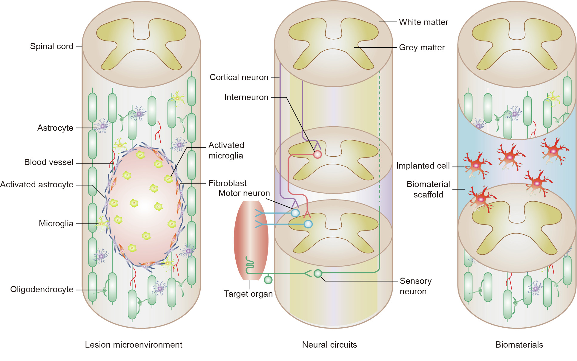
Fig. 1
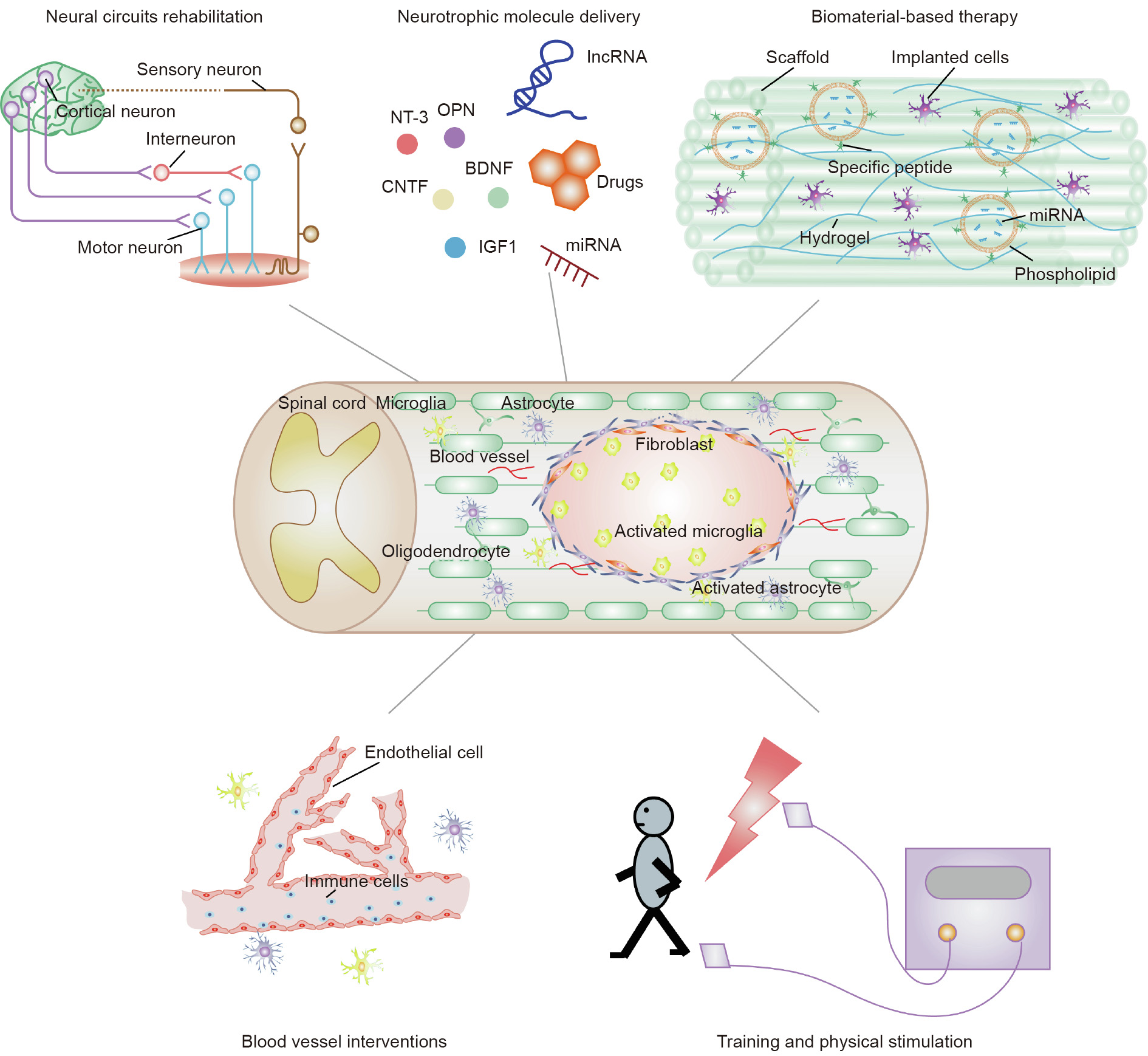
Fig. 2
References
[ 1 ] Lee BB, Cripps RA, Fitzharris M, Wing PC. The global map for traumatic spinal cord injury epidemiology: update 2011, global incidence rate. Spinal Cord 2014;52(2):110–6. link1
[ 2 ] Thuret S, Moon LDF, Gage FH. Therapeutic interventions after spinal cord injury. Nat Rev Neurosci 2006;7(8):628–43. link1
[ 3 ] Ashammakhi N, Kim HJ, Ehsanipour A, Bierman RD, Kaarela O, Xue C, et al. Regenerative therapies for spinal cord injury. Tissue Eng Part B Rev 2019;25 (6):471–91. link1
[ 4 ] Oyinbo CA. Secondary injury mechanisms in traumatic spinal cord injury: a nugget of this multiply cascade. Acta Neurobiol Exp 2011;71(2):281–99. link1
[ 5 ] Singh A, Tetreault L, Kalsi-Ryan S, Nouri A, Fehlings MG. Global prevalence and incidence of traumatic spinal cord injury. Clin Epidemiol 2014;6:309–31. link1
[ 6 ] Cao HQ, Dong ED. An update on spinal cord injury research. Neurosci Bull 2013;29(1):94–102. link1
[ 7 ] Desai J, Steiger S, Anders HJ. Molecular pathophysiology of gout. Trends Mol Med 2017;23(8):756–68. link1
[ 8 ] Tran AP, Warren PM, Silver J. The biology of regeneration failure and success after spinal cord injury. Physiol Rev 2018;98(2):881–917. link1
[ 9 ] Silva NA, Sousa N, Reis RL, Salgado AJ. From basics to clinical: a comprehensive review on spinal cord injury. Prog Neurobiol 2014;114:25–57. link1
[10] Ahuja CS, Nori S, Tetreault L, Wilson J, Kwon B, Harrop J, et al. Traumatic spinal cord injury-repair and regeneration. Neurosurgery 2017;80(3S):S9– 22.
[11] Bradbury EJ, Burnside ER. Moving beyond the glial scar for spinal cord repair. Nat Commun 2019;10(1):3879. link1
[12] Venkatesh K, Ghosh SK, Mullick M, Manivasagam G, Sen D. Spinal cord injury: pathophysiology, treatment strategies, associated challenges, and future implications. Cell Tissue Res 2019;377(2):125–51. link1
[13] Katoh H, Yokota K, Fehlings MG. Regeneration of spinal cord connectivity through stem cell transplantation and biomaterial scaffolds. Front Cell Neurosci 2019;13:248. link1
[14] Higuchi A, Suresh Kumar S, Benelli G, Ling QD, Li HF, Alarfaj AA, et al. Biomaterials used in stem cell therapy for spinal cord injury. Prog Mater Sci 2019;103:374–424. link1
[15] Assinck P, Duncan GJ, Hilton BJ, Plemel JR, Tetzlaff W. Cell transplantation therapy for spinal cord injury. Nat Neurosci 2017;20(5):637–47. link1
[16] Jones LL, Oudega M, Bunge MB, Tuszynski MH. Neurotrophic factors, cellular bridges and gene therapy for spinal cord injury. J Physiol 2001;533(1):83–9. link1
[17] Keefe KM, Sheikh IS, Smith GM. Targeting neurotrophins to specific populations of neurons: NGF, BDNF, and NT-3 and their relevance for treatment of spinal cord injury. Int J Mol Sci 2017;18(3):548. link1
[18] Hodgetts SI, Harvey AR. Neurotrophic factors used to treat spinal cord injury. Vitam Horm 2017;104:405–57. link1
[19] Boyce VS, Mendell LM. Neurotrophic factors in spinal cord injury. Handb Exp Pharmacol 2014;220:443–60. link1
[20] Schnell L, Schneider R, Kolbeck R, Barde YA, Schwab ME. Neurotrophin-3 enhances sprouting of corticospinal tract during development and after adult spinal cord lesion. Nature 1994;367(6459):170–3. link1
[21] Zhou L, Baumgartner BJ, Hill-Felberg SJ, McGowen LR, Shine HD. Neurotrophin-3 expressed in situ induces axonal plasticity in the adult injured spinal cord. J Neurosci 2003;23(4):1424–31. link1
[22] Petruska JC, Kitay B, Boyce VS, Kaspar BK, Pearse DD, Gage FH, et al. Intramuscular AAV delivery of NT-3 alters synaptic transmission to motoneurons in adult rats. Eur J Neurosci 2010;32(6):997–1005.
[23] Ruitenberg MJ, Levison DB, Lee SV, Verhaagen J, Harvey AR, Plant GW. NT-3 expression from engineered olfactory ensheathing glia promotes spinal sparing and regeneration. Brain 2005;128(4):839–53. link1
[24] Xu XM, Han Q. Neurotrophin-3-mediated locomotor recovery: a novel therapeutic strategy targeting lumbar neural circuitry after spinal cord injury. Neural Regen Res 2020;15(12):2241–2. link1
[25] Li X, Yang Z, Zhang A. The effect of neurotrophin-3/chitosan carriers on the proliferation and differentiation of neural stem cells. Biomaterials 2009;30 (28):4978–85. link1
[26] Yang Z, Duan H, Mo L, Qiao H, Li X. The effect of the dosage of NT-3/chitosan carriers on the proliferation and differentiation of neural stem cells. Biomaterials 2010;31(18):4846–54. link1
[27] Taylor SJ, Sakiyama-Elbert SE. Effect of controlled delivery of neurotrophin-3 from fibrin on spinal cord injury in a long term model. J Control Release 2006;116(2):204–10. link1
[28] Willerth SM, Sakiyama-Elbert SE. Cell therapy for spinal cord regeneration. Adv Drug Deliv Rev 2008;60(2):263–76. link1
[29] Gao L, Peng Y, Xu W, He P, Li T, Lu X, et al. Progress in stem cell therapy for spinal cord injury. Stem Cells Int 2020;2020:1–16. link1
[30] Huang L, Fu C, Xiong F, He C, Wei Q. Stem cell therapy for spinal cord injury. Cell Transplant 2021;30:963689721989266.
[31] Fischer I, Dulin JN, Lane MA. Transplanting neural progenitor cells to restore connectivity after spinal cord injury. Nat Rev Neurosci 2020;21(7):366–83. link1
[32] Ahuja CS, Mothe A, Khazaei M, Badhiwala JH, Gilbert EA, Kooy D, et al. The leading edge: emerging neuroprotective and neuroregenerative cell-based therapies for spinal cord injury. Stem Cells Transl Med 2020;9(12):1509–30. link1
[33] Zheng Y, Mao YR, Yuan TF, Xu DS, Cheng LM. Multimodal treatment for spinal cord injury: a sword of neuroregeneration upon neuromodulation. Neural Regen Res 2020;15(8):1437–50. link1
[34] Chu T, Zhou H, Li F, Wang T, Lu L, Feng S. Astrocyte transplantation for spinal cord injury: current status and perspective. Brain Res Bull 2014;107:18–30. link1
[35] David S, Aguayo A. Axonal elongation into peripheral nervous system ‘‘bridges” after central nervous system injury in adult rats. Science 1981;214(4523):931–3. link1
[36] Alizadeh A, Dyck SM, Kataria H, Shahriary GM, Nguyen DH, Santhosh KT, et al. Neuregulin-1 positively modulates glial response and improves neurological recovery following traumatic spinal cord injury. Glia 2017;65(7):1152–75. link1
[37] Orr MB, Gensel JC. Spinal cord injury scarring and inflammation: therapies targeting glial and inflammatory responses. Neurotherapeutics 2018;15 (3):541–53. link1
[38] Nagoshi N, Khazaei M, Ahlfors JE, Ahuja CS, Nori S, Wang J, et al. Human spinal oligodendrogenic neural progenitor cells promote functional recovery after spinal cord injury by axonal remyelination and tissue sparing. Stem Cells Transl Med 2018;7(11):806–18. link1
[39] Lindsay SL, Toft A, Griffin J, Emraja AMM, Barnett SC, Riddell JS. Human olfactory mesenchymal stromal cell transplants promote remyelination and earlier improvement in gait co-ordination after spinal cord injury. Glia 2017;65(4):639–56. link1
[40] Oudega M. Molecular and cellular mechanisms underlying the role of blood vessels in spinal cord injury and repair. Cell Tissue Res 2012;349(1):269–88. link1
[41] Rust R, Kaiser J. Insights into the dual role of inflammation after spinal cord injury. J Neurosci 2017;37(18):4658–60. link1
[42] Mortazavi A, Williams BA, McCue K, Schaeffer L, Wold B. Mapping and quantifying mammalian transcriptomes by RNA-seq. Nat Methods 2008;5 (7):621–8. link1
[43] Shapiro E, Biezuner T, Linnarsson S. Single-cell sequencing-based technologies will revolutionize whole-organism science. Nat Rev Genet 2013;14(9):618–30. link1
[44] Chen K, Deng S, Lu H, Zheng Y, Yang G, Kim D, et al. RNA-seq characterization of spinal cord injury transcriptome in acute/subacute phases: a resource for understanding the pathology at the systems level. PLoS ONE 2013;8(8): e72567. link1
[45] Duan H, Ge W, Zhang A, Xi Y, Chen Z, Luo D, et al. Transcriptome analyses reveal molecular mechanisms underlying functional recovery after spinal cord injury. Proc Natl Acad Sci USA 2015;112(43):13360–5. link1
[46] Luo D, Ge W, Hu X, Li C, Lee CM, Zhou L, et al. Unbiased transcriptomic analyses reveal distinct effects of immune deficiency in CNS function with and without injury. Protein Cell 2019;10(8):566–82. link1
[47] Yu B, Yao C, Wang Y, Mao S, Wang Y, Wu R, et al. The landscape of gene expression and molecular regulation following spinal cord hemisection in rats. Front Mol Neurosci 2019;12:287. link1
[48] So KF. A comprehensive study of gene expression and molecular regulation following spinal cord injury. Engineering 2020;6(4):389–90. link1
[49] Yang P, Yang Z. Enhancing intrinsic growth capacity promotes adult CNS regeneration. J Neurol Sci 2012;312(1–2):1–6. link1
[50] Yang J, Zhao L, Yi S, Ding F, Yang Y, Liu Y, et al. Developmental temporal patterns and molecular network features in the transcriptome of rat spinal cord. Engineering 2021;7(11):1592–602.
[51] Feng W, Chen L, Nguyen PK, Wu SM, Li G. Single cell analysis of endothelial cells identified organ-specific molecular signatures and heartspecific cell populations and molecular features. Front Cardiovasc Med 2019;6:165. link1
[52] Blum JA, Klemm S, Nakayama L, Kathiria A, Guttenplan KA, Hoang PT, et al. Single-cell transcriptomic analysis of the adult mouse spinal cord. Nat Neurosci 2021;24(4):572–83.
[53] Delile J, Rayon T, Melchionda M, Edwards A, Briscoe J, Sagner A. Single cell transcriptomics reveals spatial and temporal dynamics of gene expression in the developing mouse spinal cord. Development 2019;146 (12):dev173807.
[54] Milich LM, Choi J, Ryan C, Yahn SL, Tsoulfas P, Lee JK. Single cell analysis of the cellular heterogeneity and interactions in the injured mouse spinal cord. J Exp Med 2021;218(8):e20210040.
[55] Dobrott CI, Sathyamurthy A, Levine AJ. Decoding cell type diversity within the spinal cord. Curr Opin Physiol 2019;8:1–6. link1
[56] Rosenberg AB, Roco CM, Muscat RA, Kuchina A, Sample P, Yao Z, et al. Singlecell profiling of the developing mouse brain and spinal cord with split-pool barcoding. Science 2018;360(6385):176–82. link1
[57] Li Y, He X, Kawaguchi R, Zhang Y, Wang Q, Monavarfeshani A, et al. Microgliaorganized scar-free spinal cord repair in neonatal mice. Nature 2020;587 (7835):613–8. link1
[58] Kiehn O. Decoding the organization of spinal circuits that control locomotion. Nat Rev Neurosci 2016;17(4):224–38. link1
[59] Han Q, Ordaz JD, Liu NK, Richardson Z, Wu W, Xia Y, et al. Descending motor circuitry required for NT-3 mediated locomotor recovery after spinal cord injury in mice. Nat Commun 2019;10(1):5815. link1
[60] Ahuja CS, Wilson JR, Nori S, Kotter MRN, Druschel C, Curt A, et al. Traumatic spinal cord injury. Nat Rev Dis Primers 2017;3(1):17018. link1
[61] Lemon RN. Descending pathways in motor control. Annu Rev Neurosci 2008;31(1):195–218. link1
[62] Wang X, Liu Y, Li X, Zhang Z, Yang H, Zhang Y, et al. Deconstruction of corticospinal circuits for goal-directed motor skills. Cell 2017;171(2):440–55. e14. link1
[63] Chen M, Zheng B. Axon plasticity in the mammalian central nervous system after injury. Trends Neurosci 2014;37(10):583–93. link1
[64] Maier IC, Schwab ME. Sprouting, regeneration and circuit formation in the injured spinal cord: factors and activity. Philos Trans R Soc Lond B Biol Sci 2006;361(1473):1611–34. link1
[65] Liu Y, Wang X, Li W, Zhang Q, Li Y, Zhang Z, et al. A sensitized IGF1 treatment restores corticospinal axon-dependent functions. Neuron 2017;95 (4):817–33.e4. link1
[66] Han Q, Xie Y, Ordaz JD, Huh AJ, Huang N, Wu W, et al. Restoring cellular energetics promotes axonal regeneration and functional recovery after spinal cord injury. Cell Metab 2020;31(3):623–41.e8. link1
[67] Courtine G, Song B, Roy RR, Zhong H, Herrmann JE, Ao Y, et al. Recovery of supraspinal control of stepping via indirect propriospinal relay connections after spinal cord injury. Nat Med 2008;14(1):69–74. link1
[68] Benthall KN, Hough RA, McClellan AD. Descending propriospinal neurons mediate restoration of locomotor function following spinal cord injury. J Neurophysiol 2017;117(1):215–29. link1
[69] Bertuzzi M, Chang W, Ampatzis K. Adult spinal motoneurons change their neurotransmitter phenotype to control locomotion. Proc Natl Acad Sci USA 2018;115(42):E9926–33. link1
[70] Wang Y, Wu W, Wu X, Sun Y, Zhang YP, Deng LX, et al. Remodeling of lumbar motor circuitry remote to a thoracic spinal cord injury promotes locomotor recovery. elife 2018;7:e39016.
[71] Zholudeva LV, Qiang L, Marchenko V, Dougherty KJ, Sakiyama-Elbert SE, Lane MA. The neuroplastic and therapeutic potential of spinal interneurons in the injured spinal cord. Trends Neurosci 2018;41(9):625–39. link1
[72] Zholudeva LV, Abraira VE, Satkunendrarajah K, McDevitt TC, Goulding MD, Magnuson DSK, et al. Spinal interneurons as gatekeepers to neuroplasticity after injury or disease. J Neurosci 2021;41(5):845–54. link1
[73] Chen B, Li Y, Yu B, Zhang Z, Brommer B, Williams PR, et al. Reactivation of dormant relay pathways in injured spinal cord by KCC2 manipulations. Cell 2018;174(3):521–35.e13. link1
[74] Martino G, Pluchino S. The therapeutic potential of neural stem cells. Nat Rev Neurosci 2006;7(5):395–406. link1
[75] Lu P, Wang Y, Graham L, McHale K, Gao M, Wu D, et al. Long-distance growth and connectivity of neural stem cells after severe spinal cord injury. Cell 2012;150(6):1264–73. link1
[76] Kadoya K, Lu P, Nguyen K, Lee-Kubli C, Kumamaru H, Yao L, et al. Spinal cord reconstitution with homologous neural grafts enables robust corticospinal regeneration. Nat Med 2016;22(5):479–87. link1
[77] Straley KS, Foo CW, Heilshorn SC. Biomaterial design strategies for the treatment of spinal cord injuries. J Neurotrauma 2010;27(1):1–19. link1
[78] Haggerty AE, Maldonado-Lasunción I, Oudega M. Biomaterials for revascularization and immunomodulation after spinal cord injury. Biomed Mater 2018;13(4):044105. link1
[79] Li X, Liu D, Xiao Z, Zhao Y, Han S, Chen B, et al. Scaffold-facilitated locomotor improvement post complete spinal cord injury: motor axon regeneration versus endogenous neuronal relay formation. Biomaterials 2019;197:20–31. link1
[80] Yang Z, Zhang A, Duan H, Zhang S, Hao P, Ye K, et al. NT3-chitosan elicits robust endogenous neurogenesis to enable functional recovery after spinal cord injury. Proc Natl Acad Sci USA 2015;112(43): 13354–9. link1
[81] Rao JS, Zhao C, Zhang A, Duan H, Hao P, Wei RH, et al. NT3-chitosan enables de novo regeneration and functional recovery in monkeys after spinal cord injury. Proc Natl Acad Sci USA 2018;115(24):E5595–604. link1
[82] Lin H, Chen B, Wang B, Zhao Y, Sun W, Dai J. Novel nerve guidance material prepared from bovine aponeurosis. J Biomed Mater Res A 2006;79A (3):591–8. link1
[83] Han S, Wang B, Jin W, Xiao Z, Li X, Ding W, et al. The linear-ordered collagen scaffold-BDNF complex significantly promotes functional recovery after completely transected spinal cord injury in canine. Biomaterials 2015;41:89–96. link1
[84] Han Q, Sun W, Lin H, Zhao W, Gao Y, Zhao Y, et al. Linear ordered collagen scaffolds loaded with collagen-binding brain-derived neurotrophic factor improve the recovery of spinal cord injury in rats. Tissue Eng Part A 2009;15 (10):2927–35. link1
[85] Han Q, Jin W, Xiao Z, Ni H, Wang J, Kong J, et al. The promotion of neural regeneration in an extreme rat spinal cord injury model using a collagen scaffold containing a collagen binding neuroprotective protein and an EGFR neutralizing antibody. Biomaterials 2010;31 (35):9212–20. link1
[86] Li X, Fan C, Xiao Z, Zhao Y, Zhang H, Sun J, et al. A collagen microchannel scaffold carrying paclitaxel-liposomes induces neuronal differentiation of neural stem cells through Wnt/b-catenin signaling for spinal cord injury repair. Biomaterials 2018;183:114–27. link1
[87] Zhao Y, Tang F, Xiao Z, Han G, Wang N, Yin N, et al. Clinical study of neuroregen scaffold combined with human mesenchymal stem cells for the repair of chronic complete spinal cord injury. Cell Transplant 2017;26 (5):891–900. link1
[88] Lin XY, Lai BQ, Zeng X, Che MT, Ling EA, Wu W, et al. Cell transplantation and neuroengineering approach for spinal cord injury treatment: a summary of current laboratory findings and review of literature. Cell Transplant 2016;25 (8):1425–38. link1
[89] Lai BQ, Che MT, Du BL, Zeng X, Ma YH, Feng B, et al. Transplantation of tissue engineering neural network and formation of neuronal relay into the transected rat spinal cord. Biomaterials 2016;109:40–54. link1
[90] Lai BQ, Feng B, Che MT, Wang LJ, Cai S, Huang MY, et al. A modular assembly of spinal cord-like tissue allows targeted tissue repair in the transected spinal cord. Adv Sci 2018;5(9):1800261. link1
[91] Sun P, Liu DZ, Jickling GC, Sharp FR, Yin KJ. MicroRNA-based therapeutics in central nervous system injuries. J Cereb Blood Flow Metab 2018;38 (7):1125–48. link1
[92] Ghibaudi M, Boido M, Vercelli A. Functional integration of complex miRNA networks in central and peripheral lesion and axonal regeneration. Prog Neurobiol 2017;158:69–93. link1
[93] Yu B, Zhou S, Yi S, Gu X. The regulatory roles of non-coding RNAs in nerve injury and regeneration. Prog Neurobiol 2015;134:122–39. link1
[94] Dong J, Lu M, He X, Xu J, Qin J, Cheng Z, et al. Identifying the role of microRNAs in spinal cord injury. Neurol Sci 2014;35(11):1663–71. link1
[95] Shi Z, Zhou H, Lu L, Li X, Fu Z, Liu J, et al. The roles of microRNAs in spinal cord injury. Int J Neurosci 2017;127(12):1104–15. link1
[96] Ujigo S, Kamei N, Hadoush H, Fujioka Y, Miyaki S, Nakasa T, et al. Administration of microRNA-210 promotes spinal cord regeneration in mice. Spine 2014;39(14):1099–107. link1
[97] Li XQ, Lv HW, Wang ZL, Tan WF, Fang B, Ma H. MiR-27a ameliorates inflammatory damage to the blood–spinal cord barrier after spinal cord ischemia: reperfusion injury in rats by downregulating TICAM-2 of the TLR4 signaling pathway. J Neuroinflammation 2015;12(1):25. link1
[98] Hu J, Zeng L, Huang J, Wang G, Lu H. miR-126 promotes angiogenesis and attenuates inflammation after contusion spinal cord injury in rats. Brain Res 2015;1608:191–202. link1
[99] Xu W, Wang X, Li P, Qin K, Jiang X. miR-124 regulates neural stem cells in the treatment of spinal cord injury. Neurosci Lett 2012;529(1):12–7. link1
[100] Song JL, Zheng W, Chen W, Qian Y, Ouyang YM, Fan CY. Lentivirus-mediated microRNA-124 gene-modified bone marrow mesenchymal stem cell transplantation promotes the repair of spinal cord injury in rats. Exp Mol Med 2017;49(5):e332.
[101] Wang H, Moyano AL, Ma Z, Deng Y, Lin Y, Zhao C, et al. miR-219 cooperates with miR-338 in myelination and promotes myelin repair in the CNS. Dev Cell 2017;40(6):566–82.e5. link1
[102] Tang X, Sun C. The roles of microRNAs in neural regenerative medicine. Exp Neurol 2020;332:113394. link1
[103] Whetstone WD, Hsu JY, Eisenberg M, Werb Z, Noble-Haeusslein LJ. Blood– spinal cord barrier after spinal cord injury: relation to revascularization and wound healing. J Neurosci Res 2003;74(2):227–39. link1
[104] Figley SA, Khosravi R, Legasto JM, Tseng YF, Fehlings MG. Characterization of vascular disruption and blood–spinal cord barrier permeability following traumatic spinal cord injury. J Neurotrauma 2014;31(6):541–52. link1
[105] Ng MTL, Stammers AT, Kwon BK. Vascular disruption and the role of angiogenic proteins after spinal cord injury. Transl Stroke Res 2011;2 (4):474–91. link1
[106] Ni S, Luo Z, Jiang L, Guo Z, Li P, Xu X, et al. UTX/KDM6A deletion promotes recovery of spinal cord injury by epigenetically regulating vascular regeneration. Mol Ther 2019;27(12):2134–46. link1
[107] Anderson MA, O’Shea TM, Burda JE, Ao Y, Barlatey SL, Bernstein AM, et al. Required growth facilitators propel axon regeneration across complete spinal cord injury. Nature 2018;561(7723):396–400. link1
[108] Flynn JR, Graham BA, Galea MP, Callister RJ. The role of propriospinal interneurons in recovery from spinal cord injury. Neuropharmacology 2011;60(5):809–22. link1
[109] Donnelly DJ, Popovich PG. Inflammation and its role in neuroprotection, axonal regeneration and functional recovery after spinal cord injury. Exp Neurol 2008;209(2):378–88. link1
[110] Slomnicki LP, Myers SA, Saraswat Ohri S, Parsh MV, Andres KR, Chariker JH, et al. Improved locomotor recovery after contusive spinal cord injury in Bmal1–/– mice is associated with protection of the blood spinal cord barrier. Sci Rep 2020;10(1):14212. link1
[111] Rossignol S, Schwab M, Schwartz M, Fehlings MG. Spinal cord injury: time to move? J Neurosci 2007;27(44):11782–92. link1


 京公网安备 11010502051620号


