2015年 第1卷 第3期
《工程(英文)》 >> 2015年 第1卷 第3期 doi: 10.15302/J-ENG-2015081
用于细胞成像的共聚焦内窥镜
1 Britton Chance Center for Biomedical Photonics, Wuhan National Laboratory for Optoelectronics, Huazhong University of Science and Technology, Wuhan 430074, China
2 MoE Key Laboratory for Biomedical Photonics, Department of Biomedical Engineering, Huazhong University of Science and Technology, Wuhan 430074, China
3 Division of Gastroenterology, Union Hospital, Tongji Medical College, Huazhong University of Science and Technology, Wuhan 430074, China
下一篇 上一篇
摘要
使用内窥镜的目的是实现与组织病理诊断相符的即时诊断。在过去的十年里,共聚焦激光扫描显微成像技术被引入内窥镜领域。共聚焦内窥镜使得对活体组织的显微观察能够达到与组织学样品的体外显微成像相当的放大倍数和分辨率。发展这种内窥镜的主要挑战在于具有微米级分辨率的显微成像光纤探头的小型化。笔者研制了一套基于光纤束,分辨率为1.4 μm, 成像速度可达8 fps的共聚焦内窥镜系统。光纤探头的直径为2.6 mm,可与常规内窥镜的活检通道兼容。该共聚焦内窥镜的样机已经被用于观察小鼠胃肠道上皮细胞,并将进一步被用于临床试验。此外,该共聚焦内窥镜能够被用于上皮细胞功能的转化研究,以监测自然生理环境中细微颗粒的功能和细胞间的相互作用。
图片
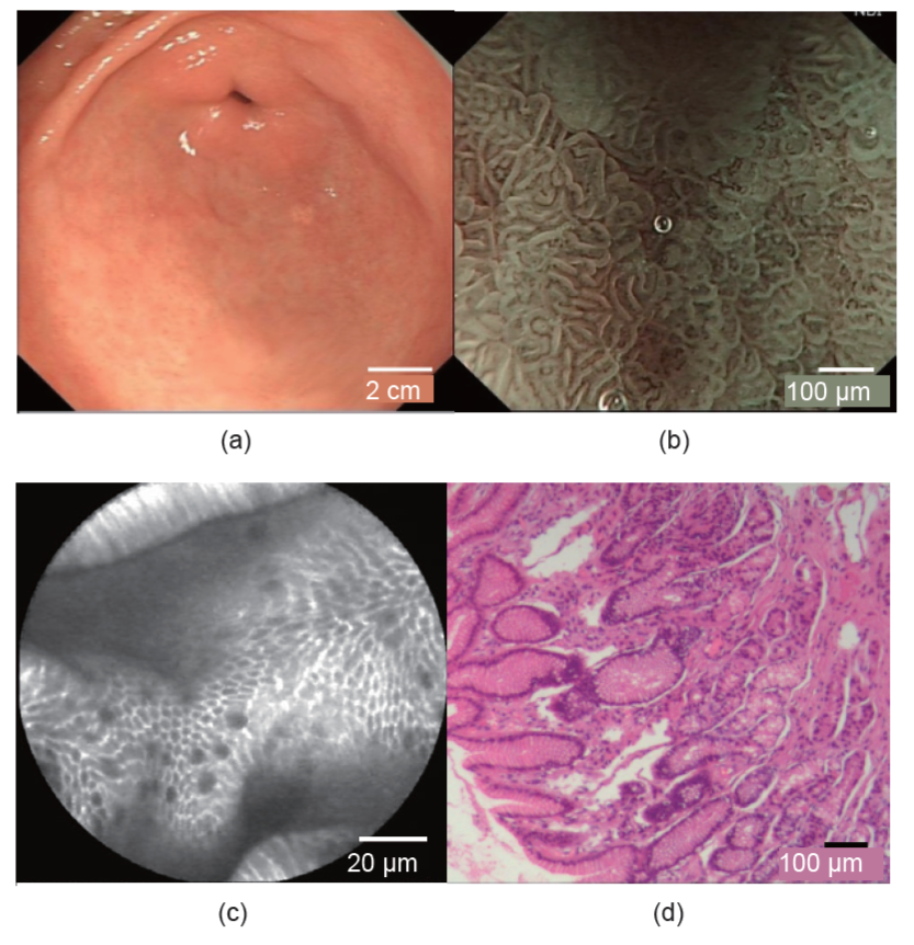
图1
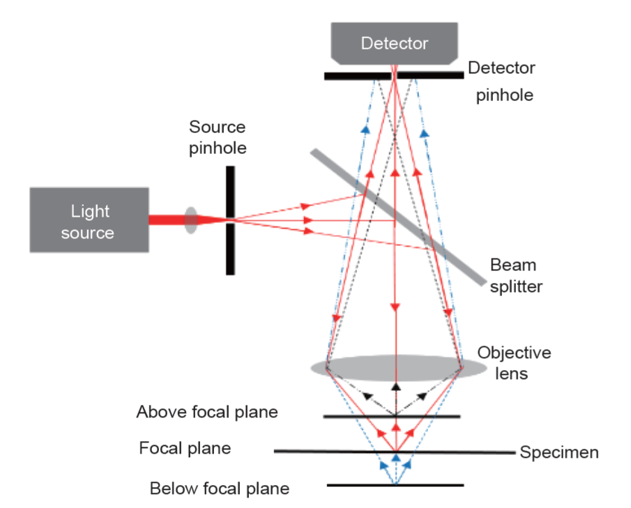
图2
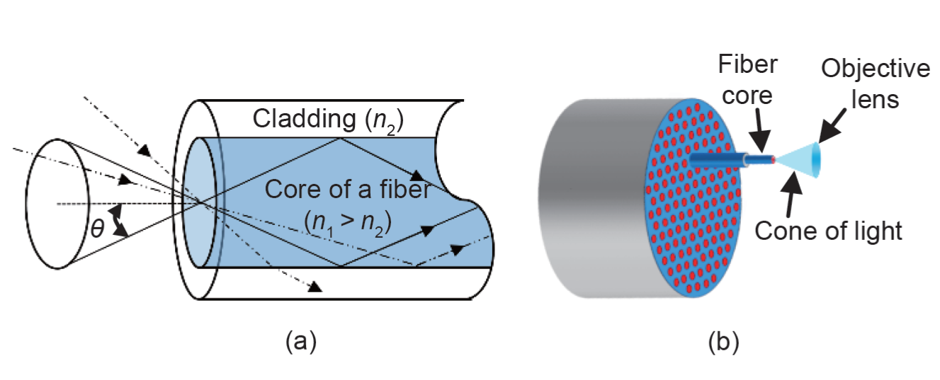
图3
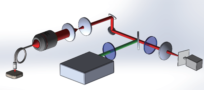
图5
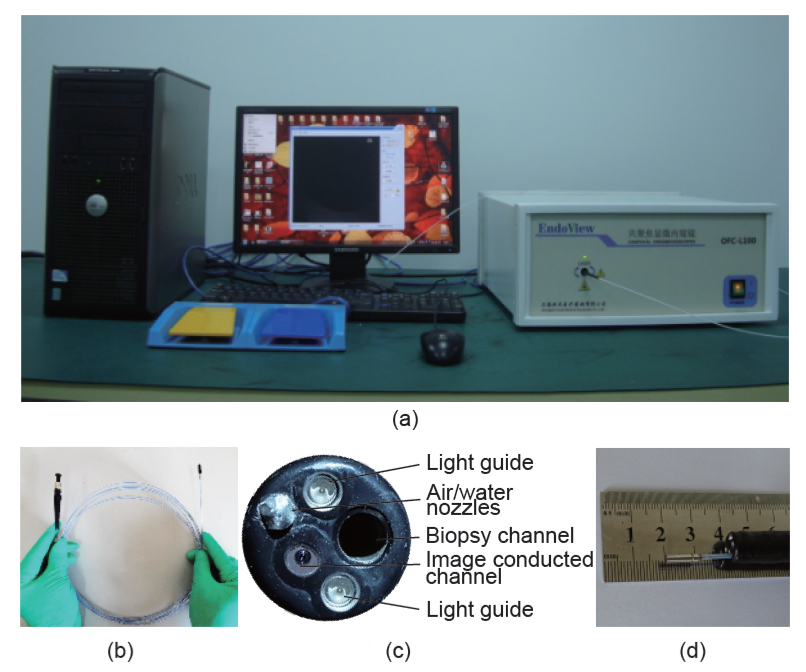
图6
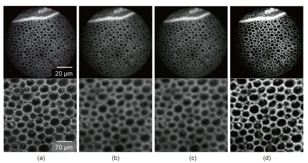
图7
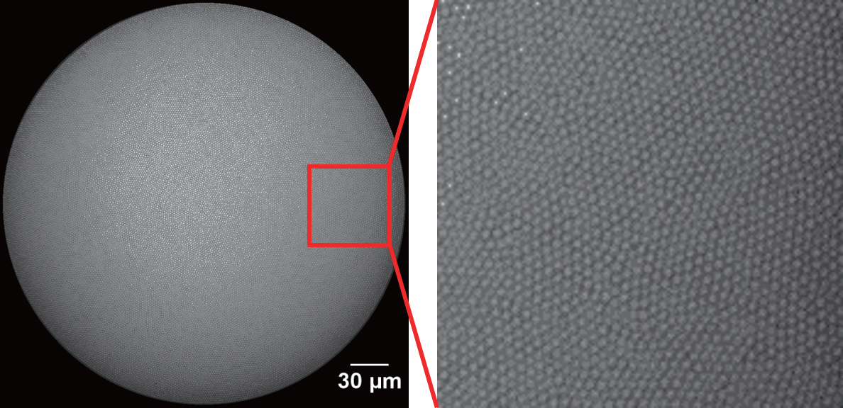
图8
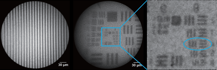
图9
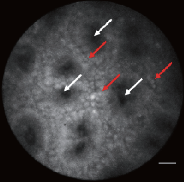
图10
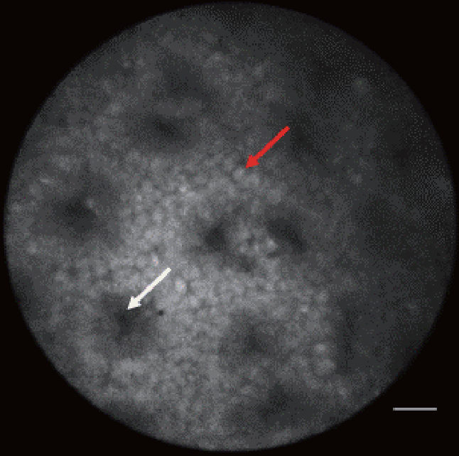
图11
参考文献
[ 1 ] B. Stewart, C. P. Wild. World Cancer Report 2014. Geneva: World Health Organization, 2014
[ 2 ] T. A. Stamey, N. Yang, A. R. Hay, J. E. McNeal, F. S. Freiha, E. Redwine. Prostate-specific antigen as a serum marker for adenocarcinoma of the prostate. N. Engl. J. Med., 1987, 317(15): 909–916
[ 3 ] J. M. Edmonson. History of the instruments for gastrointestinal endoscopy. Gastrointest. Endosc., 1991, 37(Suppl. 2): S27–S56 链接1
[ 4 ] B. I. Hirschowitz, C. W. Peters, L. E. Curtiss. Preliminary report on a long fiberscope for examination of stomach and duodenum. Med. Bull. (Ann Arbor), 1957, 23(5): 178–180
[ 5 ] B. I. Hirschowitz. A personal history of the fiberscope. Gastroenterology, 1979, 76(4): 864–869
[ 6 ] J. Pohl, Comparison of computed virtual chromoendoscopy and conventional chromoendoscopy with acetic acid for detection of neoplasia in Barrett’s esophagus. Endoscopy, 2007, 39(7): 594–598 链接1
[ 7 ] ASGE Technology Committee; L. M. Wong Kee Song, Chromoendoscopy. Gastrointest. Endosc., 2007, 66(4): 639–649 链接1
[ 8 ] K. K. Wang, N. Okoro, G. Prasad, M. Wong Kee Song, N. S. Buttar, J. Tian. Endoscopic evaluation and advanced imaging of Barrett’s esophagus. Gastrointest. Endosc. Clin. N. Am., 2011, 21(1): 39–51 链接1
[ 9 ] R. Kiesslich, Confocal laser endoscopy for diagnosing intraepithelial neoplasias and colorectal cancer in vivo. Gastroenterology, 2004, 127(3): 706–713 链接1
[10] M. Goetz, N. P. Malek, R. Kiesslich. Microscopic imaging in endoscopy: Endomicroscopy and endocytoscopy. Nat. Rev. Gastroenterol. Hepatol., 2014, 11(1): 11–18
[11] A. Meining, Direct visualization of indeterminate pancreaticobiliary strictures with probe-based confocal laser endomicroscopy: A multicenter experience. Gastrointest. Endosc., 2011, 74(5): 961–968 链接1
[12] T. Liu, H. Zheng, W. Gong, C. Chen, B. Jiang. The accuracy of confocal laser endomicroscopy, narrow band imaging, and chromoendoscopy for the detection of atrophic gastritis. J. Clin. Gastroenterol., 2015, 49(5): 379–386 链接1
[13] M. Goetz. Endomicroscopy and targeted imaging of gastric neoplasia. Gastrointest. Endosc. Clin. N. Am., 2013, 23(3): 597–606 链接1
[14] L. Ginlünas, R. Juškaitis, S. V. Shatalin. Scanning fibre-optic microscope. Electron. Lett., 1991, 27(9): 724–726 链接1
[15] M. Gu, C. J. R. Sheppard, X. Gan. Image formation in a fiber-optical confocal scanning microscope. J. Opt. Soc. Am. A, 1991, 8(11): 1755–1761
[16] S. Kimura, T. Wilson. Confocal scanning optical microscope using single-mode fiber for signal detection. Appl. Opt., 1991, 30(16): 2143–2150 链接1
[17] A. F. Gmitro, D. Aziz. Confocal microscopy through a fiber-optic imaging bundle. Opt. Lett., 1993, 18(8): 565–567 链接1
[18] M. B. Wallace, P. Fockens. Probe-based confocal laser endomicroscopy. Gastroenterology, 2009, 136(5): 1509–1513 链接1
[19] R. Kiesslich, M. Goetz, M. Vieth, P. R. Galle, M. F. Neurath. Technology insight: Confocal laser endoscopy for in vivo diagnosis of colorectal cancer. Nat. Clin. Pract. Oncol., 2007, 4(8): 480–490 链接1
[20] M. Goetz, A. Watson, R. Kiesslich. Confocal laser endomicroscopy in gastrointestinal diseases. J. Biophotonics, 2011, 4(7−8): 498–508 链接1
[21] J. Knittel, L. Schnieder, G. Buess, B. Messerschmidt, T. Possner. Endoscope-compatible confocal microscope using a gradient index-lens system. Opt. Commun., 2001, 188(5−6): 267–273 链接1
[22] J. C. Jung, M. J. Schnitzer. Multiphoton endoscopy. Opt. Lett., 2003, 28(11): 902–904 链接1
[23] A. R. Rouse, A. Kano, J. A. Udovich, S. M. Kroto, A. F. Gmitro. Design and demonstration of a miniature catheter for a confocal microendoscope. Appl. Opt., 2004, 43(31): 5763–5771 链接1
[24] C. Liang, K. B. Sung, R. R. Richards-Kortum, M. R. Descour. Design of a high-numerical-aperture miniature microscope objective for an endoscopic fiber confocal reflectance microscope. Appl. Opt., 2002, 41(22): 4603–4610 链接1
[25] M. D. Chidley, K. D. Carlson, R. R. Richards-Kortum, M. R. Descour. Design, assembly, and optical bench testing of a high-numerical-aperture miniature injection-molded objective for fiber-optic confocal reflectance microscopy. Appl. Opt., 2006, 45(11): 2545–2554 链接1
[26] R. T. Kester, T. Christenson, R. R. Kortum, T. S. Tkaczyk. Low cost, high performance, self-aligning miniature optical systems. Appl. Opt., 2009, 48(18): 3375–3384 链接1
[27] M. Kyrish, Needle-based fluorescence endomicroscopy via structured illumination with a plastic, achromatic objective. J. Biomed. Opt., 2013, 18(9): 096003 链接1
[28] W. Piyawattanametha, In vivo brain imaging using a portable 2.9 g two-photon microscope based on a microelectromechanical systems scanning mirror. Opt. Lett., 2009, 34(15): 2309–2311 链接1
[29] J. Sawinski, D. J. Wallace, D. S. Greenberg, S. Grossmann, W. Denk, J. N. Kerr. Visually evoked activity in cortical cells imaged in freely moving animals. Proc. Natl. Acad. Sci. U.S.A., 2009, 106(46): 19557–19562 链接1
[30] J. Sawinski, W. Denk. Miniature random-access fiber scanner for in vivo multiphoton imaging. J. Appl. Phys., 2007, 102(3): 034701
[31] Y. Zhang, A compact fiber-optic SHG scanning endomicroscope and its application to visualize cervical remodeling during pregnancy. Proc. Natl. Acad. Sci. U.S.A., 2012, 109(32): 12878–12883 链接1
[32] C. M. Lee, C. J. Engelbrecht, T. D. Soper, F. Helmchen, E. J. Seibel. Scanning fiber endoscopy with highly flexible, 1 mm catheterscopes for wide-field, full-color imaging. J. Biophotonics, 2010, 3(5−6): 385–407 链接1
[33] B. A. Flusberg, E. D. Cocker, W. Piyawattanametha, J. C. Jung, E. L. M. Cheung, M. J. Schnitzer. Fiber-optic fluorescence imaging. Nat. Methods, 2005, 2(12): 941–950 链接1
[34] Z. Li, Z. Yang, L. Fu. Scanning properties of a resonant fiber-optic piezoelectric scanner. Rev. Sci. Instrum., 2011, 82(12): 123707 链接1
[35] Z. Li, L. Fu. Note: A resonant fiber-optic piezoelectric scanner achieves a raster pattern by combining two distinct resonances. Rev. Sci. Instrum., 2012, 83(8): 086102 链接1
[36] R. Sjöback, J. Nygren, M. Kubista. Absorption and fluorescence properties of fluorescein. Spectrochim. Acta A Mol. Biomol. Spectrosc., 1995, 51(6): L7–L21 链接1
[37] V. K. Sharma, P. D. Sahare, R. C. Rastogi, S. K. Ghoshal, D. Mohan. Excited state characteristics of acridine dyes: Acriflavine and acridine orange. Spectrochim. Acta A Mol. Biomol. Spectrosc., 2003, 59(8): 1799–1804 链接1
[38] A. L. Polglase, W. J. McLaren, S. A. Skinner, R. Kiesslich, M. F. Neurath, P. M. Delaney. A fluorescence confocal endomicroscope for in vivo microscopy of the upper- and the lower-GI tract. Gastrointest. Endosc., 2005, 62(5): 686–695 链接1
[39] J. M. Jabbour, M. A. Saldua, J. N. Bixler, K. C. Maitland. Confocal endomicroscopy: Instrumentation and medical applications. Ann. Biomed. Eng., 2012, 40(2): 378–397 链接1
[40] S. C. Park, M. K. Park, M. G. Kang. Super-resolution image reconstruction: A technical overview. IEEE Signal Proc. Mag., 2003, 20(3): 21–36 链接1
[41] S. Lertrattanapanich, N. K. Bose. High resolution image formation from low resolution frames using Delaunay triangulation. IEEE Trans. Image Process., 2002, 11(12): 1427–1441 链接1
[42] T. Kuiper, New classification for probe-based confocal laser endomicroscopy in the colon. Endoscopy, 2011, 43(12): 1076–1081 链接1
[43] M. Goetz, In vivo molecular imaging of colorectal cancer with confocal endomicroscopy by targeting epidermal growth factor receptor. Gastroenterology, 2010, 138(2): 435–446 链接1
[44] D. Moussata, Confocal laser endomicroscopy is a new imaging modality for recognition of intramucosal bacteria in inflammatory bowel disease in vivo. Gut, 2011, 60(1): 26–33 链接1
[45] Y. Goto, H. Kiyono. Epithelial barrier: An interface for the cross-communication between gut flora and immune system. Immunol. Rev., 2012, 245(1): 147–163 链接1
[46] S. Foersch, et al. Molecular imaging of VEGF in gastrointestinal cancer in vivo using confocal laser endomicroscopy. Gut, 2010, 59(8): 1046–1055 链接1


 京公网安备 11010502051620号


