
2017, Volume 3, Issue 1
Engineering >> 2017, Volume 3, Issue 1 doi: 10.1016/J.ENG.2017.01.007
Tethering of Gly-Arg-Gly-Asp-Ser-Pro-Lys Peptides on Mg-Doped Hydroxyapatite
a Department of Engineering, University of Messina, Messina 98166, Italy
b Institute of Science and Technology for Ceramics, National Research Council of Italy, Faenza 48018, Italy
Next Previous
Abstract
Stem cell homing, namely the recruitment of mesenchymal stem cells (MSCs) to injured tissues, is highly effective for bone regeneration in vivo. In order to explore whether the incorporation of mimetic peptide sequences on magnesium-doped (Mg-doped) hydroxyapatite (HA) may regulate the homing of MSCs, and thus induce cell migration to a specific site, we covalently functionalized MgHA disks with two chemotactic/haptotactic factors: either the fibronectin fragment III1-C human (FF III1-C), or the peptide sequence Gly-Arg-Gly-Asp-Ser-Pro-Lys, a fibronectin analog that is able to bind to integrin transmembrane receptors. Preliminary biological evaluation of MSC viability, analyzed by 3-(4,5-dimethylthiazol-2-yl)-2,5-diphenyltetrazolium bromide (MTT) test, suggested that stem cells migrate to the MgHA disks in response to the grafted haptotaxis stimuli.
Keywords
Mg-doped hydroxyapatite ; Mesenchymal stem cells ; Chemotactic/haptotactic factors ; Bone tissue engineering
Figures

Fig.1

Fig.2
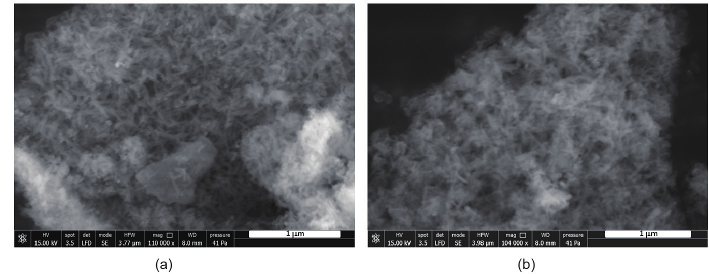
Fig.3
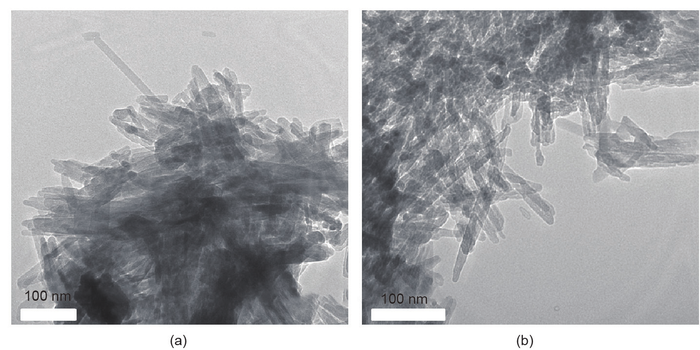
Fig.4

Fig.5

Fig.6

Fig.7

Fig.8
References
[ 1 ] Laurencin CT, Khan Y. Regenerative Engineering. Sci Transl Med? 2012;4(160):160ed9 link1
[ 2 ] Amini AR, Laurencin CT, Nukavarapu SP. Bone tissue engineering: recent advances and challenges. Crit Rev Biomed Eng? 2012;40(5):363–408 link1
[ 3 ] Dawson JI, Kanczler J, Tare R, Kassem M, Oreffo RO. Concise review: bridging the gap: bone regeneration using skeletal stem cell-based strategies—where are we now? Stem Cells 2014;32(1):35–44 link1
[ 4 ] Wang P, Zhao L, Liu J, Weir MD, Zhou X, Xu HH. Bone tissue engineering via nanostructured calcium phosphate biomaterials and stem cells. Bone Res 2014;2:14017 link1
[ 5 ] Gong T, Xie J, Liao J, Zhang T, Lin S, Lin Y. Nanomaterials and bone regeneration. Bone Res 2015;3:15029 link1
[ 6 ] Iannazzo D, Pistone A, Espro C, Galvagno S. Drug delivery strategies for bone tissue regeneration. In: Panseri S, Taraballi F, Cunha C, editors Biomimetic approaches for tissue healing. Foster City: OMICS Group eBooks; 2015. p. 1–39.
[ 7 ] Panseri S, Cunha C, D’Alessandro T, Sandri M, Russo A, Giavaresi G, et al. Magnetic hydroxyapatite bone substitutes to enhance tissue regeneration: evaluation in vitro using osteoblast-like cells and in vivo in a bone defect. PLoS One 2012;7(6):e38710 link1
[ 8 ] Cunha C, Panseri S, Iannazzo D, Piperno A, Pistone A, Fazio M, et al. Hybrid composites made of multiwalled carbon nanotubes functionalized with Fe3O4 nanoparticles for tissue engineering applications. Nanotechnology 2012;23(46):465102 link1
[ 9 ] Wang DX, He Y, Bi L, Qu ZH, Zou JW, Pan Z, et al. Enhancing the bioactivity of Poly(lactic-co-glycolic acid) scaffold with a nano-hydroxyapatite coating for the treatment of segmental bone defect in a rabbit model. Int J Nanomedicine 2013;8: 1855–65 link1
[10] Yoshikawa H, Myoui A. Bone tissue engineering with porous hydroxyapatite ceramics. J Artif Organs 2005;8(3):131–6 link1
[11] Bellucci D, Sola A, Gazzarri M, Chiellini F, Cannillo V. A new hydroxyapatite-based biocomposite for bone replacement. Mater Sci Eng C Mater Biol Appl 2013;33(3):1091–101 link1
[12] Pistone A, Iannazzo D, Panseri S, Montesi M, Tampieri A, Galvagno S. Hydroxyapatite-magnetite-MWCNT nanocomposite as a biocompatible multifunctional drug delivery system for bone tissue engineering. Nanotechnology 2014;25(42):425701 link1
[13] Laurencin D, Almora-Barrios N, de Leeuw NH, Gervais C, Bonhomme C, Mauri F, et al. Magnesium incorporation into hydroxyapatite. Biomaterials 2011;32(7):1826–37 link1
[14] Landi E, Logroscino G, Proietti L, Tampieri A, Sandri M, Sprio S. Biomimetic Mg-substituted hydroxyapatite: from synthesis to in vivo behaviour. J Mater Sci Mater Med 2008;19(1):239–47 link1
[15] Barthes J, Özçelik H, Hindié M, Ndreu-Halili A, Hasan A, Vrana NE. Cell microenvironment engineering and monitoring for tissue engineering and regenerative medicine: the recent advances. Biomed Res Int 2014;2014:921905 link1
[16] Schantz JT, Chim H, Whiteman M. Cell guidance in tissue engineering: SDF-1 mediates site-directed homing of mesenchymal stem cells within three-dimensional polycaprolactone scaffolds. Tissue Eng 2007;13(11):2615–24 link1
[17] Vo TN, Kasper FK, Mikos AG. Strategies for controlled delivery of growth factors and cells for bone regeneration. Adv Drug Deliv Rev 2012;64(12):1292–309 link1
[18] García AJ, Reyes CD. Bio-adhesive surfaces to promote osteoblast differentiation and bone formation. J Dent Res 2005;84(5):407–13 link1
[19] Yun YR, Pham BH, Yoo YR, Lee S, Kim HW, Jang JH. Engineering of self-assembled fibronectin matrix protein and its effects on mesenchymal stem cells. Int J Mol Sci 2015;16(8):19645–56 link1
[20] Liu Y, Peterson DA, Kimura H, Schubert D. Mechanism of cellular 3-(4,5-dimethylthiazol-2-yl)-2,5-diphenyltetrazolium bromide (MTT) reduction. J Neurochem 1997;69(2):581–93 link1


 京公网安备 11010502051620号


