2020, Volume 6, Issue 11
Engineering >> 2020, Volume 6, Issue 11 doi: 10.1016/j.eng.2020.02.011
Temporal Integrative Omics Reveals an Increase in Nondegradative Ubiquitylation during Primary Hepatocyte Dedifferentiation
State Key Laboratory for Diagnosis and Treatment of Infectious Diseases, National Clinical Research Center for Infectious Diseases, Collaborative Innovation Center for Diagnosis and Treatment of Infectious Diseases, The First Affiliated Hospital, College of Medicine, Zhejiang University, Hangzhou 310003, China
# These authors contributed equally to this work.
Next Previous
Abstract
Primary hepatocytes (PHCs) are widely used in various fields, but the progressive deterioration of liver-specific features in vitro significantly limits their application. While the transcriptional regulation and whole cell proteome (WCP) of PHCs have been extensively studied, only a small number of studies have addressed the role of posttranslational modifications in this process. To elucidate the underlying mechanisms that induce dedifferentiation, we carried out parallel quantifications of the transcriptome, WCP, ubiquitinome, and phosphoproteome of rat PHCs after 0, 6, 12, 24, and 48 h of in vitro culture. Our data constitute a detailed proteomic analysis of dedifferentiated PHCs including 2196 proteins, 2056 ubiquitinated sites, and 4932 phosphorylated peptides. We revealed a low correlation between the transcriptome and WCP during dedifferentiation. A combined analysis of the ubiquitinome with the corresponding WCP indicated that the dedifferentiation of PHCs led to an increase in nondegradative K27 ubiquitination. Functional analysis of the altered phosphoproteins suggested a significant enrichment in ferroptosis. In all, 404 proteins with both ubiquitination and phosphorylation were identified to be involved in critical metabolic events. Furthermore, Ptbp1, Hnrpd, Hnrnpu, and Srrm2 were identified as hub genes. Taken together, our data provide new insights into proteome dynamics during PHC dedifferentiation and potential targets to inhibit the dedifferentiation process.
Keywords
Ubiquitination ; Phosphoproteome ; Proteome ; Dedifferentiation ; Primary hepatocytes
SupplementaryMaterials
Figures

Fig. 1
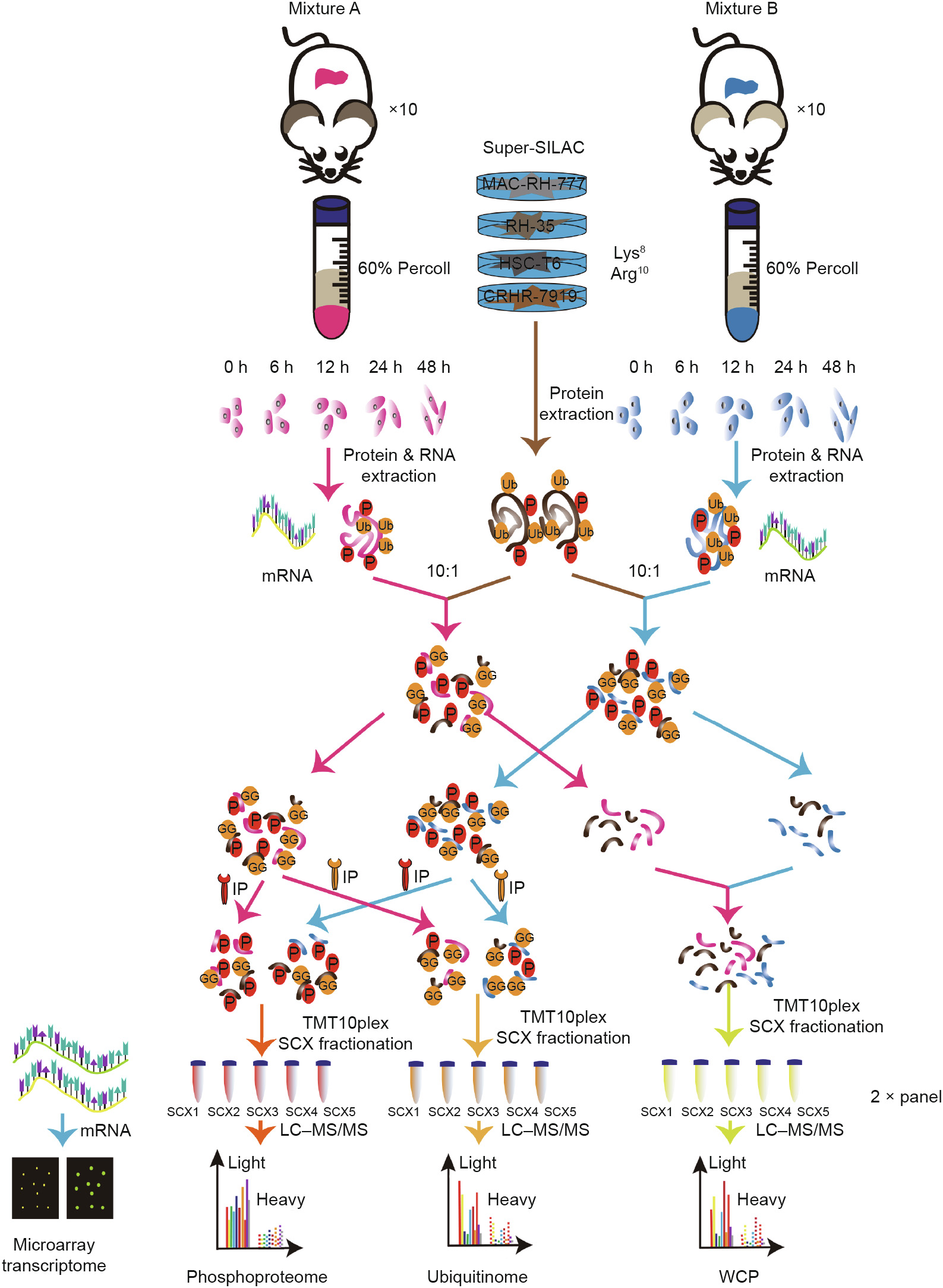
Fig. 2
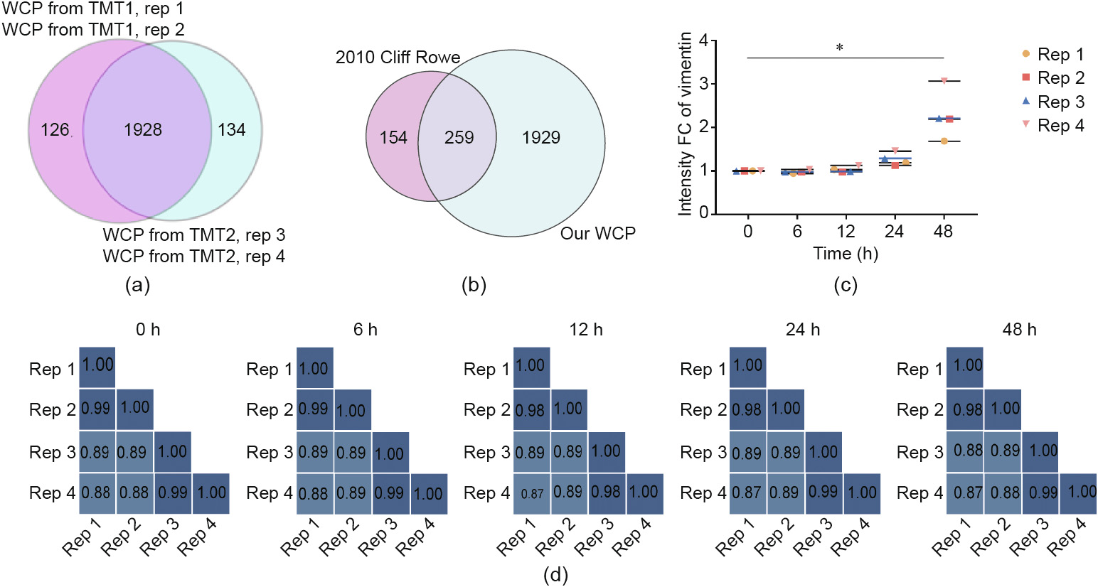
Fig. 3

Fig. 4

Fig. 5
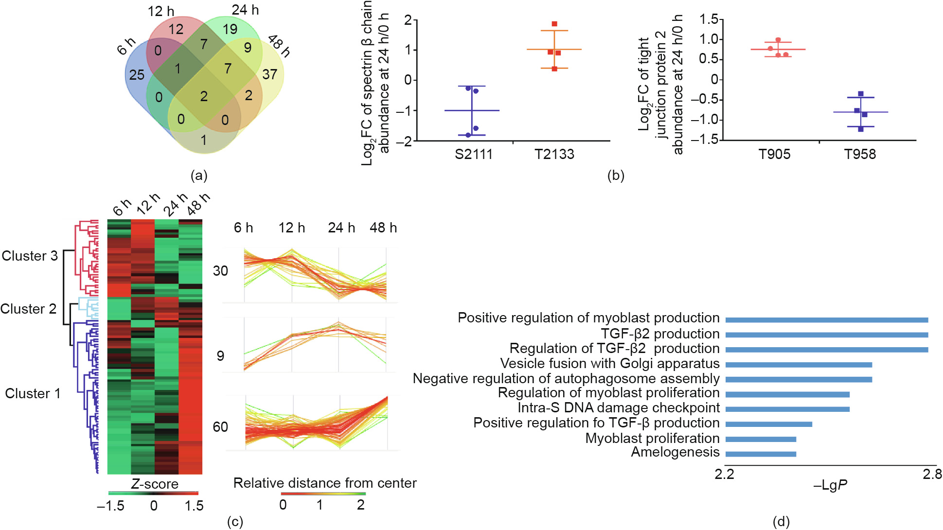
Fig. 6

Fig. 7
References
[ 1 ] Gomez-Lechon MJ, Tolosa L, Conde I, Donato MT. Competency of different cell models to predict human hepatotoxic drugs. Expert Opin Drug Metab Toxicol 2014;10(11):1553–68. link1
[ 2 ] Xia Y, Carpentier A, Cheng X, Block PD, Zhao Y, Zhang Z, et al. Human stem cellderived hepatocytes as a model for hepatitis B virus infection, spreading and virus–host interactions. J Hepatol 2017;66(3):494–503. link1
[ 3 ] Soltys KA, Setoyama K, Tafaleng EN, Gutiérrez AS, Fong J, Fukumitsu K, et al. Host conditioning and rejection monitoring in hepatocyte transplantation in humans. J Hepatol 2017;66(5):987–1000. link1
[ 4 ] Schuetz EG, Li D, Omiecinski CJ, Muller-Eberhard U, Kleinman HK, Elswick B, et al. Regulation of gene expression in adult rat hepatocytes cultured on a basement membrane matrix. J Cell Physiol 1988;134(3):309–23. link1
[ 5 ] Heslop JA, Rowe C, Walsh J, Sison-Young R, Jenkins R, Kamalian L, et al. Mechanistic evaluation of primary human hepatocyte culture using global proteomic analysis reveals a selective dedifferentiation profile. Arch Toxicol 2017;91(1):439–52. link1
[ 6 ] Cicchini C, Amicone L, Alonzi T, Marchetti A, Mancone C, Tripodi M. Molecular mechanisms controlling the phenotype and the EMT/MET dynamics of hepatocyte. Liver Int 2015;35(2):302–10. link1
[ 7 ] Mehta A, Comunale MA, Rawat S, Casciano JC, Lamontagne J, Herrera H, et al. Intrinsic hepatocyte dedifferentiation is accompanied by upregulation of mesenchymal markers, protein sialylation and core alpha 1,6 linked fucosylation. Sci Rep 2016;6:27965. link1
[ 8 ] Fraczek J, Bolleyn J, Vanhaecke T, Rogiers V, Vinken M. Primary hepatocyte cultures for pharmaco–toxicological studies: at the busy crossroad of various anti-dedifferentiation strategies. Arch Toxicol 2013;87(4):577–610. link1
[ 9 ] Odom DT, Zizlsperger N, Gordon DB, Bell GW, Rinaldi NJ, Murray HL, et al. Control of pancreas and liver gene expression by HNF transcription factors. Science 2004;303(5662):1378–81. link1
[10] Lauschke VM, Vorrink SU, Moro SM, Rezayee F, Nordling Å, Hendriks DFG, et al. Massive rearrangements of cellular microRNA signatures are key drivers of hepatocyte dedifferentiation. Hepatology 2016;64(5):1743–56. link1
[11] Ahmed HMM, Salerno S, Morelli S, Giorno L, de Bartolo L. 3D liver membrane system by co-culturing human hepatocytes, sinusoidal endothelial and stellate cells. Biofabrication 2017;9(2):025022. link1
[12] Bale SS, Geerts S, Jindal R, Yarmush ML. Isolation and co-culture of rat parenchymal and non-parenchymal liver cells to evaluate cellular interactions and response. Sci Rep 2016;6:25329. link1
[13] Hiramatsu K, Matsumoto Y, Miyazaki M, Tsubouchi H, Yamamoto I, Gohda E. Inhibition of hepatocyte growth factor production in human fibroblasts by ursodeoxycholic acid. Biol Pharm Bull 2005;28(4):619–24. link1
[14] Vinken M, Papeleu P, Snykers S, de Rop E, Henkens T, Chipman JK, et al. Involvement of cell junctions in hepatocyte culture functionality. Crit Rev Toxicol 2006;36(4):299–318. link1
[15] Lang R, Stern MM, Smith L, Liu Y, Bharadwaj S, Liu G, et al. Three-dimensional culture of hepatocytes on porcine liver tissue-derived extracellular matrix. Biomaterials 2011;32(29):7042–52. link1
[16] Xiang C, Du Y, Meng G, Soon YL, Sun S, Song N, et al. Long-term functional maintenance of primary human hepatocytes in vitro. Science 2019;64 (6438):399–402. link1
[17] Schwanhausser B, Busse D, Li N, Dittmar G, Schuchhardt J, Wolf J, et al. Global quantification of mammalian gene expression control. Nature 2011;73 (7347):337–42. link1
[18] Reyes-Turcu FE, Ventii KH, Wilkinson KD. Regulation and cellular roles of ubiquitin-specific deubiquitinating enzymes. Annu Rev Biochem 2009;78:363–97. link1
[19] Williamson A, Werner A, Rape M. The Colossus of ubiquitylation: decrypting a cellular code. Mol Cell 2013;49(4):591–600. link1
[20] Buckley SM, Aranda-Orgilles B, Strikoudis A, Apostolou E, Loizou E, MoranCrusio K, et al. Regulation of pluripotency and cellular reprogramming by the ubiquitin–proteasome system. Cell Stem Cell 2012;11(6):783–98. link1
[21] Choi J, Baek KH. Cellular functions of stem cell factors mediated by the ubiquitin–proteasome system. Cell Mol Life Sci 2018;75(11):1947–57. link1
[22] Pagani M, Rockstroh M, Schuster M, Rossetti G, Moro M, Crosti M, et al. Reference proteome of highly purified human Th1 cells reveals strong effects on metabolism and protein ubiquitination upon differentiation. Proteomics 2015;15(21):3644–7. link1
[23] Bax M, McKenna J, Do-Ha D, Stevens CH, Higginbottom S, Balez R, et al. The ubiquitin proteasome system is a key regulator of pluripotent stem cell survival and motor neuron differentiation. Cells 2019;8(6):581. link1
[24] Elia AE, Boardman AP, Wang DC, Huttlin EL, Everley RA, Dephoure N, et al. Quantitative proteomic atlas of ubiquitination and acetylation in the DNA damage response. Mol Cell 2015;59(5):867–81. link1
[25] Iesmantavicius V, Weinert BT, Choudhary C. Convergence of ubiquitylation and phosphorylation signaling in rapamycin-treated yeast cells. Mol Cell Proteomics 2014;13(8):1979–92. link1
[26] Kreamer BL, Staecker JL, Sawada N, Sattler GL, Stephen Hsia MT, Pitot HC. Use of a low-speed, iso-density percoll centrifugation method to increase the viability of isolated rat hepatocyte preparations. Vitro Cell Dev Biol 1986;22 (4):201–11. link1
[27] Deeb SJ, D’Souza RC, Cox J, Schmidt-Supprian M, Mann M. Super-SILAC allows classification of diffuse large B-cell lymphoma subtypes by their protein expression profiles. Mol Cell Proteomics 2012;11(5):77–89. link1
[28] Rappsilber J, Ishihama Y, Mann M. Stop and go extraction tips for matrixassisted laser desorption/ionization, nanoelectrospray, and LC/MS sample pretreatment in proteomics. Anal Chem 2003;75(3):663–70. link1
[29] Ma J, Chen T, Wu S, Yang C, Bai M, Shu K, et al. iProX: an integrated proteome resource. Nucleic Acids Res 2019;47(D1):D1211–7. link1
[30] Schneider TD, Stephens RM. Sequence logos: a new way to display consensus sequences. Nucleic Acids Res 1990;18(20):6097–100. link1
[31] Szklarczyk D, Gable AL, Lyon D, Junge A, Wyder S, Huerta-Cepas J, et al. STRING v11: protein–protein association networks with increased coverage, supporting functional discovery in genome-wide experimental datasets. Nucleic Acids Res 2019;47(D1):D607–13. link1
[32] Shannon P, Markiel A, Ozier O, Baliga NS, Wang JT, Ramage D, et al. Cytoscape: a software environment for integrated models of biomolecular interaction networks. Genome Res 2003;13(11):2498–504. link1
[33] Chin CH, Chen SH, Wu HH, Ho CW, Mt Ko, Lin CY, et al. cytoHubba: identifying hub objects and sub-networks from complex interactome. BMC Syst Biol 2014;8(Suppl 4):S11. link1
[34] Tyanova S, Temu T, Sinitcyn P, Carlson A, Hein MY, Geiger T, et al. The Perseus computational platform for comprehensive analysis of (prote)omics data. Nat Methods 2016;13(9):731–40. link1
[35] World Health Organization. Guidelines for the prevention, care and treatment of persons with chronic hepatitis B infection. In: WHO guidelines approved by the Guidelines Review Committee. Geneva: World Health Organization; 2015.
[36] Boersema PJ, Geiger T, Wis´niewski JR, Mann M. Quantification of the Nglycosylated secretome by super-SILAC during breast cancer progression and in human blood samples. Mol Cell Proteomics 2013;12(1):158–71. link1
[37] Beigel J, Fella K, Kramer PJ, Kroeger M, Hewitt P. Genomics and proteomics analysis of cultured primary rat hepatocytes. Toxicol In Vitro 2008;22 (1):171–81. link1
[38] Rowe C, Goldring CE, Kitteringham NR, Jenkins RE, Lane BS, Sanderson C, et al. Network analysis of primary hepatocyte dedifferentiation using a shotgun proteomics approach. J Proteome Res 2010;9(5):2658–68. link1
[39] Correia MA, Sadeghi S, Mundo-Paredes E. Cytochrome P450 ubiquitination: branding for the proteolytic slaughter? Annu Rev Pharmacol Toxicol 2005;45:439–64. link1
[40] Banerjee A, Kocarek TA, Novak RF. Identification of a ubiquitinationtarget/substrate-interaction domain of cytochrome P-450 (CYP) 2E1. Drug Metab Dispos 2000;28(2):118–24. link1
[41] Huan JY, Streicher JM, Bleyle LA, Koop DR. Proteasome-dependent degradation of cytochromes P450 2E1 and 2B1 expressed in tetracycline-regulated HeLa cells. Toxicol Appl Pharmacol 2004;199(3):332–43. link1
[42] Ravid T, Hochstrasser M. Diversity of degradation signals in the ubiquitinproteasome system. Nat Rev Mol Cell Biol 2008;9(9):679–90. link1
[43] Lai Y, Zhu M, Wu W, Rokutanda N, Togashi Y, Liang W, et al. HERC2 regulates RPA2 by mediating ATR-induced Ser33 phosphorylation and ubiquitindependent degradation. Sci Rep 2019;9(1):14257. link1
[44] Li D, Yang W, Ren J, Ru Y, Zhang K, Fu S, et al. The E3 ubiquitin ligase TBK1 mediates the degradation of multiple picornavirus VP3 proteins by phosphorylation and ubiquitination. J Virol 2019;93:e01438–19. link1
[45] Wanet A, Remacle N, Najar M, Sokal E, Amould T, Najimi M, et al. Mitochondrial remodeling in hepatic differentiation and dedifferentiation. Int J Biochem Cell Biol 2014;54:174–85. link1
[46] Otasek D, Morris JH, Boucas J, Pico AR, Demchak B. Cytoscape Automation: empowering workflow-based network analysis. Genome Biol 2019;20(1):185. link1
[47] Baker TK, Carfagna MA, Gao H, Dow ER, Li Q, Searfoss GH, et al. Temporal gene expression analysis of monolayer cultured rat hepatocytes. Chem Res Toxicol 2001;14(9):1218–31. link1
[48] Udeshi ND, Mani DR, Eisenhaure T, Mertins P, Jaffe JD, Clauser KR, et al. Methods for quantification of in vivo changes in protein ubiquitination following proteasome and deubiquitinase inhibition. Mol Cell Proteomics 2012;11(5):148–59. link1
[49] Wagner SA, Beli P, Weinert BT, Nielsen ML, Cox J, Mann M, et al. A proteomewide, quantitative survey of in vivo ubiquitylation sites reveals widespread regulatory roles. Mol Cell Proteomics 2011;10(10). link1
[50] van der Wal L, Bezstarosti K, Sap KA, Dekkersa DHW, Rijkersa E, Mientjes E, et al. Improvement of ubiquitylation site detection by Orbitrap mass spectrometry. J Proteomics 2018;172:49–56. link1
[51] Sap KA, Bezstarosti K, Dekkers DHW, Voets O, Demmers JAA. Quantitative proteomics reveals extensive changes in the ubiquitinome after perturbation of the proteasome by targeted dsRNA-mediated subunit knockdown in Drosophila. J Proteome Res 2017;16(8):2848–62. link1
[52] Quadroni M, Potts A, Waridel P. Hsp90 inhibition induces both protein-specific and global changes in the ubiquitinome. J Proteomics 2015;120:215–29. link1
[53] Fiskin E, Bionda T, Dikic I, Behrends C. Global analysis of host and bacterial ubiquitinome in response to salmonella typhimurium infection. Mol Cell 2016;62(6):967–81. link1
[54] Dybas JM, O’Leary CE, Ding H, Spruce LA, Seeholzer SH, Oliver PM. Integrative proteomics reveals an increase in non-degradative ubiquitylation in activated CD4+ T cells. Nat Immunol 2019;20(6):747–55. link1
[55] Noya SB, Colameo D, Bruning F, Spinnler A, Mircsof D, Opitz L, et al. The forebrain synaptic transcriptome is organized by clocks but its proteome is driven by sleep. Science 2019;366(6462):eaav2642. link1
[56] Vogel C, Marcotte EM. Insights into the regulation of protein abundance from proteomic and transcriptomic analyses. Nat Rev Genet 2012;3(4):227–32. link1
[57] Xu G, Paige JS, Jaffrey SR. Global analysis of lysine ubiquitination by ubiquitin remnant immunoaffinity profiling. Nat Biotechnol 2010;8(8):868–73. link1
[58] Dixon SJ, Lemberg KM, Lamprecht MR, Skouta R, Zaitsev EM, Gleason CE, et al. Ferroptosis: an iron-dependent form of nonapoptotic cell death. Cell 2012;49 (5):1060–72. link1
[59] Dolma S, Lessnick SL, Hahn WC, Stockwell BR. Identification of genotypeselective antitumor agents using synthetic lethal chemical screening in engineered human tumor cells. Cancer Cell 2003;3(3):285–96. link1
[60] Minguez P, Parca L, Diella F, Mende DR, Kumar R, Helmer-Citterich M, et al. Deciphering a global network of functionally associated post-translational modifications. Mol Syst Biol 2012;8:599. link1


 京公网安备 11010502051620号


