2022, Volume 10, Issue 3
Engineering >> 2022, Volume 10, Issue 3 doi: 10.1016/j.eng.2021.05.023
High-Throughput Powder Diffraction Using White X-Ray Beam and a Simulated Energy-Dispersive Array Detector
a Materials Genome Initiative Center & School of Materials Science and Engineering, Shanghai Jiao Tong University, Shanghai 200240, China
b Institute of High Energy Physics, Chinese Academy of Sciences, Beijing 100049, China
c Academy for Advanced Interdisciplinary Studies, Southern University of Science and Technology, Shenzhen 518055, China
d Department of Materials Science and Engineering & Department of Physics, Southern University of Science and Technology, Shenzhen 518055, China
e Shanghai Advanced Research Institute, Chinese Academy of Sciences, Shanghai 201204, China
f Guangdong Provincial Key Laboratory of Energy Materials for Electric Power, Southern University of Science and Technology, Shenzhen 518055, China
g Guangdong–Hong Kong–Macao Joint Laboratory for Photonic–Thermal–Electrical Energy Materials and Devices, Southern University of Science and Technology, Shenzhen 518055, China
Next Previous
Abstract
High-throughput powder X-ray diffraction (XRD) with white X-ray beam and an energy-dispersive detector array are demonstrated in this work on a CeO2 powder sample on a bending magnet synchrotron beamline at the Shanghai Synchrotron Radiation Facility (SSRF), using a simulated energy-dispersive array detector consisting of a spatially scanning silicon-drift detector (SDD). Careful analysis and corrections are applied to account for various experimental hardware-related and diffraction angle-related factors. The resulting diffraction patterns show that the relative strength between different diffraction peaks from energy-dispersive XRD (EDXRD) spectra is consistent with that from angle-resolved XRD (ARXRD), which is necessary for analyzing crystal structures for unknown samples. The X-ray fluorescence (XRF) signal is collected simultaneously. XRF counts from all pixels are integrated directly by energy, while the diffraction spectra are integrated by d-spacing, resulting in a much improved peak strength and signal-to-noise (S/N) ratio for the array detector. In comparison with ARXRD, the diffraction signal generated by a white X-ray beam over monochromic light under the experimental conditions is about 104 times higher. The full width at half maximum (FWHM) of the peaks in q-space is found to be dependent on the energy resolution of the detector, the angle span of the detector, and the diffraction angle. It is possible for EDXRD to achieve the same or even smaller FWHM as ARXRD under the energy resolution of the current detector if the experimental parameters are properly chosen.
Keywords
High-throughput experiment ; White beam X-ray diffraction ; Energy-dispersive array detector ; Energy-dispersive X-ray diffraction ; Angle-resolved X-ray diffraction
Figures
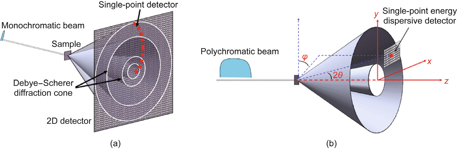
Fig. 1

Fig. 2

Fig. 3

Fig. 4

Fig. 5
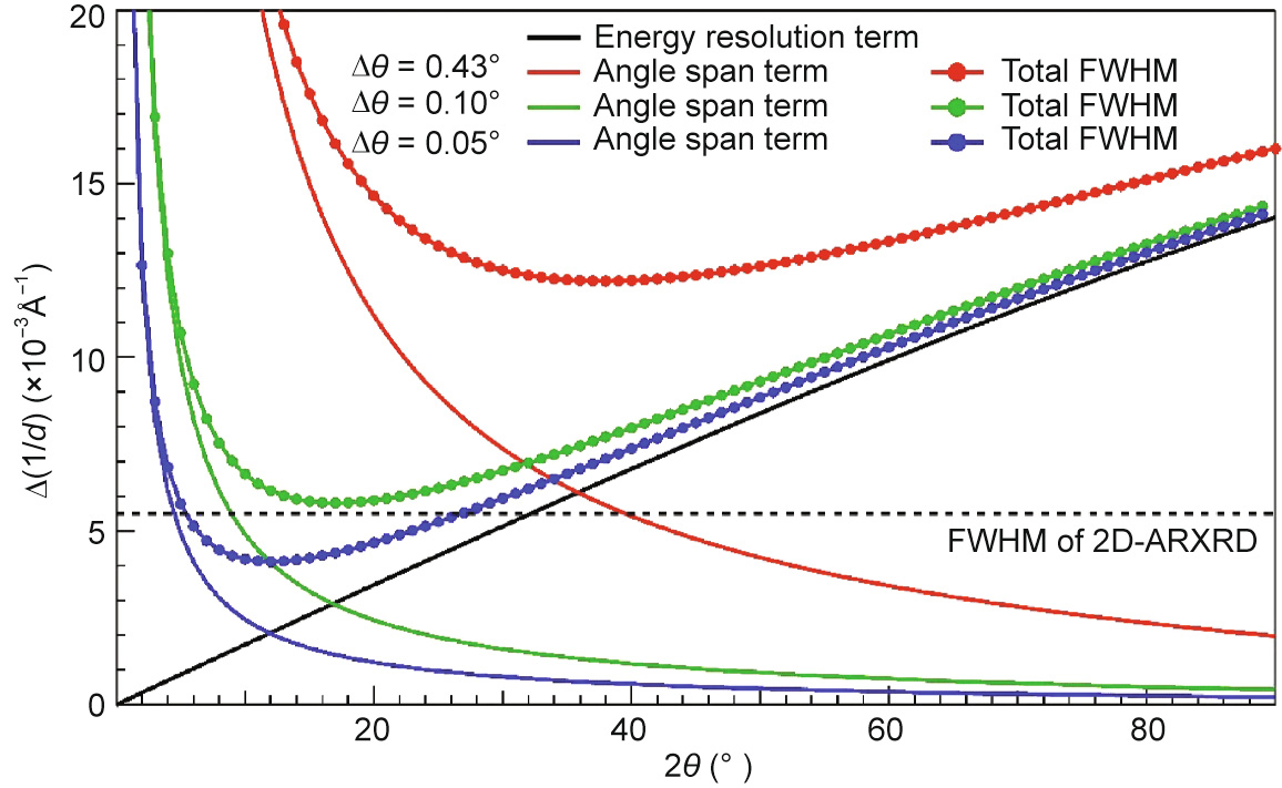
Fig. 6
References
[ 1 ] Cullity BD, Stock SR. Elements of X-ray diffraction. 3rd ed. London: Pearson; 2014. link1
[ 2 ] Xiang XD, Sun X, Briceño G, Lou Y, Wang KA, Chang H, et al. A combinatorial approach to materials discovery. Science 1995;268(5218):1738–40. link1
[ 3 ] Xiang XD, Wang G, Zhang X, Xiang Y, Wang H. Individualized pixel synthesis and characterization of combinatorial materials chips. Engineering 2015;1 (2):225–33. link1
[ 4 ] Gregoire JM, Dale D, Kazimirov A, DiSalvo FJ, van Dover RB. High energy X-ray diffraction/X-ray fluorescence spectroscopy for high-throughput analysis of composition spread thin films. Rev Sci Instrum 2009;80(12):123905. https:// doi.org/10.1063/1.3274179. link1
[ 5 ] Xing H, Zhao B, Wang Y, Zhang X, Ren Y, Yan N, et al. Rapid construction of Fe– Co–Ni composition-phase map by combinatorial materials chip approach. ACS Comb Sci 2018;20(3):127–31. link1
[ 6 ] Rodriguez-Alvarez H, Weber A, Lauche J, Kaufmann CA, Rissom T, Greiner D, et al. Formation of CuInSe2 and CuGaSe2 thin films deposited by three-stage thermal Co-evaporation: a real-time X-ray diffraction and fluorescence study. Adv Energy Mater 2013;3(10):1381–7. link1
[ 7 ] Nielsen MB, Ceresoli D, Parisiades P, Prakapenka VB, Yu T, Wang Y, et al. Phase stability of the SrMnO3 hexagonal perovskite system at high pressure and temperature. Phys Rev B 2014;90(21):214101. link1
[ 8 ] Giessen BC, Gordon GE. X-ray diffraction: new high-speed technique based on X-ray spectrography. Science 1968;159(3818):973–5. link1
[ 9 ] Buras B, Olsen JS, Gerward L. White beam, X-ray, energy-dispersive diffractometry using synchrotron radiation. Nucl Instrum Methods 1978;152 (1):293–6. link1
[10] Luo Z, Geng B, Bao J, Liu C, Liu W, Gao C, et al. High-throughput X-ray characterization system for combinatorial materials studies. Rev Sci Instrum 2005;76(9):095105. link1
[11] Mendoza Cuevas A, Bernardini F, Gianoncelli A, Tuniz C. Energy dispersive Xray diffraction and fluorescence portable system for cultural heritage applications. X-Ray Spectrom 2015;44(3):105–15. link1
[12] Drakopoulos M, Connolley T, Reinhard C, Atwood R, Magdysyuk O, Vo N, et al. I12: the joint engineering, environment and processing (JEEP) beamline at diamond light source. J Synchrotron Radiat 2015;22(3):828–38. link1
[13] O’Flynn D, Reid C, Christodoulou C, Wilson M, Veale MC, Seller P, et al. Pixelated diffraction signatures for explosive detection. In: Broach JT, Holloway JH Jr, editors. Proceedings of SPIE 8357: detection and sensing of mines, explosive objects, and obscured targets XVII; 2012 Apr 23–27; Baltimore, MD, USA; 2012.
[14] O’Flynn D, Crews C, Drakos I, Christodoulou C, Wilson MD, Veale MC, et al. Materials identification using a small-scale pixellated X-ray diffraction system. J Phys D 2016;49(17):175304. link1
[15] Nakai I, Abe Y. Portable X-ray powder diffractometer for the analysis of art and archaeological materials. Appl Phys A 2012;106(2):279–93. link1
[16] Chiari G, Sarrazin P, Heginbotham A. Non-conventional applications of a noninvasive portable X-ray diffraction/fluorescence instrument. Appl Phys A 2016;122(11):990. link1
[17] FAST SDD ultra high performance silicon drift detector [Internet]. Bedford: AMPTEK, Inc.; c2019 [cited Jan. 18, 2022]. Available from: https://www. amptek.com/products/x-ray-detectors/fastsdd-x-ray-detectors-for-xrfeds/fastsdd-silicon-drift-detector.
[18] Rebuffi L, del Rio MS. OASYS (orange synchrotron suite): an open-source graphical environment for X-ray virtual experiments. In: Chubar O, Sawhney K, editors. Proceedings Volume 10388: advances in computational methods for X-ray optics IV; 2017 Aug 6–10; San Diego, CA, USA; 2017.
[19] Salavati-Niasari M, Davar F, Loghman-Estarki MR. Long chain polymer assisted synthesis of flower-like cadmium sulfide nanorods via hydrothermal process. J Alloys Compd 2009;481(1-2):776–80. link1
[20] Scarlett NVY, Madsen IC, Evans JSO, Coelho AA, McGregor K, Rowles M, et al. Energy-dispersive diffraction studies of inert anodes. J Appl Cryst 2009;42 (3):502–12. link1
[21] Wollman DA, Irwin KD, Hilton GC, Dulcie LL, Newbury DE, Martinis JM. Highresolution, energy-dispersive microcalorimeter spectrometer for X-ray microanalysis. J Microsc 1997;188(3):196–223. link1
[22] Ordavo I, Ihle S, Arkadiev V, Scharf O, Soltau H, Bjeoumikhov A, et al. A new pnCCD-based color X-ray camera for fast spatial and energy-resolved measurements. Nucl Instrum Meth A 2011;654(1):250–7. link1
[23] Ryan CG, Siddons DP, Kirkham R, Li ZY, de Jonge MD, Paterson DJ, et al. Maia Xray fluorescence imaging: capturing detail in complex natural samples. J Phys Conf Ser 2014;499:012002. link1
[24] Schroeder G. Summary of NSLS-II Source Properties [Internet]. Upton: Brookhaven National Laboratory; [cited Jan.18, 2022]. Available from: https://www.bnl.gov/nsls2/docs/PDF/Summary_of_NSLS-II_Source_ Properties.pdf. link1


 京公网安备 11010502051620号


