2023, Volume 20, Issue 1
Engineering >> 2023, Volume 20, Issue 1 doi: 10.1016/j.eng.2022.09.010
Hydrogen Sulfide Promotes Adipocyte Differentiation, Hyperplasia, and Hypertrophy
a Cardiovascular and Metabolic Research Unit, Laurentian University, Sudbury, ON, P3E2C6, Canada
b Department of Biology, York University, Toronto, ON, M3J 1P3, Canada
Next Previous
Abstract
Hydrogen sulfide (H2S) is endogenously produced in adipocytes and fat tissues and stimulates adipogenesis. The integrated pathogenic effects of H2S on the development of obesity and the underlying mechanisms, however, have been unclear. Here, we find that a decreased endogenous H2S level lowered lipid accumulation in mouse adipocytes. Exogenous H2S treatment significantly increased the adipogenesis of primary mouse preadipocytes after six days of adipogenic induction. In the early phase of adipogenesis, H2S increased cell proliferation and prepared cells to go through hyperplasia. After H2S treatment for ten days, preadipocytes exhibited significantly greater cell surface area and diameter, indicating cell hypertrophy. Although it stimulated lipid accumulation and adipogenesis, H2S had no effect on lipolysis. With nutrition overload and high glucose/insulin incubation, H2S further stimulated glucose consumption and deteriorated adipocyte hypertrophy. H2S upregulated hyperplasia genes (CCAAT/enhancer-binding protein (C/EBPβ), cell division cycle 25 (Cdc25), minichromosome maintenance 3 (Mcm3), and cell division cycle (Cdc45)) and cyclin-dependent kinase 2 protein (Cdk2), which regulates cell proliferation. H2S also upregulated the insulin receptor β (Irβ)-activated mitogen-activated protein kinase (MAPK) and protein kinase B (Akt) pathways, leading to adipogenesis. In conclusion, H2S increases adipocyte differentiation, hypertrophy, and hyperplasia, implying that it plays a pathogenic role in obesity disorder.
Keywords
Adipogenesis ; Adipocytes ; Gasotransmitter ; Glucose ; H2S ; Insulin ; Lipid ; Obesity
Figures
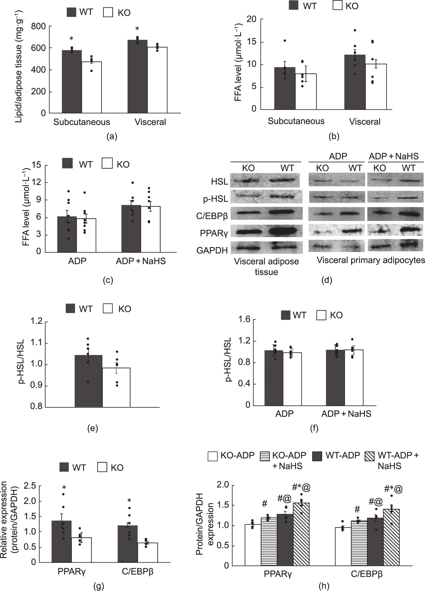
Fig. 1
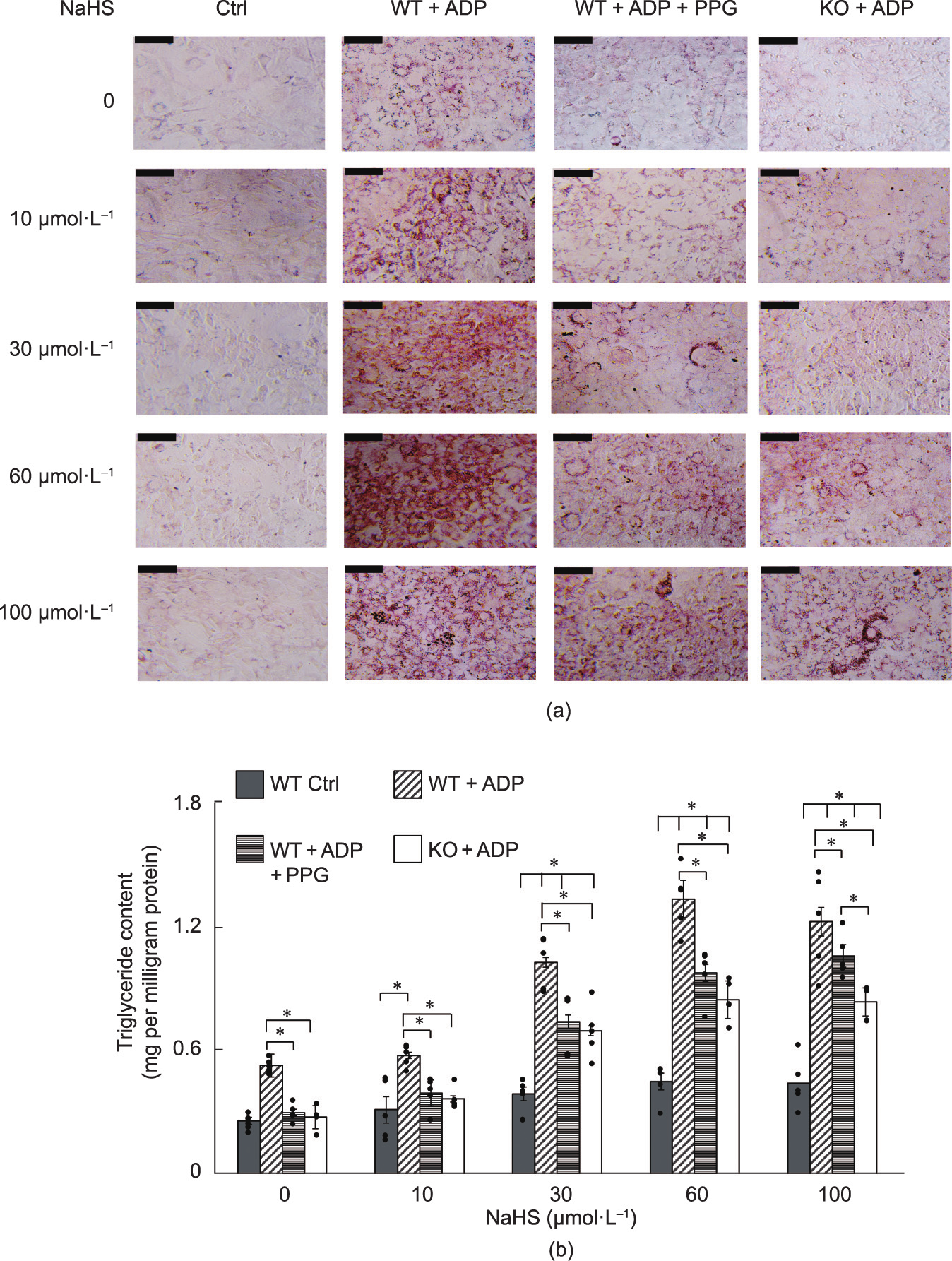
Fig. 2
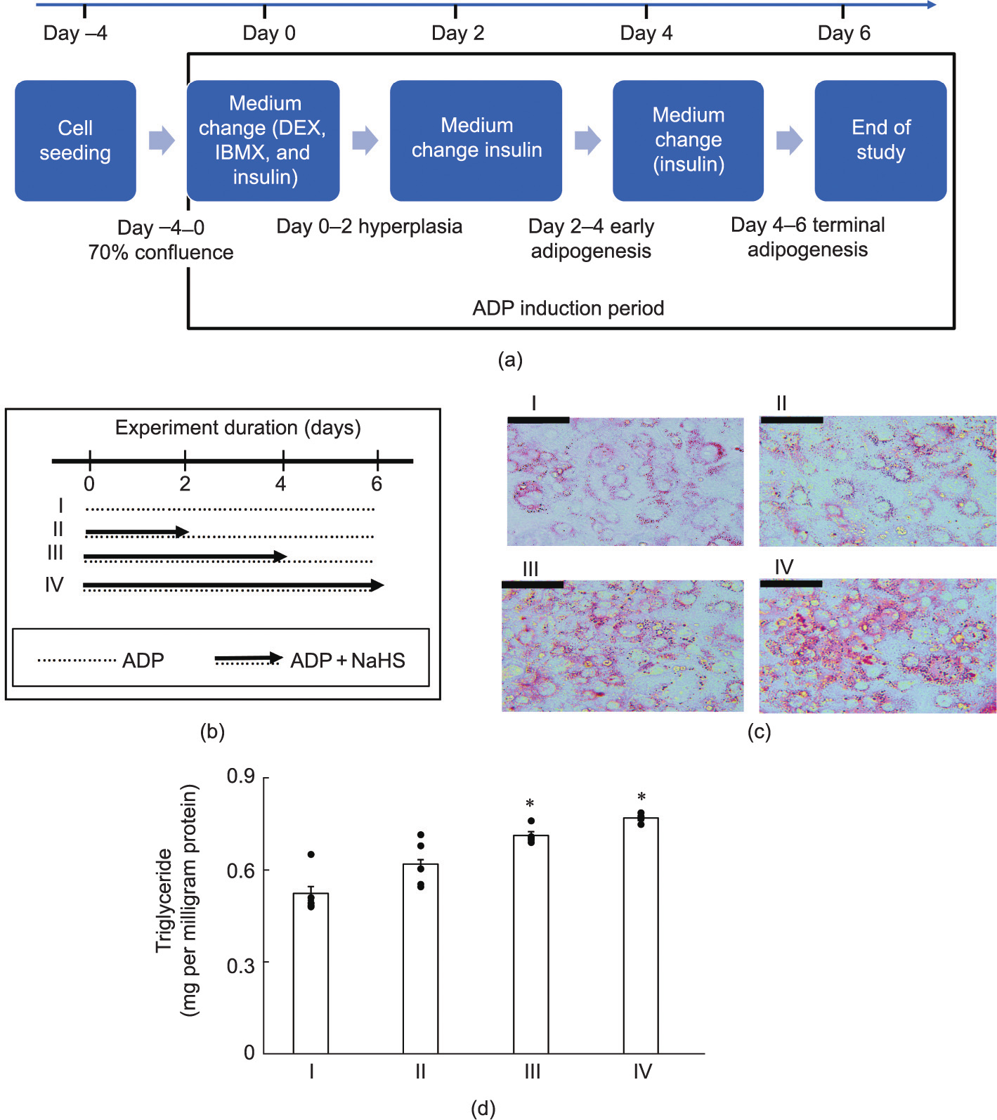
Fig. 3
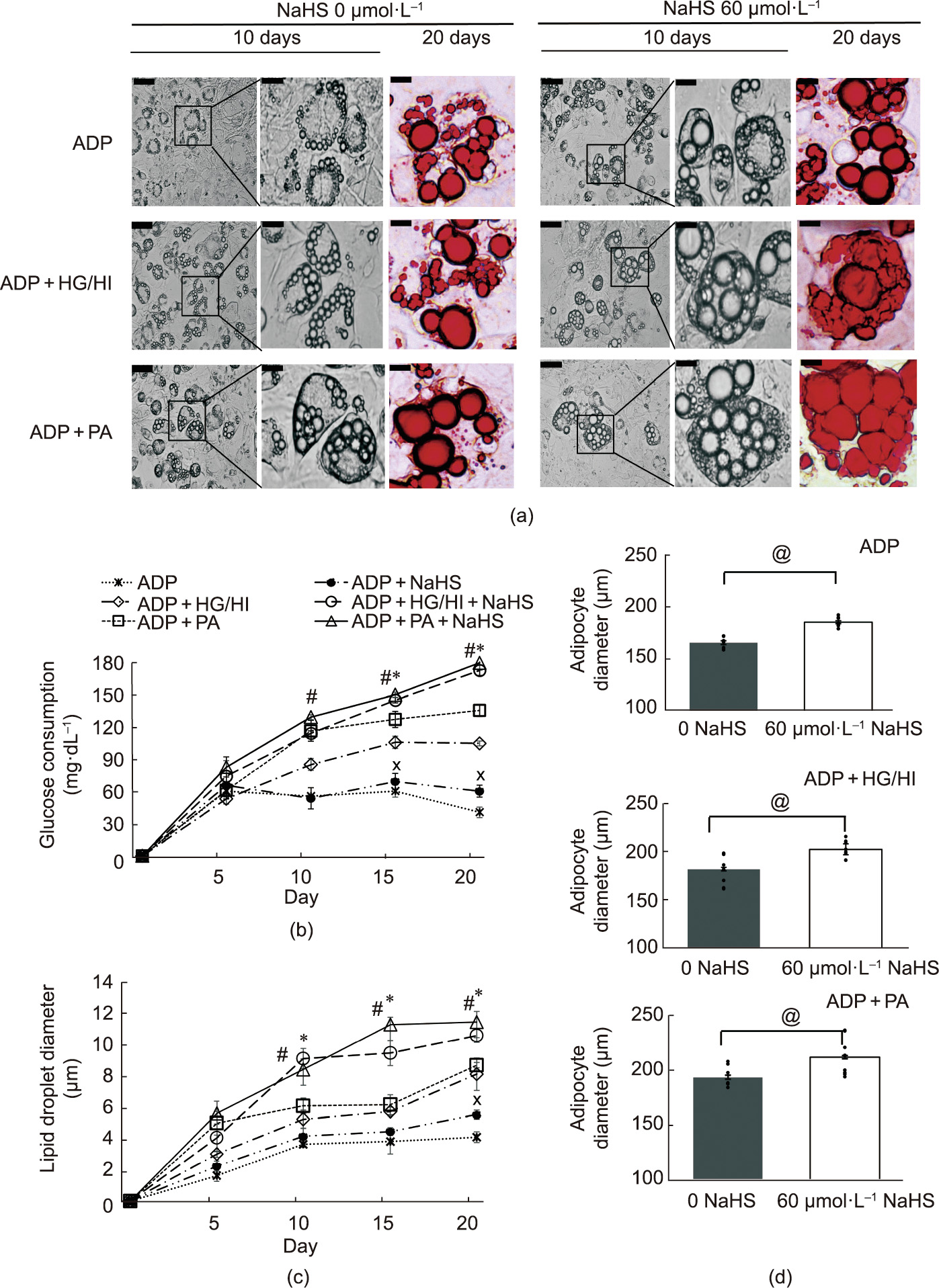
Fig. 4
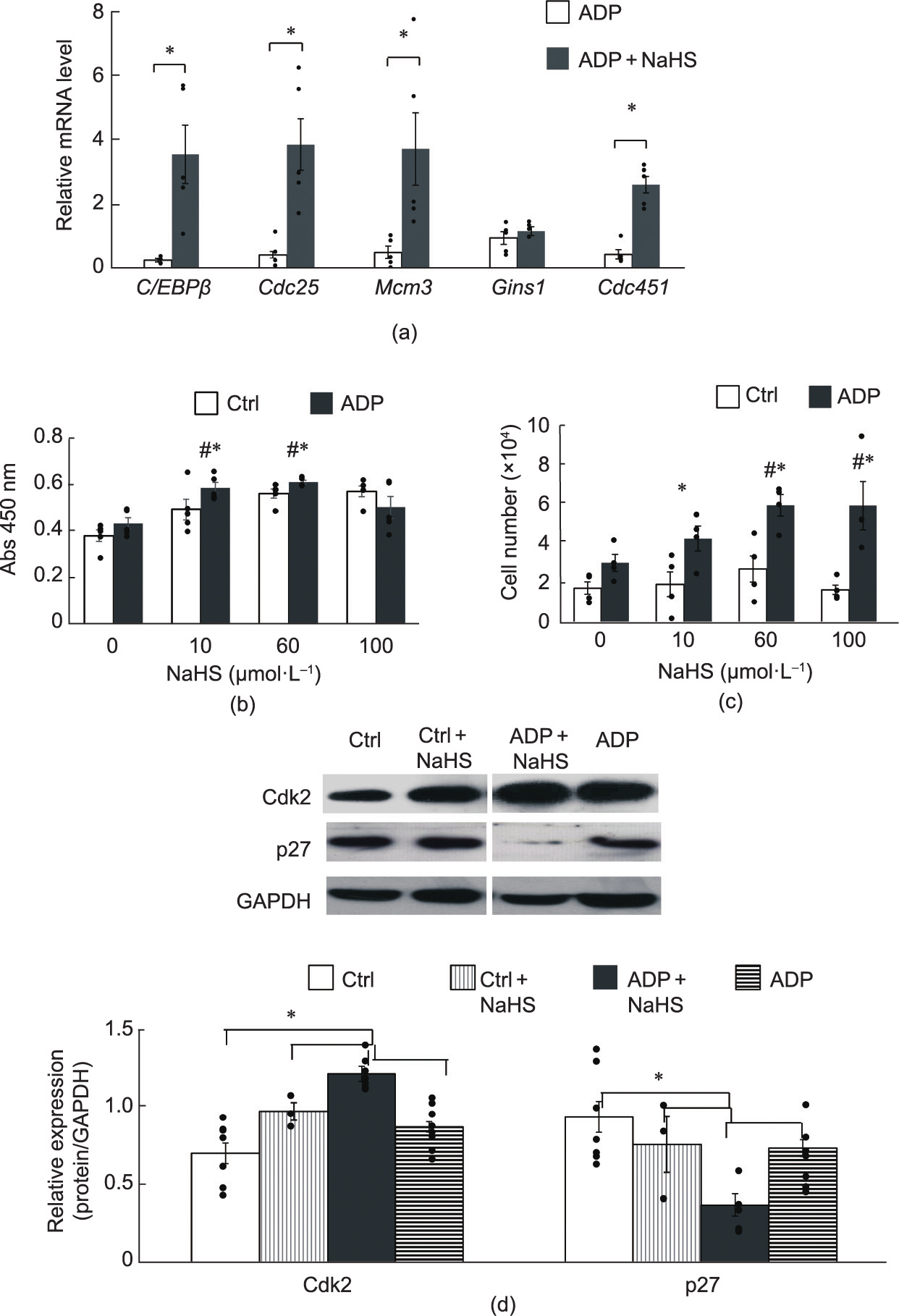
Fig. 5
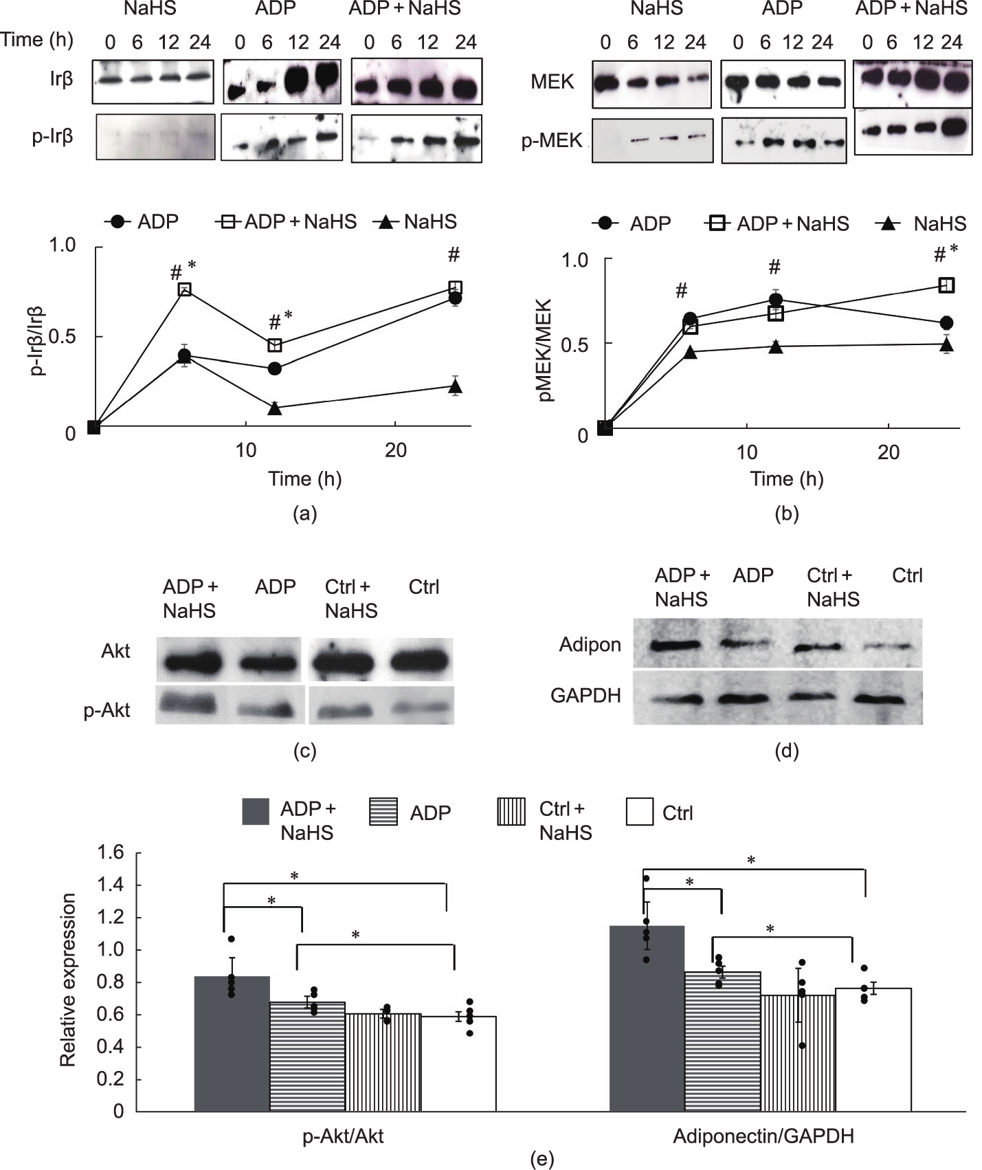
Fig. 6

Fig. 7

Fig. 8
References
[ 1 ] Wang R. Two’s company, three’s a crowd: can H2S be the third endogenous gaseous transmitter? FASEB J 2002;16(13):1792–8. link1
[ 2 ] Wang R. Physiological implications of hydrogen sulfide: a whiff exploration that blossomed. Physiol Rev 2012;92(2):791–896. link1
[ 3 ] Wallace JL, Wang R. Hydrogen sulfide-based therapeutics: exploiting a unique but ubiquitous gasotransmitter. Nat Rev Drug Discov 2015;14(5):329–45. link1
[ 4 ] Yang G, Wu L, Jiang B, Yang W, Qi J, Cao K, et al. H2S as a physiologic vasorelaxant: hypertension in mice with deletion of cystathionine c-lyase. Science 2008;322(5901):587–90. link1
[ 5 ] Sun Y, Huang Y, Zhang R, Chen Q, Chen J, Zong Y, et al. Hydrogen sulfide upregulates KATP channel expression in vascular smooth muscle cells of spontaneously hypertensive rats. J Mol Med (Berl) 2015;93(4):439–55. link1
[ 6 ] Guo C, Liang F, Shah Masood W, Yan X. Hydrogen sulfide protected gastric epithelial cell from ischemia/reperfusion injury by Keap1 S-sulfhydration, MAPK dependent anti-apoptosis and NF-jB dependent anti-inflammation pathway. Eur J Pharmacol 2014;725(725):70–8. link1
[ 7 ] Du J, Huang Y, Yan H, Zhang Q, Zhao M, Zhu M, et al. Hydrogen sulfide suppresses oxidized low-density lipoprotein (ox-LDL)-stimulated monocyte chemoattractant protein 1 generation from macrophages via the nuclear factor jB (NF-jB) pathway. J Biol Chem 2014;289(14):9741–53. link1
[ 8 ] Zheng J, Zhao T, Yuan Y, Hu N, Tang X. Hydrogen sulfide (H2S) attenuates uranium-induced acute nephrotoxicity through oxidative stress and inflammatory response via Nrf2–NF-jB pathways. Chem Biol Interact 2015;242:353–62. link1
[ 9 ] Wu L, Yang W, Jia X, Yang G, Duridanova D, Cao K, et al. Pancreatic islet overproduction of H2S and suppressed insulin release in Zucker diabetic rats. Lab Invest 2009;89(1):59–67. link1
[10] World Health Organization. Obesity and overweight [Internet]. Geneva: WHO; 2021 Jun 9 [cited on 2021 Nov 14]. Available from: https://www.who.int/ news-room/fact-sheets/detail/obesity-and-overweight. link1
[11] Trayhurn P. Adipocyte biology. Clinical obesity in adults and children. 3rd ed. Singapore: Wiley-Blackwell; 2010. link1
[12] Lee MJ, Wu Y, Fried SK. A modified protocol to maximize differentiation of human preadipocytes and improve metabolic phenotypes. Obesity 2012;20 (12):2334–40. link1
[13] Tsai CY, Peh MT, Feng W, Dymock BW, Moore PK. Hydrogen sulfide promotes adipogenesis in 3T3L1 cells. PLoS One 2015;10(3):e0119511. link1
[14] Cai J, Shi X, Wang H, Fan J, Feng Y, Lin X, et al. Cystathionine c lyase–hydrogen sulfide increases peroxisome proliferator-activated receptor c activity by sulfhydration at C139 site thereby promoting glucose uptake and lipid storage in adipocytes. Biochim Biophys Acta 2016;1861(5):419–29. link1
[15] Yang G, Ju Y, Fu M, Zhang Y, Pei Y, Racine M, et al. Cystathionine gamma-lyase/ hydrogen sulfide system is essential for adipogenesis and fat mass accumulation in mice. Biochim Biophys Acta Mol Cell Biol Lipids 2018;1863 (2):165–76. link1
[16] Khan T, Muise ES, Iyengar P, Wang ZV, Chandalia M, Abate N, et al. Metabolic dysregulation and adipose tissue fibrosis: role of collagen VI. Mol Cell Biol 2009;29(6):1575–91. link1
[17] Kim JI, Huh JY, Sohn JH, Choe SS, Lee YS, Lim CY, et al. Lipid-overloaded enlarged adipocytes provoke insulin resistance independent of inflammation. Mol Cell Biol 2015;35(10):1686–99. link1
[18] Shepherd PR, Crowther NJ, Desai M, Hales CN, Ozanne SE. Altered adipocyte properties in the offspring of protein malnourished rats. Br J Nutr 1997;78 (1):121–9. link1
[19] Longo M, Zatterale F, Naderi J, Parrillo L, Formisano P, Raciti GA, et al. Adipose tissue dysfunction as determinant of obesity-associated metabolic complications. Int J Mol Sci 2019;20(9):2358. link1
[20] Hirsch J, Fried SK, Edens NK, Leibel RL. The fat cell. Med Clin N 1989;73 (1):83–96. link1
[21] Björntorp P, Karlsson M, Pettersson P. Expansion of adipose tissue storage capacity at different ages in rats. Metabolism 1982;31(4):366–73. link1
[22] De Ferranti S, Mozaffarian D. The perfect storm: obesity, adipocyte dysfunction, and metabolic consequences. Clin Chem 2008;54(6):945–55. link1
[23] Spalding KL, Arner E, Westermark PO, Bernard S, Buchholz BA, Bergmann O, et al. Dynamics of fat cell turnover in humans. Nature 2008;453(7196):783–7. link1
[24] Pellegrinelli V, Carobbio S, Vidal-Puig A. Adipose tissue plasticity: how fat depots respond differently to pathophysiological cues. Diabetologia 2016;59 (6):1075–88. link1
[25] Muir LA, Neeley CK, Meyer KA, Baker NA, Brosius AM, Washabaugh AR, et al. Adipose tissue fibrosis, hypertrophy, and hyperplasia: correlations with diabetes in human obesity. Obesity 2016;24(3):597–605. link1
[26] Iverson SJ, Lang SL, Cooper MH. Comparison of the Bligh and Dyer and Folch methods for total lipid determination in a broad range of marine tissue. Lipids 2001;36(11):1283–7. link1
[27] Manirakiza P, Covaci A, Schepens P. Comparative study on total lipid determination using Soxhlet, Roese-Gottlieb, Bligh & Dyer, and modified Bligh & Dyer extraction methods. J Food Compos Anal 2001;14(1):93–100. link1
[28] Yang G, Tang G, Zhang L, Wu L, Wang R. The pathogenic role of cystathionine c-lyase/hydrogen sulfide in streptozotocin-induced diabetes in mice. Am J Pathol 2011;179(2):869–79. link1
[29] Yang R, Liu Y, Shi S. Hydrogen sulfide regulates homeostasis of mesenchymal stem cells and regulatory T cells. J Dent Res 2016;95(13):1445–51. link1
[30] Hausman DB, Park HJ, Hausman GJ. Isolation and culture of preadipocytes from rodent white adipose tissue. In: Adipose tissue protocols. Totowa: Humana Press; 2018. p. 201–19. link1
[31] Waldhart AN, Dykstra H, Peck AS, Boguslawski EA, Madaj ZB, Wen J, et al. Phosphorylation of TXNIP by AKT mediates acute influx of glucose in response to insulin. Cell Rep 2017;19(10):2005–13. link1
[32] Du Q, Zhang S, Li A, Mohammad IS, Liu B, Li Y. Astragaloside IV inhibits adipose lipolysis and reduces hepatic glucose production via Akt dependent PDE3B expression in HFD-fed mice. Front Physiol 2018;9:15. link1
[33] Betzi S, Alam R, Martin M, Lubbers DJ, Han H, Jakkaraj SR, et al. Discovery of a potential allosteric ligand binding site in CDK2. ACS Chem Biol 2011;6 (5):492–501. link1
[34] Lane ME, Yu B, Rice A, Lipson KE, Liang C, Sun L, et al. A novel CDK2-selective inhibitor, SU9516, induces apoptosis in colon carcinoma cells. Cancer Res 2001;61(16):6170–7. link1
[35] Ramirez-Zacarias JL, Castro-Munozledo F, Kuri-Harcuch W. Quantitation of adipose conversion and triglycerides by staining intracytoplasmic lipids with oil red O. Histochemistry 1992;97(6):493–7. link1
[36] Singh P, Garg R, Goand UK, Riyazuddin M, Reza MI, Syed AA, et al. Combination of pancreastatin inhibitor PSTi8 with metformin inhibits Fetuin-A in type 2 diabetic mice. Heliyon 2020;6(10):e05133. link1
[37] Avinash RG, Kotresh AM, Anantha Krishna LR, Shambulingappa BE, Rudresh BH, Ramesh D, et al. Association studies on biochemical parameters and uterine health in crossbred cows of central dry zone of Karnataka. J Pharm Innov 2021;10(12):1740–3. link1
[38] Rao X, Huang X, Zhou Z, Lin X. An improvement of the 2ˆ(–delta delta CT) method for quantitative real-time polymerase chain reaction data analysis. Biostat Bioinforma Biomath 2013;3(3):71–85. link1
[39] Kraemer FB, Shen WJ. Hormone-sensitive lipase knockouts. Nutr Metab 2006;3 (1):12. link1
[40] Auwerx J, Martin G, Guerre-Millo M, Staels B. Transcription, adipocyte differentiation, and obesity. J Mol Med 1996;74(7):347–52. link1
[41] Bost F, Aouadi M, Caron L, Binétruy B. The role of MAPKs in adipocyte differentiation and obesity. Biochimie 2005;87(1):51–6. link1
[42] Gregoire FM, Smas CM, Sul HS. Understanding adipocyte differentiation. Physiol Rev 1998;78(3):783–809. link1
[43] Fang L, Zhao J, Chen Y, Ma T, Xu G, Tang C, et al. Hydrogen sulfide derived from periadventitial adipose tissue is a vasodilator. J Hypertens 2009;27 (11):2174–85. link1
[44] Feng X, Chen Y, Zhao J, Tang C, Jiang Z, Geng B. Hydrogen sulfide from adipose tissue is a novel insulin resistance regulator. Biochem Biophys Res Commun 2009;380(1):153–9. link1
[45] Galmozzi A, Kok BP, Saez E. Isolation and differentiation of primary white and brown preadipocytes from newborn mice. J Vis Exp 2021;167(167):e62005. link1
[46] Ruiz-Ojeda FJ, Rupérez AI, Gomez-Llorente C, Gil A, Aguilera CM. Cell models and their application for studying adipogenic differentiation in relation to obesity: a review. Int J Mol Sci 2016;17(7):1040. link1
[47] Lee MJ, Wu Y, Fried SK. Adipose tissue heterogeneity: implication of depot differences in adipose tissue for obesity complications. Mol Aspects Med 2013;34(1):1–11. link1
[48] Chu DT, Malinowska E, Gawronska-Kozak B, Kozak LP. Expression of adipocyte biomarkers in a primary cell culture models reflects preweaning adipobiology. J Biol Chem 2014;289(26):18478–88. link1
[49] Xue R, Hao DD, Sun JP, Li WW, Zhao MM, Li XH, et al. Hydrogen sulfide treatment promotes glucose uptake by increasing insulin receptor sensitivity and ameliorates kidney lesions in type 2 diabetes. Antioxid Redox Signal 2013;19(1):5–23. link1
[50] Patel YM, Lane MD. Mitotic clonal expansion during preadipocyte differentiation: calpain-mediated turnover of p27. J Biol Chem 2000;275 (23):17653–60. link1
[51] Drolet R, Richard C, Sniderman AD, Mailloux J, Fortier M, Huot C, et al. Hypertrophy and hyperplasia of abdominal adipose tissues in women. Int J Obes 2008;32(2):283–91. link1
[52] Jo J, Gavrilova O, Pack S, Jou W, Mullen S, Sumner AE, et al. Hypertrophy and/or hyperplasia: dynamics of adipose tissue growth. PLoS Comput Biol 2009;5(3): e1000324. link1
[53] Rosen ED, Spiegelman BM. Molecular regulation of adipogenesis. Annu Rev Cell Dev Biol 2000;16(1):145–71. link1
[54] Prusty D, Park BH, Davis KE, Farmer SR. Activation of MEK/ERK signaling promotes adipogenesis by enhancing peroxisome proliferator-activated receptor c (PPARc) and C/EBPa gene expression during the differentiation of 3T3-L1 preadipocytes. J Biol Chem 2002;277(48):46226–32. link1
[55] Tang QQ, Otto TC, Lane MD. Mitotic clonal expansion: a synchronous process required for adipogenesis. Proc Natl Acad Sci USA 2003;100(1):44–9. link1
[56] Zhang YY, Li SF, Qian SW, Zhang YY, Liu Y, Tang QQ, et al. Phosphorylation prevents C/EBPb from the calpain-dependent degradation. Biochem Biophys Res Commun 2012;419(3):550–5. link1
[57] Rangwala SM, Lazar MA. Transcriptional control of adipogenesis. Annu Rev Nutr 2000;20(1):535–59. link1
[58] Cottineau J, Kottemann MC, Lach FP, Kang YH, Vély F, Deenick EK, et al. Inherited Gins1 deficiency underlies growth retardation along with neutropenia and NK cell deficiency. J Clin Invest 2017;127(5):1991–2006. link1
[59] Seo YS, Kang YH. The human replicative helicase, the CMG complex, as a target for anti-cancer therapy. Front Mol Biosci 2018;5:26. link1
[60] Guo L, Li X, Huang JX, Huang HY, Zhang YY, Qian SW, et al. Histone demethylase Kdm4b functions as a co-factor of C/EBPb to promote mitotic clonal expansion during differentiation of 3T3-L1 preadipocytes. Cell Death Differ 2012;19(12):1917–27. link1
[61] Zhao Y, Wei H, Kong G, Shim W, Zhang G. Hydrogen sulfide augments the proliferation and survival of human induced pluripotent stem cell-derived mesenchymal stromal cells through inhibition of BKCa. Cytotherapy 2013;15 (11):1395–405. link1
[62] Bian JS, Yong QC, Pan TT, Feng ZN, Ali MY, Zhou S, et al. Role of hydrogen sulfide in the cardioprotection caused by ischemic preconditioning in the rat heart and cardiac myocytes. J Pharmacol Exp Ther 2006;316(2):670–8. link1
[63] Pyiochou A, Papapetropoulos A, Olah G, Wintner E, Jeschke M, Branski L, et al. The hydrogen sulfide donor IK-1001 stimulates neovascularization and improves wound healing. FASEB J 2008;22(1):912–42. link1
[64] Papapetropoulos A, Pyriochou A, Altaany Z, Yang G, Marazioti A, Zhou Z, et al. Hydrogen sulfide is an endogenous stimulator of angiogenesis. Proc Natl Acad Sci USA 2009;106(51):21972–7. link1
[65] Shao M, Vishvanath L, Busbuso NC, Hepler C, Shan B, Sharma AX, et al. De novo adipocyte differentiation from Pdgfrb+ preadipocytes protects against pathologic visceral adipose expansion in obesity. Nat Commun 2018;9 (1):1–16. link1
[66] Saltiel AR, Kahn CR. Insulin signalling and the regulation of glucose and lipid metabolism. Nature 2001;414(6865):799–806. link1
[67] Hernandez R, Teruel T, Lorenzo M. Akt mediates insulin induction of glucose uptake and up-regulation of GLUT4 gene expression in brown adipocytes. FEBS Lett 2001;494(3):225–31. link1
[68] Kotani K, Ogawa W, Matsumoto M, Kitamura T, Sakaue H, Hino Y, et al. Requirement of atypical protein kinase Ck for insulin stimulation of glucose uptake but not for Akt activation in 3T3-L1 adipocytes. Mol Cell Biol 1998;18 (12):6971–82. link1
[69] Manna P, Jain SK. Vitamin D up-regulates glucose transporter 4 (GLUT4) translocation and glucose utilization mediated by cystathionine-c-lyase (CSE) activation and H2S formation in 3T3L1 adipocytes. J Biol Chem 2012;287 (50):42324–32. link1


 京公网安备 11010502051620号


